. 2001:1:7.
doi: 10.1186/1471-2210-1-7. Epub 2001 Oct 2.

Effect of a short-term in vitro exposure to the marine toxin domoic acid on viability, tumor necrosis factor-alpha, matrix metalloproteinase-9 and superoxide anion release by rat neonatal microglia

Affiliations

Effect of a short-term in vitro exposure to the marine toxin domoic acid on viability, tumor necrosis factor-alpha, matrix metalloproteinase-9 and superoxide anion release by rat neonatal microglia

A M Mayer et al. BMC Pharmacol. 2001.

Abstract

Background: The excitatory amino acid domoic acid, a glutamate and kainic acid analog, is the causative agent of amnesic shellfish poisoning in humans. No studies to our knowledge have investigated the potential contribution to short-term neurotoxicity of the brain microglia, a cell type that constitutes circa 10% of the total glial population in the brain. We tested the hypothesis that a short-term in vitro exposure to domoic acid, might lead to the activation of rat neonatal microglia and the concomitant release of the putative neurotoxic mediators tumor necrosis factor-alpha (TNF-alpha), matrix metalloproteinases-2 and-9 (MMP-2 and -9) and superoxide anion (O2-).

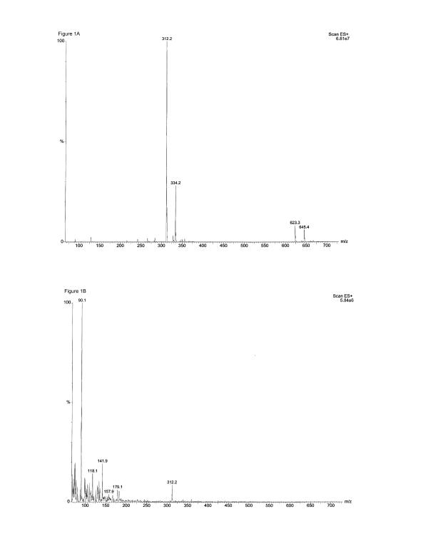
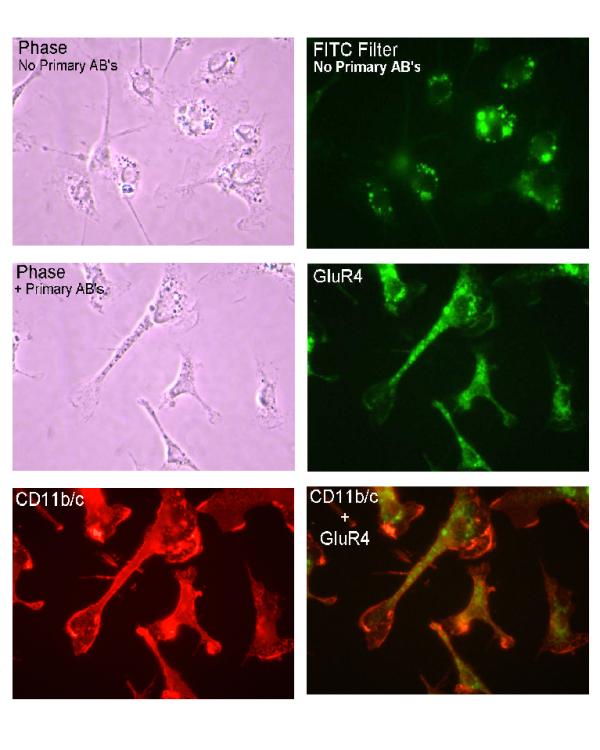
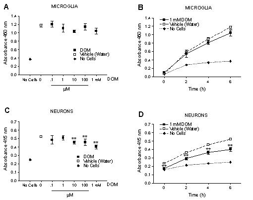
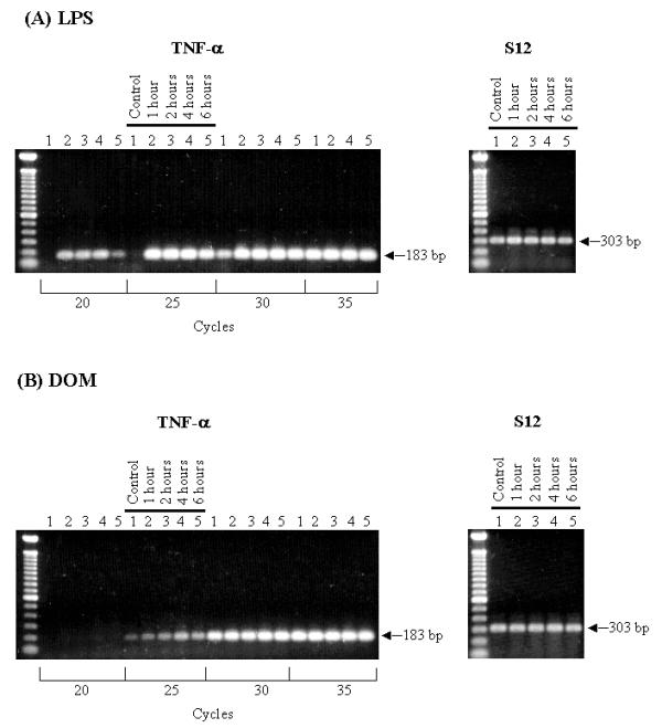
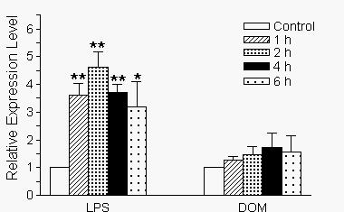
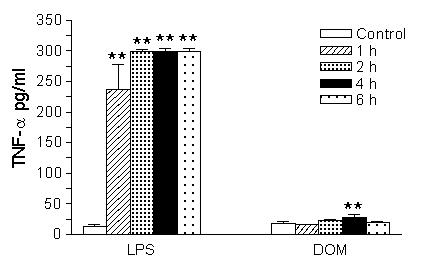
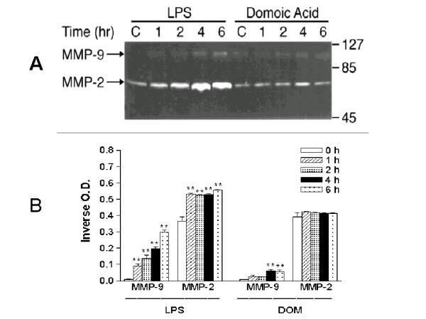
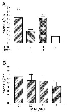
Results: In vitro, domoic acid [10 microM-1 mM] was significantly neurotoxic to primary cerebellar granule neurons. Although neonatal rat microglia expressed ionotropic glutamate GluR4 receptors, exposure during 6 hours to domoic acid [10 microM-1 mM] had no significant effect on viability. By four hours, LPS (10 ng/mL) stimulated an increase in TNF-alpha mRNA and a 2,233 % increase in TNF-alpha protein In contrast, domoic acid (1 mM) induced a slight rise in TNF-alpha expression and a 53 % increase (p < 0.01) of immunoreactive TNF-alpha protein. Furthermore, though less potent than LPS, a 4-hour treatment with domoic acid (1 mM) yielded a 757% (p < 0.01) increase in MMP-9 release, but had no effect on MMP-2. Finally, while PMA (phorbol 12-myristate 13-acetate) stimulated O2- generation was elevated in 6 hour LPS-primed microglia, a similar pretreatment with domoic acid (1 mM) did not prime O2- release.

Conclusions: To our knowledge this is the first experimental evidence that domoic acid, at in vitro concentrations that are toxic to neuronal cells, can trigger a release of statistically significant amounts of TNF-alpha and MMP-9 by brain microglia. These observations are of considerable pathophysiological significance because domoic acid activates rat microglia several days after in vivo administration.


Figures

Figure 1
Figure 1
Determination of Domoic acid by electrospray mass spectrometry.(A) The electrospray mass spectrometry spectrum of 12.5 mM Domoic acid shows that the dominate ion in the spectrum is m/z 312.2, the pseudomolecular ion [M+H+] of Domoic acid, demonstrating that undegraded Domoic acid was present in the 12.5 mM stock solution; (B) The electrospray mass spectrometry spectrum of 0.0025 mM Domoic acid, the highest dilution used in the experiments, also shows the presence of undegraded Domoic acid.
Figure 2
Figure 2
Immunofluorescent visualization of CD11b/c and ionotropic glutamate receptor subunit GluR4 in neonatal rat brain microglia. The integrin CD11b/c and the ionotropic glutamate receptor subunit GluR4 were visualized using the dual-labeling indirect immunofluorescent technique (See Materials and Methods). (Top left) bright field, phase contrast view of microglia incubated in the absence of the primary antibodies against GluR4 and CD11b/c. (Top right) same microglia viewed using FITC filter configuration. Note vesicular structures exhibiting intense autofluorescence, but little or no fluorescence over most of the microglia cell bodies or the elongated processes. (Middle left) bright field, phase contrast view of microglia labeled with anti-glutamate receptor GluR4 subunit and the leukocyte marker CD11b/c. (Middle right)(green) same field viewed using the FITC filter configuration. Microglia not only contain fluorescent vesicles, but exhibit a prominent speckled pattern of fluorescence over the cell bodies and the elongated processes. Absence of speckled pattern of labeling in cells incubated without primary GluR4 antibody (compare with top right photo) suggests the speckled pattern represents specific labeling of GluR4 subunit. (Lower left) same microglia exhibited intense labeling of CD11b/c (red), which confirms microglia are of leukocytic origin. (Bottom right) GluR4 and CD11b/c superimposed labeling (yellow). Approximate magnification of the original was × 525.
Figure 3
Figure 3
Effect of a 6 hour exposure of Domoic acid on neonatal rat brain microglia and primary cerebellar granule cell viability as determined by the WST-1 assay (See Materials and Methods). Domoic acid did not affect viability of neonatal rat brain microglia in either a (A) concentration or (B) time-dependent manner. Domoic acid [10 μM – 1 mM] significantly affected viability of primary cerebellar granule cells in both a (C) concentration and (D) time-dependent manner. Data (absorbance of formazan formation at 450 nM) are expressed as mean ± SD of values obtained from one representative experiment (n = 4–8). *p < 0.05 vs vehicle control.
Figure 4
Figure 4
RT-PCR transcript analysis of TNF-α gene expression. Neonatal rat brain microglia (2.8–5 × 10⁶ cells/culture dish) were treated with (A) LPS [10 ng/mL] or (B) Domoic acid [1 mM] for 1 to 6 hours. (See Materials and Methods). Amplification of TNF-α and S12 genes shows the predicted fragment size after separation on a 1.5 % agarose gel and visualization by SYBRR Gold nucleic acid staining. The S12 rRNA gene product was amplified for 25 cycles and demonstrates equal loading of the gels. Quantification of the TNF-α product was done after 20 cycles (LPS) or 25 cycles (Domoic acid). The cycle numbers used for quantification were shown to be in the linear range.
Figure 5
Figure 5
Alterations in the time-dependent expression levels of TNF-α and ribosomal S12 genes in neonatal rat brain microglia after treatment with LPS or Domoic acid. Levels of gene expression were quantified as described in Materials and Methods and were normalized to sl2 rRNA. Relative expression levels were calculated by dividing the experimental level at each time point by the level observed in the control. Expression is relative to steady-state levels in control (1.00). Data (relative expression level) is expressed as mean ± SE of 3 independent experiments. *p < 0.05 or **p < 0.01 vs. vehicle control.
Figure 6
Figure 6
The time-dependent effect of LPS and Domoic acid on neonatal rat brain microglia TNF-α release. Neonatal rat brain microglia (2.8–5 × 10⁶ cells/culture dish) were treated with Domoic acid [1 mM] or LPS [10 ng/mL] for 1–6 hours. TNF-α was determined as described under Materials and Methods. Data (pg/mL) are expressed as mean ± SE of 2 independent experiments. **p < 0.01 vs. vehicle control.
Figure 7
Figure 7
The time-course of MMP-2 and MMP-9 expression in neonatal rat brain microglia cultured in the presence of LPS or Domoic acid. MMP-2 and MMP-9 were determined as described in Materials and Methods. (A) SDS-PAGE zymography (B) bar-graph depicting the quantitated results. Neonatal rat brain microglia (2.8–5 × 10⁶ cells/culture dish) were treated with LPS [10 ng/ml] or Domoic acid [1 mM] for 1–6 hours. Data (inverse o.d. units) are expressed as mean ± SE of values obtained from 3 independent experiments. **p < 0.01 vs. vehicle control.
Figure 8
Figure 8
The effect of LPS and Domoic acid on neonatal rat brain microglia O2- release. O2- generation was determined as described in Materials and Methods. (A) Neonatal rat brain microglia (250,000 cells/well) were pretreated with LPS [10 ng/mL] and/ or Domoic acid [1 mM] for 6 hours and then stimulated with PMA [1 μM] for 2 hours. (B) Neonatal rat brain microglia (250,000 cells/well) were first pretreated with LPS [10 ng/mL] for 6 hours and then stimulated with PMA [1 μM] ± Domoic acid [0–1 mM] for 2 hours. Data (O2- nanomoles/2 hours) are expressed as mean ± SD of values obtained from three culture wells of one representative experiment. **p < 0.01 vs. untreated control.

References

    1. Lassmann H, Schmied M, Vass K, Hickey WF. Bone marrow derived elements and resident microglia in brain inflammation. GLIA. 1993;7:19–24. - PubMed
    1. Gehrmann J, Matsumoto Y, Kreutzberg GW. Microglia: intrinsic immuneffector cell of the brain. Brain Res Brain Res Rev. 1995;20:269–287. doi: 10.1016/0165-0173(94)00015-H. - DOI - PubMed
    1. Mayer AM. Therapeutic implications of microglia activation by lipopolysaccharide and reactive oxygen species generation in septic shock and central nervous system pathologies: a review. Medicina (B Aires) 1998;58:377–385. - PubMed
    1. Perry VH, Gordon S. Macrophages and the nervous system. Int Rev Cytol. 1991;125:203–244. - PubMed
    1. Flavin MP, Zhao G, Ho LT. Microglial tissue plasminogen activator (tPA) triggers neuronal apoptosis in vitro. GLIA. 2000;29:347–354. doi: 10.1002/(SICI)1098-1136(20000215)29:4<347::AID-GLIA5>3.0.CO;2-8. - DOI - PubMed

Publication types

MeSH terms

Associated data

LinkOut - more resources