

Early events in macrophage killing of Aspergillus fumigatus conidia: new flow cytometric viability assay
- PMID: 11687470
- PMCID: PMC96256
- DOI: 10.1128/CDLI.8.6.1240-1247.2001
Early events in macrophage killing of Aspergillus fumigatus conidia: new flow cytometric viability assay
Abstract
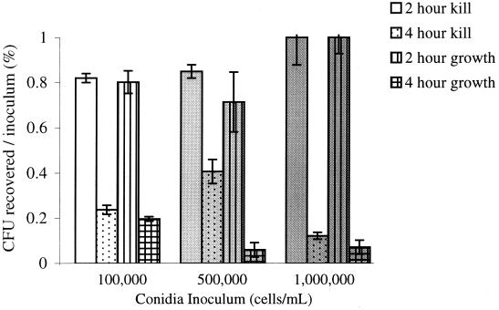
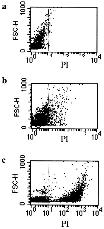
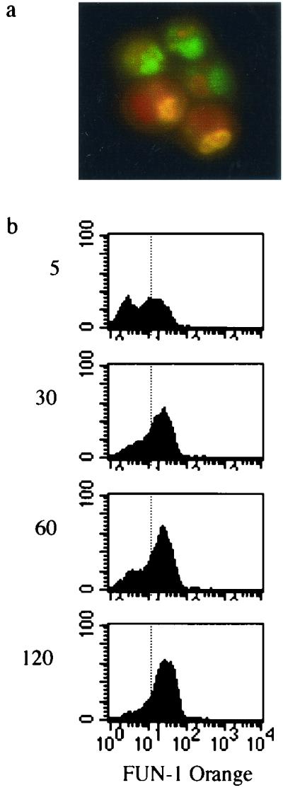
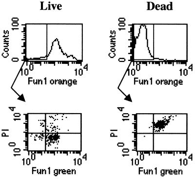
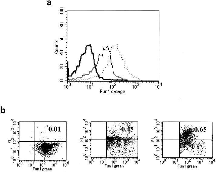
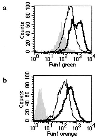
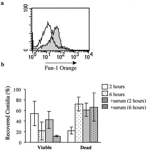
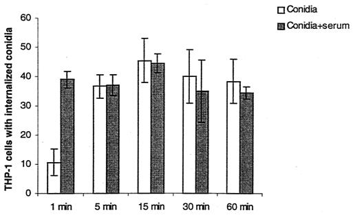
Detailed investigations of macrophage phagocytosis and killing of Aspergillus fumigatus conidia have been limited by technical difficulties in quantifying fungal uptake and viability. In order to study early events in cell pathogen ingestion and killing, we developed a new flow cytometry assay that utilizes the fungus-specific viability dye FUN-1. Metabolically active A. fumigatus conidia accumulate orange fluorescence in vacuoles, while dormant or dead conidia stain green. After incubation within THP-1 cells, recovered conidia are costained with propidium iodide (PI) to discriminate between dormant and dead cells. Flow cytometric measurements of FUN-1 metabolism and PI uptake provide indicators of conidial viability, dormancy, and death. Conidial phagocytosis and killing are also assessed by measurement of green and orange FUN-1 fluorescence within the THP-1 cell population. Compared to previously described methods, this assay has less error introduced by membrane permeability changes and serial dilution of filamentous fungal forms. Results suggest that the THP-1 cells kill conidia rapidly (within 6 h) after exposure. Conidia that are preexposed to human serum are ingested and killed more quickly than are nonopsonized conidia.
Figures
Similar articles
-
Characterisation of the phagocytic uptake of Aspergillus fumigatus conidia by macrophages.Microbes Infect. 2008 Feb;10(2):175-84. doi: 10.1016/j.micinf.2007.11.001. Epub 2007 Nov 17. Microbes Infect. 2008. PMID: 18248765
-
Accumulation of amphotericin B in human macrophages enhances activity against Aspergillus fumigatus conidia: quantification of conidial kill at the single-cell level.Antimicrob Agents Chemother. 1998 Oct;42(10):2569-75. doi: 10.1128/AAC.42.10.2569. Antimicrob Agents Chemother. 1998. PMID: 9756757 Free PMC article.
-
Fast and Quantitative Evaluation of Human Leukocyte Interaction with Aspergillus fumigatus Conidia by Flow Cytometry.Cytometry A. 2019 Mar;95(3):332-338. doi: 10.1002/cyto.a.23653. Epub 2018 Nov 19. Cytometry A. 2019. PMID: 30450827
-
Intracellular and extracellular growth of Aspergillus fumigatus.Med Mycol. 2005 May;43 Suppl 1:S27-30. doi: 10.1080/13693780400029247. Med Mycol. 2005. PMID: 16110789 Review.
-
Phagocyte responses towards Aspergillus fumigatus.Int J Med Microbiol. 2011 Jun;301(5):436-44. doi: 10.1016/j.ijmm.2011.04.012. Epub 2011 May 14. Int J Med Microbiol. 2011. PMID: 21571589 Review.
Cited by
-
Conidial viability assay for rapid susceptibility testing of Aspergillus species.J Clin Microbiol. 2002 Aug;40(8):2741-5. doi: 10.1128/JCM.40.8.2741-2745.2002. J Clin Microbiol. 2002. PMID: 12149322 Free PMC article.
-
Development of an in vitro model for the multi-parametric quantification of the cellular interactions between Candida yeasts and phagocytes.PLoS One. 2012;7(3):e32621. doi: 10.1371/journal.pone.0032621. Epub 2012 Mar 30. PLoS One. 2012. PMID: 22479332 Free PMC article.
-
Impaired bactericidal but not fungicidal activity of polymorphonuclear neutrophils in patients with chronic lymphocytic leukemia.Leuk Lymphoma. 2013 Aug;54(8):1730-3. doi: 10.3109/10428194.2012.750723. Epub 2013 Feb 20. Leuk Lymphoma. 2013. PMID: 23163595 Free PMC article.
-
Immunostimulatory effects of the anionic alkali mineral complex Barodon on equine lymphocytes.Clin Vaccine Immunol. 2006 Nov;13(11):1255-66. doi: 10.1128/CVI.00150-06. Epub 2006 Aug 30. Clin Vaccine Immunol. 2006. PMID: 16943344 Free PMC article.
-
Persistence versus escape: Aspergillus terreus and Aspergillus fumigatus employ different strategies during interactions with macrophages.PLoS One. 2012;7(2):e31223. doi: 10.1371/journal.pone.0031223. Epub 2012 Feb 3. PLoS One. 2012. PMID: 22319619 Free PMC article.
References
-
- Epstein J, Eichbaum Q, Sheriff S, Ezekowitz R A. The collectins in innate immunity. Curr Opin Immunol. 1996;8:29–35. - PubMed
Publication types
MeSH terms
Grants and funding
LinkOut - more resources
Full Text Sources
Research Materials
Miscellaneous
