Review
. 2001:17:463-516.
doi: 10.1146/annurev.cellbio.17.1.463.

How matrix metalloproteinases regulate cell behavior

Affiliations
Review

How matrix metalloproteinases regulate cell behavior

M D Sternlicht et al. Annu Rev Cell Dev Biol. 2001.

Abstract

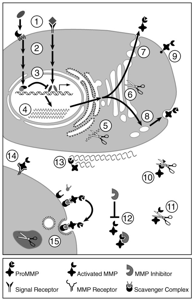
The matrix metalloproteinases (MMPs) constitute a multigene family of over 25 secreted and cell surface enzymes that process or degrade numerous pericellular substrates. Their targets include other proteinases, proteinase inhibitors, clotting factors, chemotactic molecules, latent growth factors, growth factor-binding proteins, cell surface receptors, cell-cell adhesion molecules, and virtually all structural extracellular matrix proteins. Thus MMPs are able to regulate many biologic processes and are closely regulated themselves. We review recent advances that help to explain how MMPs work, how they are controlled, and how they influence biologic behavior. These advances shed light on how the structure and function of the MMPs are related and on how their transcription, secretion, activation, inhibition, localization, and clearance are controlled. MMPs participate in numerous normal and abnormal processes, and there are new insights into the key substrates and mechanisms responsible for regulating some of these processes in vivo. Our knowledge in the field of MMP biology is rapidly expanding, yet we still do not fully understand how these enzymes regulate most processes of development, homeostasis, and disease.


Figures

Figure 1
Figure 1
Domain structure of the MMPs. Pre, signal sequence; Pro, propeptide with a free zinc-ligating thiol (SH) group; F, furin-susceptible site; Zn, zinc-binding site; II, collagen-binding fibronectin type II inserts; H, hinge region; TM, transmembrane domain; C, cytoplasmic tail; GPI, glycophosphatidyl inositol-anchoring domain; C/P, cysteine/proline; IL-1R, interleukin-1 receptor. The hemopexin/vitronectin-like domain contains four repeats with the first and last linked by a disulfide bond.
Figure 2
Figure 2
Regulation of the MMPs. MMP regulatory mechanisms include inductive and suppressive signaling (1), intracellular signal transduction (2), transcriptional activation and repression (3), post-transcriptional mRNA processing (4), mRNA degradation (5), intracellular activation of furin-susceptible MMPs (6), constitutive secretion (7), regulated secretion (8), cell surface expression (9), proteolytic activation (10), proteolytic processing and inactivation (11), protein inhibition (12), ECM localization (13), cell surface localization (14), and endocytosis and intracellular degradation (15).
Figure 3
Figure 3
Cell surface activation of MMP2. A ProMT-MMP is activated during transport to the cell surface by an intracellular furin-like serine proteinase, at the cell surface by plasmin, or by non-proteolytic conformational changes. The activated MT-MMP is then inhibited by TIMP2 and the hemopexin domain of ProMMP2 binds to the C-terminal portion of TIMP2 to form a trimolecular complex. An uninhibited MT-MMP then partially activates the ProMMP2 by removing most of the MMP2 propeptide. The remaining portion of the propeptide is removed by a separate MMP2 molecule at the cell surface to yield fully active mature MMP2. Mature MMP2 can then be released from the cell surface or bound by another cell surface MMP2-docking protein. It can also be inhibited by another TIMP molecule or left in an uninhibited active state depending on local MMP:TIMP molar ratios.
Figure 4
Figure 4
Potential mechanisms of MMP-mediated cellular signaling.
Figure 5
Figure 5
BP180 cleavage in bullous pemphigoid. Infiltrating neutrophils (PMNs) in and around bullous pemphigoid lesions release substantial amounts of neutrophil elastase (NE) and gelatinase B (MMP9) from large azurophilic primary granules and smaller tertiary granules, respectively. In addition, α1-proteinase inhibitor (α1-PI) is elevated, but because it is inactivated by MMP9, its ability to inhibit NE is abolished, thus enabling NE to cleave hemidesmosomal type XVII collagen (BP180) in an unrestricted manner. Furthermore, cleaved α1-PI fragments may exacerbate this process by enhancing PMN recruitment. Although MMP9 can also cleave BP180 in vitro, its direct contribution to BP180 cleavage in vivo is minimal or non-existent.

References

    1. Abbaszade I, Liu RQ, Yang F, Rosenfeld SA, Ross OH, et al. Cloning and characterization of ADAMTS11, an aggrecanase from the ADAMTS family. J. Biol. Chem. 1999;274:23443–23450. - PubMed
    1. Alexander CM, Howard EW, Bissell MJ, Werb Z. Rescue of mammary epithelial cell apoptosis and entactin degradation by a tissue inhibitor of metalloproteinase-1 transgene. J. Cell Biol. 1996;135:1669–1677. - PMC - PubMed
    1. Alexander CM, Selvarajan S, Mudgett J, Werb Z. Stromelysin-1 regulates adipogenesis during mammary gland involution. J. Cell Biol. 2001;152:693–703. - PMC - PubMed
    1. Andrews KL, Betsuyaku T, Rogers S, Shipley JM, Senior RM, Miner JH. Gelatinase B (MMP-9) is not essential in the normal kidney and does not influence progression of renal disease in a mouse model of Alport syndrome. Am. J. Pathol. 2000;157:303–311. - PMC - PubMed
    1. Ashworth JL, Murphy G, Rock MJ, Sherratt MJ, Shapiro SD, et al. Fibrillin degradation by matrix metalloproteinases: implications for connective tissue remodelling. Biochem. J. 1999;340:171–181. - PMC - PubMed

Publication types

MeSH terms

Substances

LinkOut - more resources