. 2001 Nov 6;98(23):13060-5.
doi: 10.1073/pnas.181479798. Epub 2001 Oct 30.

Specificity in intracellular protein aggregation and inclusion body formation

Affiliations

Specificity in intracellular protein aggregation and inclusion body formation

R S Rajan et al. Proc Natl Acad Sci U S A. .

Abstract

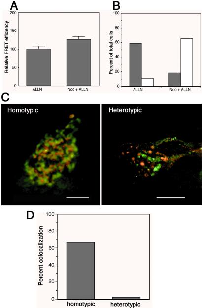
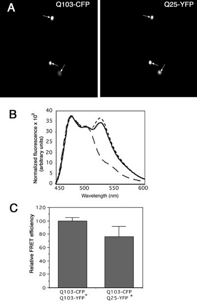
Protein aggregation is widely considered to be a nonspecific coalescence of misfolded proteins, driven by interactions between solvent-exposed hydrophobic surfaces that are normally buried within a protein's interior. Accordingly, abnormal interactions between misfolded proteins with normal cellular constituents has been proposed to underlie the toxicity associated with protein aggregates in many neurodegenerative disorders. Here we have used fluorescence resonance energy transfer and deconvolution microscopy to investigate the degree to which unrelated misfolded proteins expressed in the same cells coaggregate with one another. Our data reveal that in cells, protein aggregation exhibits exquisite specificity even among extremely hydrophobic substrates expressed at very high levels.


Figures

Figure 1
Figure 1
Colocalization of aggregated proteins in cytoplasmic inclusion bodies. (AF) Visualization of inclusion bodies in cells cotransfected with P23H-YFP and CFP-ΔF508 (A), TCRα (C), or Q103-CFP (E) by conventional epifluorescence microscopy. TCRα was detected by indirect immunofluorescence against an HA epitope bar. Inclusion bodies are denoted by arrows. (GI) Visualization of inclusion bodies by digital deconvolution microscopy. (G) P23H-YFP (red) and ΔF508-CFP (green; single cell). (H) P23H-YFP (red) and Q103 (green; single cell). (I) Q103 -CFP (red) and ΔF508-YFP (green; two cells, each with one inclusion body). All cells were treated with ALLN to maximize protein aggregation. (Scale bars: AF, 15 μm; G, 5 μm; H and I, 7.5 μm.)
Figure 2
Figure 2
Specificity of protein aggregation measured by FRET. (A) FRET in cells containing P23H inclusion bodies. Fluorescence emission spectra (excitation 425 nm) of a homogeneous suspension of cells cotransfected with equal amounts of P23H-CFP and P23H-YFP plasmid (solid line) compared with the spectrum of a suspension of a mixture of cells transfected with only P23H-CFP and cells transfected with only P23H-YFP (dashed line). Arrows indicate the emission peaks for CFP (476) and YFP (525) used to calculate the FRET value as described in the text. (B) P23H (open squares), but not ΔF508 (filled squares), can compete out FRET between P23H-CFP and P23H-YFP. The ratio of unlabeled competitor plasmid to the total labeled plasmid is plotted on the x axis. (C) Other aggregation-prone proteins (at a 4:1 plasmid ratio) do not decrease FRET between P23H-CFP and P23H-YFP.
Figure 3
Figure 3
Protein aggregation is independent of inclusion body formation. (A) Cells coexpressing P23H-CFP and P23H-YFP were treated with the proteasome inhibitor ALLN to enhance protein aggregation in the presence or absence of nocodazole to disrupt microtubules. Data represent mean ± SEM from three independent trials. (B) The fraction of cells from the experiment in A with a single inclusion body (shaded bars) or dispersed foci (open bars). A total of 400 cells were counted for each treatment group. (C) Intracellular localization (deconvolved microscopic images) of fusion proteins in dispersed foci from nocodazole + ALLN treated cells expressing the heterotypic aggregation pair YFP-ΔF508 (red) + P23H CFP (green) or the homotypic pair P23H-CFP (red) + P23H-YFP (green). (D) Quantification of colocalization in the experiment from C.
Figure 4
Figure 4
Huntingtin Q103 coaggregates with a huntingtin fragment containing a nonpathogenic polyQ tract. (A) Inclusion bodies of Q103 colocalize with Q25. (B) fluorescence emission spectra, as in Fig. 2A, from cells cotransfected with equal amounts of Q25-CFP + Q25-YFP (large dashes), Q103-CFP + Q103-YFP (small dashes), or Q103-CFP + Q25-YFP (solid line). Spectra were normalized for CFP fluorescence. (C) Quantification of FRET efficiency between huntingtin fragments containing different glutamine repeat lengths.

References

    1. Fink A L. Folding Des. 1998;3:R9–R23. - PubMed
    1. Trojanowski J Q, Lee V M. Ann NY Acad Sci. 2000;924:62–67. - PubMed
    1. Perutz M F, Johnson T, Suzuki M, Finch J T. Proc Natl Acad Sci USA. 1994;91:5355–5358. - PMC - PubMed
    1. Bruijn L I, Houseweart M K, Kato S, Anderson K L, Anderson S D, Ohama E, Reaume A G, Scott R W, Cleveland D W. Science. 1998;281:1851–1854. - PubMed
    1. Wetzel R. Cell. 1996;86:699–702. - PubMed

Publication types

Substances

LinkOut - more resources