. 2001 Nov 6;98(23):13420-4.
doi: 10.1073/pnas.221315298. Epub 2001 Oct 30.

Ouabain, a steroid hormone that signals with slow calcium oscillations

Affiliations

Ouabain, a steroid hormone that signals with slow calcium oscillations

O Aizman et al. Proc Natl Acad Sci U S A. .

Abstract

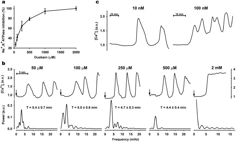
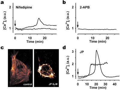
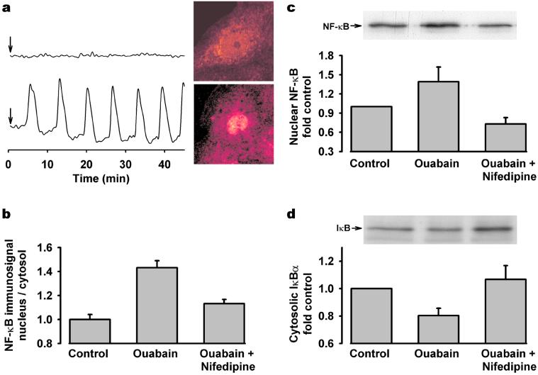
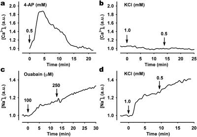
The plant-derived steroid, digoxin, a specific inhibitor of Na,K-ATPase, has been used for centuries in the treatment of heart disease. Recent studies demonstrate the presence of a digoxin analog, ouabain, in mammalian tissue, but its biological role has not been elucidated. Here, we show in renal epithelial cells that ouabain, in doses causing only partial Na,K-ATPase inhibition, acts as a biological inducer of regular, low-frequency intracellular calcium ([Ca(2+)](i)) oscillations that elicit activation of the transcription factor, NF-kappa B. Partial inhibition of Na,K-ATPase using low extracellular K(+) and depolarization of cells did not have these effects. Incubation of cells in Ca(2+)-free media, inhibition of voltage-gated calcium channels, inositol triphosphate receptor antagonism, and redistribution of actin to a thick layer adjacent to the plasma membrane abolished [Ca(2+)](i) oscillations, indicating that they were caused by a concerted action of inositol triphosphate receptors and capacitative calcium entry via plasma membrane channels. Blockade of ouabain-induced [Ca(2+)](i) oscillations prevented activation of NF-kappa B. The results demonstrate a new mechanism for steroid signaling via plasma membrane receptors and underline a novel role for the steroid hormone, ouabain, as a physiological inducer of [Ca(2+)](i) oscillations involved in transcriptional regulation in mammalian cells.


Figures

Figure 1
Figure 1
Effect of ouabain on [Ca2+]i in primary culture of RPT cells. (a) Na,K-ATPase activity measured as ouabain-sensitive 86Rb+ uptake (mean ± SE). (b Upper) Representative single cell [Ca2+]i tracings in response to indicated ouabain concentrations. At time 0 (indicated by arrow), cells were exposed to ouabain concentrations ranging from 50 μM to 2 mM, and recordings were made every 30 s. Arbitrary units (a.u.) represent ratio values corresponding to [Ca2+]i changes. (Lower) Spectral analysis of ouabain-induced [Ca2+]i oscillations. Each plot corresponds to the single-cell recording above. [Ca2+]i oscillation periodicity (T) of each ouabain concentration was calculated as mean ± SE from ≈50 cells from at least three separate experiments. (c) Representative single-cell [Ca2+]i tracings observed in cells superfused for 3 h at a slow rate (100 μl/min) with nanomolar ouabain.
Figure 2
Figure 2
Characterization of ouabain-induced [Ca2+]i response in RPT cells. Typical [Ca2+]i response in cells preincubated with 50 μM nifedipine for 5 min (a), 50 μM 2-aminoethoxydiphenyl borate for 5 min (b), and 6 μM JP for 45 min (d) before 250 μM ouabain treatment. In studies using nifedipine (a) and JP (d), two different types of responses were observed. Each experiment was repeated at least four times by using individual cell preparations. (c) Confocal microscopy images of cellular F-actin distribution in actin-GFP transiently transfected cells treated without (Left) or with (Right) 6 μM JP for 30 min. JP caused polymerization and reorganization of F-actin subjacent to the cell membrane.
Figure 3
Figure 3
Effect of [Ca2+]i oscillations on ouabain-induced NF-κB activation. (a) A cell cluster was treated with 250 μM ouabain (indicated by arrow), and individual cells were analyzed for both [Ca2+]i and NF-κB immunofluorescence. (Upper) Typical nonoscillating [Ca2+]i response (Left) and its corresponding cellular NF-κB localization (Right). (Lower) Typical oscillating [Ca2+]i response (Left) and its corresponding cellular NF-κB localization (Right). (b) Semiquantitative analysis of NF-κB immunofluorescence signal showing translocation from cytosol to nucleus in cells exposed to 250 μM ouabain, in the absence or presence of 50 μM nifedipine. Values are mean ± SE 50–150 cells. Representative Western blot and densitometric analysis of three to five experiments showing changes in (c) nuclear NF-κB and (d) cytosolic IκBα protein in cells exposed to 250 μM ouabain in the presence or absence of nifedipine.
Figure 4
Figure 4
Effect of membrane depolarization and low K+ on ouabain-mediated changes in [Ca2+]i and [Na+]i. [Ca2+]i response in cells exposed (arrow) to 5 mM 4-aminopyridine (a) or reductions in extracellular K+ (b) from 4.0 mM to 1.0 or 0.5 mM. Effect of ouabain (c) and low extracellular K+ (d) on representative single-cell [Na+]i measurements. SBFI/AM-loaded cells were treated with 100 μM and 250 μM ouabain or low extracellular K+, and ratio images were recorded every 30 s. Arbitrary units (a.u.) represent ratio values corresponding to changes in [Ca2+]i (a and b) and [Na+]i (c and d).

References

    1. Berridge M J, Bootman M D, Lipp P. Nature (London) 1998;395:645–648. - PubMed
    1. Gomez T M, Spitzer N C. Nature (London) 1999;397:350–355. - PubMed
    1. Putney J W. Science. 1998;279:191–192. - PubMed
    1. Berridge M J. Nature (London) 1997;386:759–760. - PubMed
    1. De Koninck P, Schulman H. Science. 1998;279:227–230. - PubMed

Publication types

MeSH terms

LinkOut - more resources