. 2001 Nov 6;98(23):13385-90.
doi: 10.1073/pnas.231177098. Epub 2001 Oct 30.

c-Jun mediates axotomy-induced dopamine neuron death in vivo

Affiliations

c-Jun mediates axotomy-induced dopamine neuron death in vivo

S J Crocker et al. Proc Natl Acad Sci U S A. .

Abstract

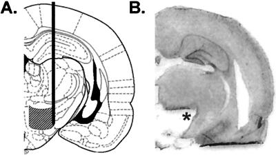
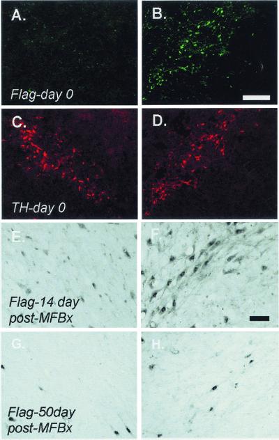
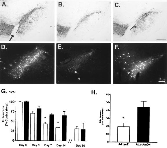
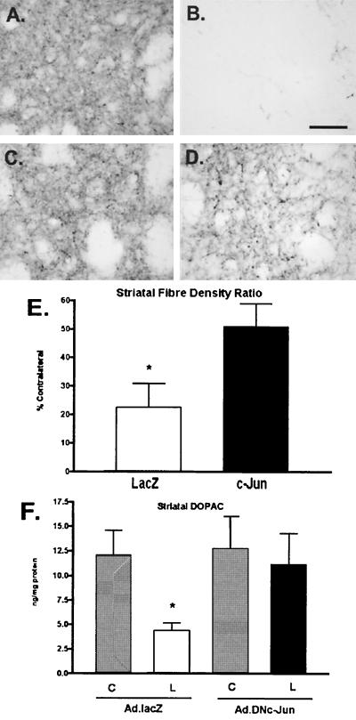
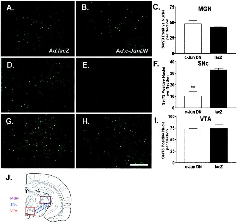
Expression of the transcription factor c-Jun is induced in neurons of the central nervous system (CNS) in response to injury. Mechanical transection of the nigrostriatal pathway at the medial forebrain bundle (MFB) results in the delayed retrograde degeneration of the dopamine neurons in the substantia nigra pars compacta (SNc) and induces protracted expression and phosphorylation of c-Jun. However, the role of c-Jun after axotomy of CNS neurons is unclear. Here, we show that adenovirus-mediated expression of a dominant negative form of c-Jun (Ad.c-JunDN) inhibited axotomy-induced dopamine neuron death and attenuated phosphorylation of c-Jun in nigral neurons. Ad.c-JunDN also delayed the degeneration of dopaminergic nigral axons in the striatum after MFB axotomy. Taken together, these findings suggest that activation of c-Jun mediates the loss of dopamine neurons after axotomy injury.


Figures

Figure 1
Figure 1
(A) Schematic line drawing of a coronal section from rat brain indicating the site and extent of mechanical injury made by the Scouten wire knife to transect the MFB. Verification of anatomical MFB transection was accomplished by hematoxylin and eosin staining of sections from every animal (−3.6 to −4.16 mm caudal to bregma; ref. 24), as demonstrated by representative section shown (B). Line drawing schematic of coronal brain section adapted from atlas of Paxinos and Watson (24).
Figure 2
Figure 2
Intrastriatal adenovirus administration results in retrograde labeling of dopamine neurons in the SNc. Expression of c-JunDN was detected 1 week later in the SNc ipsilateral to the adenovirus injection (B), but not in the contralateral hemisphere (A). Immunohistochemical detection of FLAG (Ad.c-JunDN) was predominantly in SNc neurons expressing TH (C and D). Fourteen days after MFB axotomy, FLAG immunoreactivity was still detectable in SNc neurons ipsilateral to injection of Ad.c-JunDN (F), yet by 50 days postinjury, adenovirus expression was not detectable in the SNc (H). FLAG expression was not detected in the contralateral SNc at these time points (E and G, respectively). Scale bars = 160 μm (AD) and 40 μm (EH).
Figure 3
Figure 3
Ad.c-JunDN attenuates loss of nigral dopamine neurons after MFB axotomy. Representative TH immunoreactivity in the SNc from unlesioned (A), and rats injected with either Ad.lacZ (B), or Ad.c-JunDN (C), 14 days after MFB axotomy. Arrow indicates the medial terminal nucleus (MTN), an anatomical landmark used to ensure for comparisons between sections (see Materials and Methods). Retrograde labeling of neurons in the SNc after intrastriatal injection of FluoroGold (FG) from unlesioned (D) Ad.lacZ or Ad.c-JunDN 14 days after MFB axotomy (E and F, respectively). (G) Quantification of TH neurons in the SNc at varying times after MFB axotomy: Ad.lacZ (white bars), Ad.c-JunDN (black bars) (P < 0.0001, ANOVA; *, P < 0.001, Newman-Keuls Multiple Comparison test; n = 3–4 per group, days 0–7, 50, n = 6 day 14). (H) Quantification of FG-labeled neurons in the SNc confirmed increased survival of SNc neurons after MFB axotomy in Ad.c-JunDN-treated rats, when compared with lesioned Ad.lacZ-treated animals (n = 6 per group; *, P < 0.02 t test). Scale bar in C = 350 μm (A-C) and 100 μm in F (DF).
Figure 4
Figure 4
Degeneration of dopaminergic axon fibers in the striatum is attenuated by c-JunDN. Immunohistochemical detection of dopamine fibers in the striatum of adenovirus-treated rats was examined 2 weeks after MFB axotomy. Immunostaining for TH in the unlesioned hemisphere of Ad.lacZ and Ad.c-JunDN axotomized rats revealed dense afferent fibrous staining in the striatum (A and C; respectively). A profound reduction in striatal TH fiber staining in Ad.lacZ was observed (B), whereas Ad.c-JunDN-treated animals exhibited a marked attenuation of this loss (D). Quantification of striatal densities is shown in E (*, P < 0.02). Axotomized striatal afferents in Ad.c-JunDN-treated animals released dopamine in response to administration of amphetamine (2.0 mg/kg, s.c.), whereas Ad.lacZ-treated animals exhibited a marked deficit in responsiveness (F, P < 0.02). Scale bar = 60 μm.
Figure 5
Figure 5
MFB axotomy-induced phosphorylation of c-Jun Ser73 is significantly attenuated by Ad.c-JunDN in the SNc. Increased c-Jun-Ser73 was detected in the MGN (A, B, and C), the SNc (D, E, and F), and VTA (G, H, and I), of Ad.lacZ (A, D, and G), and Ad.c-JunDN (B, E, and H) animals after MFB axotomy. Quantification of Ser73 immunoreactive cells (C, F, and I) in the MGN, SNc, and VTA is shown (P < 0.001 ANOVA; **, P < 0.01 Newman-Keuls test). (J) Schematic of coronal rat brain section indicating square area used for quantitative analyses of c-Jun phosphorylation (adapted from Paxinos and Watson, ref. 24). Scale bar = 180 μm.

Similar articles

Cited by

References

    1. Herdegen T, Waetzig V. Oncogene. 2001;20:2424–2437. - PubMed
    1. Horner P J, Gage F H. Nature (London) 2000;407:963–970. - PubMed
    1. Park D S, Stefanis L, Greene L A. Trends Cardiovasc Med. 1997;7:294–301. - PubMed
    1. Dragunow M, Young D, Hughes P, MacGibbon G, Lawlor P, Singleton K, Sirimanne E, Beilharz E, Gluckman P. Brain Res Mol Brain Res. 1993;18:347–352. - PubMed
    1. Vaudano E, Rosenblad C, Bjorklund A. Eur J Neurosci. 2001;13:1–14. - PubMed

Publication types

LinkOut - more resources