. 2001 Nov 6;98(23):13025-30.
doi: 10.1073/pnas.231323598. Epub 2001 Oct 30.

The NMR structure of the 47-kDa dimeric enzyme 3,4-dihydroxy-2-butanone-4-phosphate synthase and ligand binding studies reveal the location of the active site

Affiliations

The NMR structure of the 47-kDa dimeric enzyme 3,4-dihydroxy-2-butanone-4-phosphate synthase and ligand binding studies reveal the location of the active site

M J Kelly et al. Proc Natl Acad Sci U S A. .

Abstract

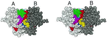
Recent developments in NMR have extended the size range of proteins amenable to structural and functional characterization to include many larger proteins involved in important cellular processes. By applying a combination of residue-specific isotope labeling and protein deuteration strategies tailored to yield specific information, we were able to determine the solution structure and study structure-activity relationships of 3,4-dihydroxy-2-butanone-4-phosphate synthase, a 47-kDa enzyme from the Escherichia coli riboflavin biosynthesis pathway and an attractive target for novel antibiotics. Our investigations of the enzyme's ligand binding by NMR and site-directed mutagenesis yields a conclusive picture of the location and identity of residues directly involved in substrate binding and catalysis. Our studies illustrate the power of state-of-the-art NMR techniques for the structural characterization and investigation of ligand binding in protein complexes approaching the 50-kDa range in solution.


Figures

Figure 1
Figure 1
Two-dimensional 100-ms NOESY spectra of DHBPS. The regions show the aromatic protons (Lower) and long range correlations from aromatics to methyl protons (Upper). Spectra from a sample with 75% random fractional deuteration, 13C/15N labeling (A) and a sample with protonated Phe, Tyr, Thr, Ile, and Val in a fully protonated and 15N-labeled background (B) are shown.
Figure 2
Figure 2
(A) Ensemble of the eight lowest energy structures of the DHBPS monomer. Three loops (31–43, 84–94, and 176–185) and the N and C termini for which no long range NOE correlations were observed are marked in black. (B) Ribbon (MOLMOL; ref. 31) diagram of the DHBPS monomer.
Figure 3
Figure 3
Sequence alignments of four divergent members of the DHBPS family. Residues strictly conserved in 20 sequences are shown in bold (Ec, E. coli; Mj, M. jannaschii; Mt, Methanobacterium thermoautotrophicum; Af, Archaeoglobus fulgidus; A, Aeropyrum). The secondary structure of the E. coli enzyme is shown above the sequence. Results from NMR chemical shift mapping experiments investigating the binding of ligands is indicated by bars (red, blue, and green) above the sequence. The combined chemical shift perturbations (ΔδTOTAL) of all 1H and 15N resonances were weighted according to ΔδTOTAL = Δδ [1H] + 0.2 Δδ [15N] (20) and grouped into two classes (moderate, short bars ΔδTOTAL = 0.06–0.12 ppm and strong, tall bars ΔδTOTAL > 0.12 ppm). Red bars show the location and magnitude of chemical shift changes on the addition of the substrate, ribulose-5-phosphate, in the presence of Mg2+ ions. Blue bars indicate signals affected after the addition of substrate in the absence of Mg2+. Green bars indicate perturbations after the addition of Mg2+. The backbone nuclei of the residues that were not assigned in NMR spectra are marked with a dashed line in the secondary structure. Amino acids that, when substituted in the M. jannaschii sequence by site-directed mutagenesis, led to a loss of catalytic activity are highlighted in red, and those leading to significantly reduced activity are highlighted in cyan.
Figure 4
Figure 4
Data from chemical shift mapping experiments investigating ligand binding and mutations with reduced activity mapped onto the three-dimensional structure depicted as a ribbon diagram (MOLMOL; ref. 31). Residues with strong (ΔδTOTAL > 0.12 ppm) or moderate (moderate ΔδTOTAL = 0.06–0.12 ppm) chemical shift perturbations after addition of ligands and complete or significant loss in activity after mutation are marked in dark and light colors, respectively. The conserved acidic loop joining strands β1 and β2 is indicated by green stripes. (A) The reaction catalyzed by DHBPS. (B) Chemical shift mapping of substrate binding to the enzyme in the absence of Mg2+ (nonturnover conditions). Strong and moderate shifts (see Fig. 3) are shown in blue and cyan, respectively. (C) Chemical shift mapping of product binding to the enzyme and catalysis in the presence of Mg2+ (turnover conditions). Strong and moderate shifts (see Fig. 3) are shown in red and yellow, respectively. (D) Results of site-directed mutagenesis experiments. Residues that, when substituted in the Methanococcus sequence, led to a loss of catalytic activity and those with a significant reduction in activity are shown in magenta and pink, respectively (see Table 2).
Figure 5
Figure 5
Space-filled representation (MOLMOL; ref. 31) of the dimer model built using the monomeric NMR structure. The model satisfies the intermolecular contacts reported by Liao et al. (6). The active site shown on the front of the dimer is formed by the regions showing large chemical shift changes on ligand binding: I, 32–42, green; II, 59–71, red; V, 150–155, magenta; VI, 170–176, cyan, from monomer A; III, 105–113, yellow; IV, 135–138, blue, from monomer B.

References

    1. Bacher A, Eberhardt S, Fischer M, Kis K, Richter G. Annu Rev Nutr. 2000;20:153–167. - PubMed
    1. Götze E, Kis K, Eisenreich W, Yamauchi N, Kakinuma K, Bacher A. J Org Chem. 1998;63:6456–6457.
    1. Richter G, Kelly M, Krieger C, Yu Y, Bermel W, Karlsson G, Bacher A, Oschkinat H. Eur J Biochem. 1999;261:57–65. - PubMed
    1. Volk R, Bacher A. J Am Chem Soc. 1988;110:3651–3653.
    1. Volk R, Bacher A. J Biol Chem. 1990;265:19479–19485. - PubMed

Publication types

Substances

Associated data

LinkOut - more resources