. 2001 Nov 6;98(23):13102-7.
doi: 10.1073/pnas.231364598. Epub 2001 Oct 30.

Phosphorylation of serines 635 and 645 of human Rad17 is cell cycle regulated and is required for G(1)/S checkpoint activation in response to DNA damage

Affiliations

Phosphorylation of serines 635 and 645 of human Rad17 is cell cycle regulated and is required for G(1)/S checkpoint activation in response to DNA damage

S Post et al. Proc Natl Acad Sci U S A. .

Abstract

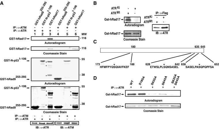
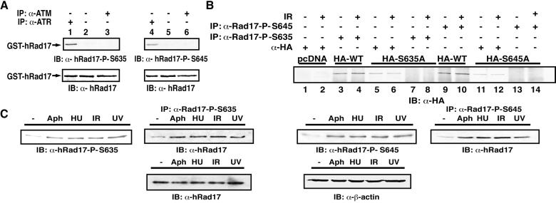
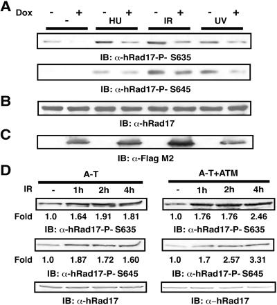
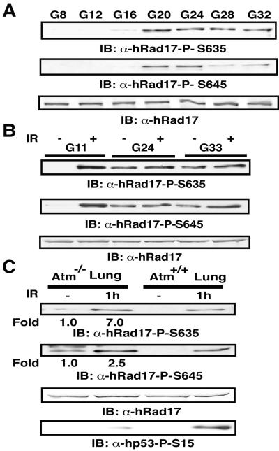
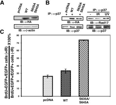
ATR [ataxia-telangiectasia-mutated (ATM)- and Rad3-related] is a protein kinase required for both DNA damage-induced cell cycle checkpoint responses and the DNA replication checkpoint that prevents mitosis before the completion of DNA synthesis. Although ATM and ATR kinases share many substrates, the different phenotypes of ATM- and ATR-deficient mice indicate that these kinases are not functionally redundant. Here we demonstrate that ATR but not ATM phosphorylates the human Rad17 (hRad17) checkpoint protein on Ser(635) and Ser(645) in vitro. In undamaged synchronized human cells, these two sites were phosphorylated in late G(1), S, and G(2)/M, but not in early-mid G(1). Treatment of cells with genotoxic stress induced phosphorylation of hRad17 in cells in early-mid G(1). Expression of kinase-inactive ATR resulted in reduced phosphorylation of these residues, but these same serine residues were phosphorylated in ionizing radiation (IR)-treated ATM-deficient human cell lines. IR-induced phosphorylation of hRad17 was also observed in ATM-deficient tissues, but induction of Ser(645) was not optimal. Expression of a hRad17 mutant, with both serine residues changed to alanine, abolished IR-induced activation of the G(1)/S checkpoint in MCF-7 cells. These results suggest ATR and hRad17 are essential components of a DNA damage response pathway in mammalian cells.


Figures

Figure 1
Figure 1
Phosphorylation of hRad17 on Ser635 and Ser645 in vitro. (A) Kinase assays using immunoprecipitated (IP) ATM and ATR. GST-hRad17 was incubated with ATM (Top Two Panels, lane 6) and ATR (Top Two Panels, lane 3) immunoprecipitated from HeLa cells treated with 10 Gy of IR. GST-N-p531–106, a known substrate of the two kinases, was incubated with ATM (Bottom Two Panels, lane 5) or ATR (Bottom Two Panels, lane 2). GST-hRad9255–295, a known substrate of ATM, was incubated with ATM (Bottom Two Panels, lane 4) or ATR (Bottom Two Panels, lane 1). Three micrograms of substrate was used in each reaction. The kinase reaction products were separated by SDS/PAGE and analyzed by Coomassie staining and autoradiography. Levels of ATR and ATM in the kinase reactions were determined by immunoprecipitation followed by Western blotting (IB). MW, molecular weight × 10−3. (B) Kinase assays using recombinant ATR protein. Human kidney 293 cells were transiently transfected with Flag-tagged ATRWt or ATRKi. Cells were treated with 10 Gy of IR 36 h after transfection and lysed 1 h after IR. GST-hRad17 fusion proteins were incubated with recombinant ATR immunoprecipitated with α-Flag antibodies. Immunoprecipitation with α-Flag antibodies followed by immunoblotting with α-ATR antibodies confirmed the presence of recombinant ATR. (C) Schematic representation of mutant hRad17 proteins. Site-specific mutation of serine to alanine was confirmed by DNA sequencing. (D) Ser635 and Ser645 of hRad17 are substrate sites of ATR in vitro. Wild-type and mutant hRad17 fusion proteins were incubated with ATR and the resultant proteins were analyzed as described in B.
Figure 2
Figure 2
In vivo phosphorylation of hRad17. (A and B) Specificity of the α-hRad17 phosphospecific antibodies. GST-hRad17 was incubated with ATR (A, Top Two Panels, lanes 1 and 4) or ATM (Top Two Panels, lanes 3 and 6), immunoprecipitated from HeLa cells, and immunoblotted with phosphopeptide antibodies. Human kidney 293 cells transfected with HA-tagged hRad17 plasmid as indicated (B). Cells were mock-treated or treated with 10 Gy of IR. Cells were lysed 1 h after treatment, and soluble proteins were immunoprecipitated with α-HA and phosphopeptide antibodies, as indicated. Proteins in the immunoprecipitates were separated by SDS/PAGE and immunoblotted with α-HA antibodies. (C) Phosphorylation of hRad17 on Ser635 and Ser645 in vivo. Human fibroblast VA-13 cells were mock-treated, treated with 5 μg/ml aphidicolin (Aph) for 20 h, 1 mM hydroxyurea (HU) for 24 h, 10 Gy of IR, or 50 J/m2 UV irradiation. Cells lysates were subjected to SDS/PAGE, and immunoblotting was performed by using the indicated antibodies, or lysates were immunoprecipitated with α-hRad17-PS635 or α-hRad17-PS645 antibodies. Immunoblotting of the immunoprecipitated lysates was performed by using α-hRad17 antibody, 31E9. Western blotting analysis of hRad17 protein in the whole cell extracts. Levels of hRad17 and β-actin remain constant in untreated and treated cells.
Figure 3
Figure 3
ATR-dependent phosphorylation of Ser635 and Ser645 of hRad17. (A) Analysis of Ser635 and Ser645 phosphorylation in cells expressing ATRKi. Cells expressing ATRKi under tetracycline regulation were grown in the presence of doxycycline for 72 h. Cells were treated as in Fig. 2C. Soluble proteins were prepared and cell extracts were separated by SDS/PAGE. Immunoblotting was performed by using indicated antibodies. (B) Immunoblotting analysis of hRad17 before and after DNA damage and replication block. Soluble proteins from treated and untreated cells were subjected to SDS/PAGE and immunoblotted with α-hRad17 antibody 31E9. (C) Immunoblotting analysis of recombinant ATRKi expression. Expression of ATRKi was determined by SDS/PAGE followed by immunoblotting with α-Flag-M2. (D) ATM-independent phosphorylation of Ser635 and Ser645 of hRad17. EBS and YZ5 cells were mock-treated or treated with 30 Gy of IR and harvested 1, 2, or 4 h after treatment. (Bottom) Western blotting analysis of hRad17 in the whole cell extract.
Figure 4
Figure 4
Cell cycle-dependent phosphorylation of Ser635 and Ser645 of hRad17. (A) Immunoblot analysis of phosphorylation at Ser635 and Ser645 of hRad17 during the cell cycle. Density-arrested T24 cells were released and harvested at indicated time points G8, G12, G16, G24, G28, and G33 representing 8 h, 12 h, etc. after density release, respectively. Sample analysis was as described in Fig. 2C. (B) Immunoblot analysis of hRad17 phosphorylation during different cell cycle phases. Density-arrested T24 cells were released for 11, 24, and 33 h and harvested 1 h after treatment. (C) Phosphorylation of MmRad17 in mouse tissues. Atm−/− and Atm+/+ mice were mock-treated or treated with 10 Gy of IR and killed 1 h after treatment.
Figure 5
Figure 5
Effects of expression of hRad17S635A and hRad17S645A on G1/S checkpoint activation. (A) Immunoblotting analysis of recombinant HA-hRad17 expression in the transfected cells. Proteins in the lysates were separated by SDS/PAGE followed by immunoblotting analysis using α-HA antibody. (B) Recombinant HA-hRad17 and endogenous hRad17 interact with p37/RFC. Immunoprecipitation was carried out by using α-p37/RFC antibodies, and Western blotting was with antibodies as indicated. (C) G1/S checkpoint activation in response to IR. The ratio of BrdUrd and EGFP double-positive cells to EGFP-positive cells was determined in mock- and IR-treated cells, respectively. At least 350 cells were counted from each plate. The mean and SD were calculated from three separate plates.

Similar articles

Cited by

References

    1. Weinert T. Curr Opin Genet Dev. 1998;8:185–193. - PubMed
    1. Longhese M P, Foiani M, Muzi-Falconi M, Lucchini G, Plevani P. EMBO J. 1998;17:5525–5528. - PMC - PubMed
    1. Dasika G K, Lin S C, Zhao S, Sung P, Tomkinson A, Lee E Y. Oncogene. 1999;18:7883–7899. - PubMed
    1. Caspari T, Carr A M. Biochimie. 1999;81:173–181. - PubMed
    1. Zhou B, Elledge S. Nature (London) 2000;408:433–439. - PubMed

Publication types