

Duodenal calcium absorption in vitamin D receptor-knockout mice: functional and molecular aspects
- PMID: 11687634
- PMCID: PMC60869
- DOI: 10.1073/pnas.231474698
Duodenal calcium absorption in vitamin D receptor-knockout mice: functional and molecular aspects
Abstract
Rickets and hyperparathyroidism caused by a defective vitamin D receptor (VDR) can be prevented in humans and animals by high calcium intake, suggesting that intestinal calcium absorption is critical for 1,25(OH)(2) vitamin D [1,25(OH)(2)D(3)] action on calcium homeostasis. We assessed the rate of serum (45)Ca accumulation within 10 min of oral gavage in two strains of VDR-knockout (KO) mice (Leuven and Tokyo KO) and observed a 3-fold lower area under the curve in both KO strains. Moreover, we evaluated the expression of intestinal candidate genes involved in transcellular calcium transport. The calcium transport protein1 (CaT1) was more abundantly expressed at mRNA level than the epithelial calcium channel (ECaC) in duodenum, but both were considerably reduced (CaT1>90%, ECaC>60%) in the two VDR-KO strains on a normal calcium diet. Calbindin-D(9K) expression was decreased only in the Tokyo KO, whereas plasma membrane calcium ATPase (PMCA(1b)) expression was normal in both VDR-KOs. In Leuven wild-type mice, a high calcium diet inhibited (>90%) and 1,25(OH)(2)D(3) injection or low calcium diet induced (6-fold) duodenal CaT1 expression and, to a lesser degree, ECaC and calbindin-D(9K) expression. In Leuven KO mice, however, high or low calcium intake decreased calbindin-D(9K) and PMCA(1b) expression, whereas CaT1 and ECaC expression remained consistently low on any diet. These results suggest that the expression of the novel duodenal epithelial calcium channels (in particular CaT1) is strongly vitamin D-dependent, and that calcium influx, probably interacting with calbindin-D(9K), should be considered as a rate-limiting step in the process of vitamin D-dependent active calcium absorption.
Figures
 ; intron sequences
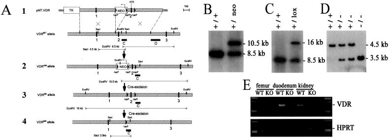
  ; D external (1.5-kb XhoI fragment) and C internal (1-kb EcoRV-SacI fragment) hybridization probe. The observed size of the diagnostic restriction fragments, used to distinguish the WT and mutant alleles, corresponds to their expected size. (B) Southern analysis of EcoRV-digested genomic DNA from VDRWT and VDRneo embryonic stem cell clones by using the external 3′ probe D, which generates a 8.5-kb VDRWT and a 10.5-kb VDRneo fragment. (C) Southern analysis of EcoRV-digested genomic DNA from VDRWT and VDR+/lox mice by using the external 3′ probe D, which generates a 8.5-kb VDRWT and a 16-kb VDRlox fragment. (D) Southern analysis of SacI-digested genomic DNA from VDRWT, VDR+/− and VDRKO mice by using the internal probe C. (E) RT-PCR analysis of RNA isolated from duodenum, kidney, and femur of VDRWT and VDRKO mice for VDR and HPRT RNA level expression.
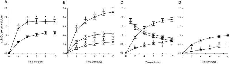
 , calbindin-D9K , and PMCA
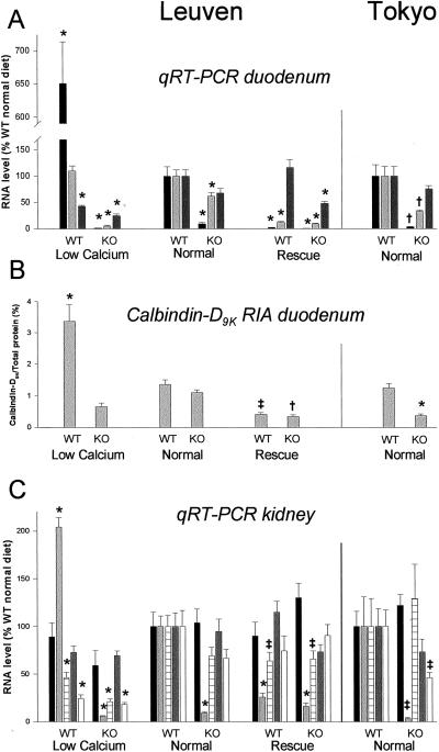
  , assessed by qRT-PCR analysis, is calculated as a ratio to the HPRT RNA level and is expressed relatively to levels of WT mice on normal diet. (B) Calbindin-D9K protein content in duodenum, measured by RIA, is expressed as % (mg/mg) of total duodenal protein. (C) Renal gene expression of ECaC
  , calbindin-D9K
  , calbindin-D28K
  , PMCA
  , and NCX , assessed by qRT-PCR analysis, is calculated as a ratio to the HPRT RNA level and expressed relatively to levels of WT mice on normal diet. *, P < 0.001, †, P < 0.01, and ‡, P < 0.05 vs. WT on normal diet.
References
-
- Reichel H, Koeffler H P, Norman A W. N Engl J Med. 1989;320:980–991. - PubMed
-
- Haussler M R, Whitfield G K, Haussler C A, Hsieh J C, Thompson P D, Selznick S H, Dominguez C E, Jurutka P W. J Bone Miner Res. 1998;13:325–349. - PubMed
-
- Hochberg Z, Tiosano D, Even L. J Pediatr. 1992;121:803–808. - PubMed
-
- Yoshizawa T, Handa Y, Uematsu Y, Takeda S, Sekine K, Yoshihara Y, Kawakami T, Arioka K, Sato H, Uchiyama Y, et al. Nat Genet. 1997;16:391–396. - PubMed
Publication types
MeSH terms
Substances
LinkOut - more resources
Full Text Sources
Molecular Biology Databases
Research Materials
Miscellaneous
