. 2001 Nov 6;98(23):12984-9.
doi: 10.1073/pnas.241175798. Epub 2001 Oct 30.

Enzyme interactions in heparan sulfate biosynthesis: uronosyl 5-epimerase and 2-O-sulfotransferase interact in vivo

Affiliations

Enzyme interactions in heparan sulfate biosynthesis: uronosyl 5-epimerase and 2-O-sulfotransferase interact in vivo

M A Pinhal et al. Proc Natl Acad Sci U S A. .

Abstract

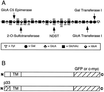
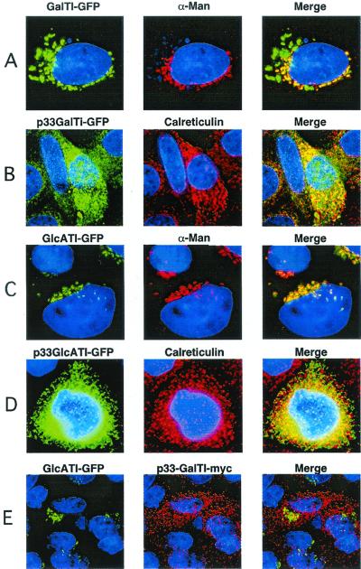
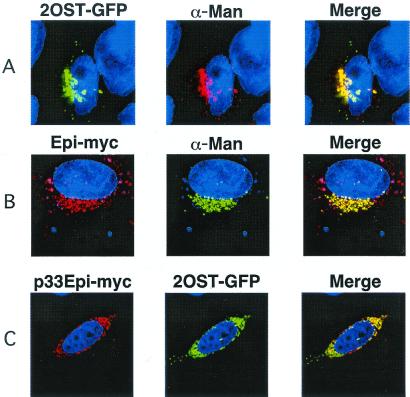
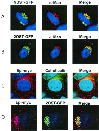
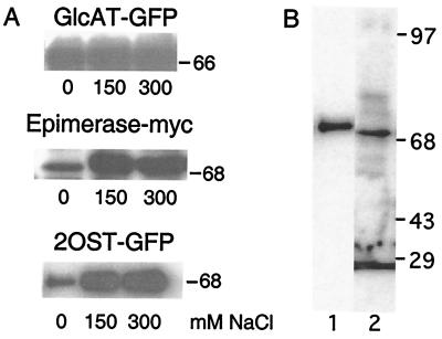
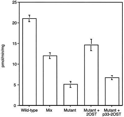
The formation of heparan sulfate occurs within the lumen of the endoplasmic reticulum-Golgi complex-trans-Golgi network by the concerted action of several glycosyltransferases, an epimerase, and multiple sulfotransferases. In this report, we have examined the location and interaction of tagged forms of five of the biosynthetic enzymes: galactosyltransferase I and glucuronosyltransferase I, required for the formation of the linkage region, and GlcNAc N-deacetylase/N-sulfotransferase 1, uronosyl 5-epimerase, and uronosyl 2-O-sulfotransferase, the first three enzymes involved in the modification of the chains. All of the enzymes colocalized with the medial-Golgi marker alpha-mannosidase II. To study whether any of these enzymes interacted with each other, they were relocated to the endoplasmic reticulum (ER) by replacing their cytoplasmic N-terminal tails with an ER retention signal derived from the cytoplasmic domain of human invariant chain (p33). Relocating either galactosyltransferase I or glucuronosyltransferase I had no effect on the other's location or activity. However, relocating the epimerase to the ER caused a parallel redistribution of the 2-O-sulfotransferase. Transfected epimerase was also located in the ER in a cell mutant lacking the 2-O-sulfotransferase, but moved to the Golgi when the cells were transfected with 2-O-sulfotransferase cDNA. Epimerase activity was depressed in the mutant, but increased upon restoration of 2-O-sulfotransferase, suggesting that their physical association was required for both epimerase stability and translocation to the Golgi. These findings provide in vivo evidence for the formation of complexes among enzymes involved in heparan sulfate biosynthesis. The functional significance of these complexes may relate to the rapidity of heparan sulfate formation.


Figures

Figure 1
Figure 1
Heparan sulfate and chimeric enzymes. (A) Schematic representation of heparan sulfate. The arrows indicate the sites of action for the enzymes examined in this study. NDST, N-deacetylase/N-sulfotransferase 1. (B) To localize the enzymes, green fluorescent protein (GFP) was spliced on the C termini of the enzymes. To relocate the enzymes, the cytoplasmic tail of the enzymes was replaced with the endoplasmic reticulum (ER)-retention domain from human invariant chain (p33). TM, transmembrane domain.
Figure 2
Figure 2
Localization of chimeric enzymes involved in the formation of linkage region. Wild-type CHO cells were transiently transfected with the indicated chimeric enzymes and imaged by deconvolution microscopy (Materials and Methods). (A and C) Golgi localization of GalTI-GFP and GlcATI-GFP, respectively. (B and D) ER localization of p33-GalTI-GFP and p33-GlcATI-GFP, respectively. (E) Coexpression of GlcATI-GFP (green) and p33-GalTI-Myc (red). α-Man, α-mannosidase II, the medial Golgi marker; Calreticulin, ER marker; Merge, colocalization (yellow) of the chimeric enzymes with each other or the markers.
Figure 3
Figure 3
ER-localized Epi relocates 2OST. (A and B) Wild-type CHO cells were transiently transfected with chimeric 2OST-GFP or Epi-Myc and their location was determined by deconvolution microscopy (Materials and Methods). (C) Cells were cotransfected with p33-Epi-Myc and 2OST-GFP. The two enzymes colocalized in the ER, in the compartments containing the calreticulin marker. α-Man, α-mannosidase II, the medial Golgi marker; Calreticulin, the ER marker; Merge, colocalization (yellow) of the chimeric enzymes with each other or the markers.
Figure 4
Figure 4
Golgi localization of Epi depends on the presence of 2OST. pgsF17 cells lacking endogenous 2OST were transiently transfected with NDST1-GFP (A), 2OST-GFP (B), or Epi-Myc (C). NDST1 and 2OST colocalized with the α-mannosidase marker, whereas Epi-Myc was found in the ER. In D, the mutant was stably transfected with 2OST cDNA to the correct the enzyme deficiency, and then transiently transfected with 2OST-GFP or Epi-Myc.
Figure 5
Figure 5
Physical interaction of Epi and 2OST. (A) CHO cells expressing GlcATI-GFP, Epi-Myc, or 2OST-GFP were extracted in buffer containing 50 mM Mes (pH 6.5), 0.5% Triton X-100, and 0, 150, or 300 mM NaCl as indicated. The cell extracts were centrifuged, separated by SDS/gel electrophoresis, and analyzed by immunoblotting (Materials and Methods). (B) Samples of cells containing Epi-Myc and 2OST-GFP were immunoprecipitated with antibody to GFP and analyzed by SDS/PAGE. Lane 1, Western blot using mouse anti-Myc mAb. Lane 2, Western blot using mouse anti-GFP mAb.
Figure 6
Figure 6
Decreased epimerase activity in 2OST-deficient cells. Wild-type, pgsF17, and pgsF17 cells stably transfected with 2OST or p33–2OST were assayed for Epi activity. “Mix” refers to a sample containing equal amounts of wild-type and mutant extracts at half the level in samples assayed alone.

Similar articles

Cited by

References

    1. Lindahl U, Kusche-Gullberg M, Kjellén L. J Biol Chem. 1998;273:24979–24982. - PubMed
    1. Esko J D, Lindahl U. J Clin Invest. 2001;108:169–173. - PMC - PubMed
    1. Shworak N W, Liu J A, Petros L M, Zhang L J, Kobayashi M, Copeland N G, Jenkins N A, Rosenberg R D. J Biol Chem. 1999;274:5170–5184. - PubMed
    1. Aikawa J, Grobe K, Tsujimoto M, Esko J D. J Biol Chem. 2001;276:5876–5882. - PubMed
    1. Habuchi H, Tanaka M, Habuchi O, Yoshida K, Suzuki H, Ban K, Kimata K. J Biol Chem. 2000;275:2859–2868. - PubMed

Publication types

Substances