. 2001 Aug;128(16):3117-31.
doi: 10.1242/dev.128.16.3117.

The interplay of matrix metalloproteinases, morphogens and growth factors is necessary for branching of mammary epithelial cells

Affiliations

The interplay of matrix metalloproteinases, morphogens and growth factors is necessary for branching of mammary epithelial cells

M Simian et al. Development. 2001 Aug.

Abstract

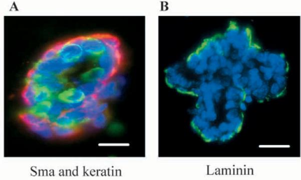
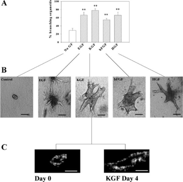
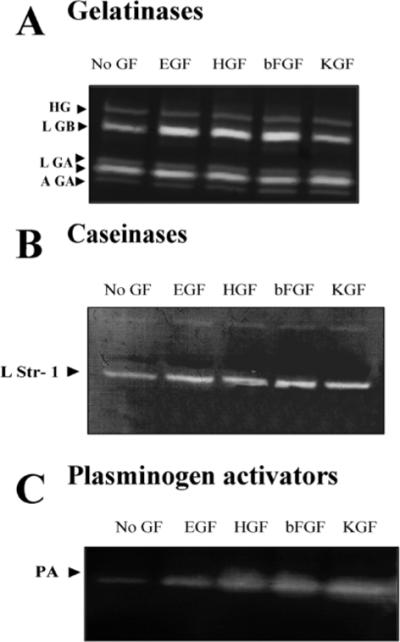
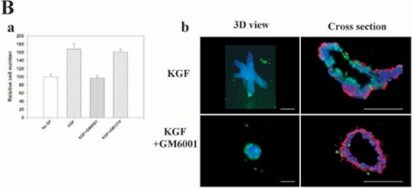
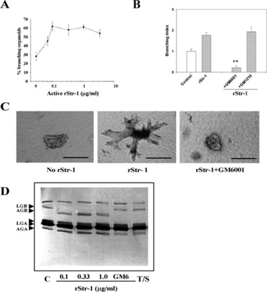
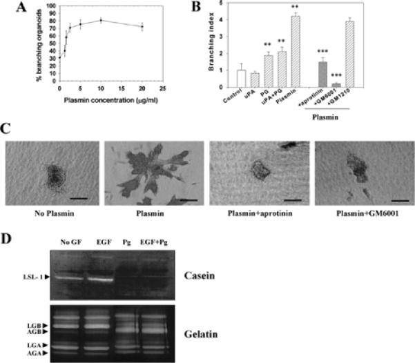
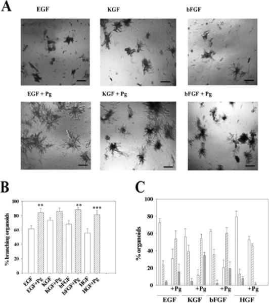
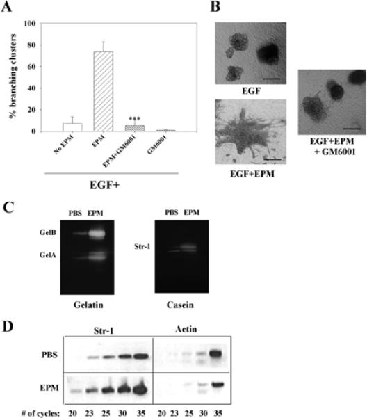
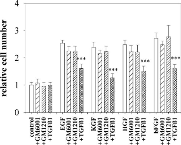
The mammary gland develops its adult form by a process referred to as branching morphogenesis. Many factors have been reported to affect this process. We have used cultured primary mammary epithelial organoids and mammary epithelial cell lines in three-dimensional collagen gels to elucidate which growth factors, matrix metalloproteinases (MMPs) and mammary morphogens interact in branching morphogenesis. Branching stimulated by stromal fibroblasts, epidermal growth factor, fibroblast growth factor 7, fibroblast growth factor 2 and hepatocyte growth factor was strongly reduced by inhibitors of MMPs, indicating the requirement of MMPs for three-dimensional growth involved in morphogenesis. Recombinant stromelysin 1/MMP3 alone was sufficient to drive branching in the absence of growth factors in the organoids. Plasmin also stimulated branching; however, plasmin-dependent branching was abolished by both inhibitors of plasmin and MMPs, suggesting that plasmin activates MMPs. To differentiate between signals for proliferation and morphogenesis, we used a cloned mammary epithelial cell line that lacks epimorphin, an essential mammary morphogen. Both epimorphin and MMPs were required for morphogenesis, but neither was required for epithelial cell proliferation. These results provide direct evidence for a crucial role of MMPs in branching in mammary epithelium and suggest that, in addition to epimorphin, MMP activity is a minimum requirement for branching morphogenesis in the mammary gland.


Figures

Fig. 1
Fig. 1
Primary organoids retain myoepithelial cells and basement membrane. (A) Detection of keratin-positive and α-smooth muscle actin-positive cells in primary mammary organoids at the time of preparation. Frozen sections of mammary organoids embedded in OCT were immunostained (A) simultaneously with mouse anti-α-smooth muscle actin to detect myoepithelial cells (red) and rabbit anti-keratin antibodies (green) as described in Materials and Methods. (B) Organoids were stained with rabbit anti-laminin antibodies (green). Scale bars: 50 μm. DAPI nuclear counterstain is in blue.
Fig. 2
Fig. 2
Growth factors induce branching morphogenesis in mammary organoids. Primary mammary organoids were embedded in collagen I gels and treated for 4 to 6 days with EGF, KGF, bFGF or HGF at a concentration of 50 ng/ml. (A) Quantification of the percentage of branching organoids for each of the treatments. The means±s.d. of three independent experiments are shown. The differences between the control group (no GF) and the growth factor-treated groups was statistically significant (**P≤0.01). (B) Appearance of control and growth factor-treated primary mammary organoids. (C) Cross sections of organoids showing the existence of a lumen at the day of preparation (day 0) and after 4 days of treatment with KGF. Scale bars: 200 μm in B; 100 μm in C.
Fig. 3
Fig. 3
Growth factors increase the level but not the activation status of proteinases secreted by primary mammary organoids. Mammary organoids were treated for 4 to 6 days in collagen I gels with no growth factor (No GF) or with EGF, HGF, bFGF or KGF. Conditioned medium was collected, concentrated and run on substrate zymograms. (A) Gelatin zymogram. Latent gelatinase B (LGB), active and latent gelatinase A (AGA, LGA) and a high molecular weight gelatinase (HG) were detected. (B) Casein zymogram. A band corresponding to the molecular weight of latent stromelysin-1 (LStr1) was detected. (C) Plasminogen casein zymogram. A band corresponding to the molecular weight of uPA was detected. There were no differences in the activation status of the gelatinases, caseinase or plasminogen activator between growth factor treated organoids and untreated controls.
Fig. 4
Fig. 4
Proteinase inhibitors suppress branching morphogenesis induced by growth factors. (A, part a) Quantification of the percentage of branching organoids treated with (i) KGF or (ii) EGF in the presence of protease inhibitors E64, pepstatin, leupeptin, aprotinin, GM6001 and GM1210. Only the MMP inhibitor GM6001 had a statistically significant inhibitory effect on branching morphogenesis of both basal (*P≤0.05) and growth factor-treated organoids (***P≤0.001). The mean±s.d. of six samples in two independent experiments are shown. (A, part b) Primary organoids after 6 days of treatment with KGF and EGF with or without GM6001. Note the absence of branches in the organoids treated with the MMP inhibitor. Similar results were obtained with bFGF and HGF (not shown). (A, part c) Quantification of branching and (A, part d) appearance of the organoids treated with TIMP1 and TIMP2 in the presence of KGF. The mean±s.d. of six (TIMP1) and five (TIMP2) samples in two independent experiments are shown; (***P≤0.001). (A, part e) Quantification of the effect of proteinase inhibitors on branching morphogenesis induced by co-culturing the organoids with primary fibroblasts from virgin animals (Fv). Only GM6001 had a statistically significant inhibitory effect (**P≤0.01). The mean±s.d. of six samples in two independent experiments are shown. (B) GM6001 inhibits increase in cell number in organoids (B, part a), but does not alter apoptosis. Primary organoids were embedded in collagen I gels and treated for 6 days with KGF in the presence and absence of GM6001. (B, part a) Cell number was measured using Alamar Blue. The mean±s.d. of six samples in two independent experiments are shown. (B, part b) Whole organoids (3D) and cross sections are shown. Apoptotic nuclei were stained in green (FITC). Cross sections were also stained for α-smooth muscle actin-positive cells (red). There were no statistical differences in the number of apoptotic cells in control and treated groups. DAPI nuclear counterstain is in blue. Scale bars: 400 μm in A; 200 μm in B.
Fig. 4
Fig. 4
Proteinase inhibitors suppress branching morphogenesis induced by growth factors. (A, part a) Quantification of the percentage of branching organoids treated with (i) KGF or (ii) EGF in the presence of protease inhibitors E64, pepstatin, leupeptin, aprotinin, GM6001 and GM1210. Only the MMP inhibitor GM6001 had a statistically significant inhibitory effect on branching morphogenesis of both basal (*P≤0.05) and growth factor-treated organoids (***P≤0.001). The mean±s.d. of six samples in two independent experiments are shown. (A, part b) Primary organoids after 6 days of treatment with KGF and EGF with or without GM6001. Note the absence of branches in the organoids treated with the MMP inhibitor. Similar results were obtained with bFGF and HGF (not shown). (A, part c) Quantification of branching and (A, part d) appearance of the organoids treated with TIMP1 and TIMP2 in the presence of KGF. The mean±s.d. of six (TIMP1) and five (TIMP2) samples in two independent experiments are shown; (***P≤0.001). (A, part e) Quantification of the effect of proteinase inhibitors on branching morphogenesis induced by co-culturing the organoids with primary fibroblasts from virgin animals (Fv). Only GM6001 had a statistically significant inhibitory effect (**P≤0.01). The mean±s.d. of six samples in two independent experiments are shown. (B) GM6001 inhibits increase in cell number in organoids (B, part a), but does not alter apoptosis. Primary organoids were embedded in collagen I gels and treated for 6 days with KGF in the presence and absence of GM6001. (B, part a) Cell number was measured using Alamar Blue. The mean±s.d. of six samples in two independent experiments are shown. (B, part b) Whole organoids (3D) and cross sections are shown. Apoptotic nuclei were stained in green (FITC). Cross sections were also stained for α-smooth muscle actin-positive cells (red). There were no statistical differences in the number of apoptotic cells in control and treated groups. DAPI nuclear counterstain is in blue. Scale bars: 400 μm in A; 200 μm in B.
Fig. 5
Fig. 5
Recombinant stromelysin-1 induces branching morphogenesis of primary mammary organoids. (A) Dose response to rStr1. Mean±s.d. of three independent experiments is shown. (B) GM6001 inhibits rStr1-induced branching. rStr1 (1μg/ml) induced-branching morphogenesis compared with the control (**P<0.01). Control samples were treated with trypsin and soy bean trypsin inhibitor (SBTI), GM6001, (***P≤0.001). The means±s.d. of three independent experiments are shown. To calculate the branching index, control values were equated to 1. (C) Appearance of organoids in collagen I gels after 6 days with or without Str1 and GM6001 treatments. (D) Gelatin zymogram of conditioned media of organoids treated with buffer (Control, C), or different concentration of rStr1, with or without GM6001 (GM6) or trypsin+SBTI (T/S). Note the activation of gelatinase B in the presence of rStr1. This activation is inhibited in the presence of GM6001, as expected. Treatment with trypsin+SBTI (T/S) did not activate gelatinase B. Scale bars: 200 μm in C.
Fig. 6
Fig. 6
Plasmin induces branching morphogenesis of primary mammary organoids. (A) Dose response to increasing concentrations of plasmin. Mean±s.d. of three independent experiments is shown. (B) Effect of protease inhibitors on plasmin-induced branching morphogenesis. The difference with the control group (buffer) was statistically significant (**P≤ 0.01). Inhibition by both aprotinin and GM6001 were statistically significant (***P≤ 0.001). Mean±s.d. of three independent experiments is shown. To calculate the branching index, control values were taken as equal to 1. (C) Appearance of organoids treated for 6 days with no plasmin, or plasmin, with and without inhibitors. (D) Activation of MMPs by plasminogen. Casein zymogram (top panel) of conditioned media from organoids treated with buffer only (no GF), EGF, plasminogen (Pg) and EGF+plasminogen (EGF+Pg). When plasminogen was added to the treatment, the band corresponding to the latent form of Str1 disappeared. The bottom panel is a gelatin zymogram, using the same set of conditioned media as for the casein zymogram. Gelatinase B was activated in the presence of plasminogen. Scale bars: 150 μm in C.
Fig. 7
Fig. 7
Plasminogen and plasmin synergize with growth factors. (A) Appearance of organoids treated with growth factors with or without added plasminogen (Pg). Note the increase in the size and number of branches of the organoids when plasminogen was added together with the growth factors. (B) Effect of plasminogen on the percentage of organoids with branches. Shown are the percentage of branching organoids in the presence of growth factors alone (white bars) and growth factors+Pg (hatched bars). The increase in the number of organoids with branches when plasminogen was added together with the growth factor, compared with the group without plasminogen was statistically significant in all cases except for the KGF group, (**, P≤ 0.01) and (***, P≤ 0.001). (C) Effect of plasminogen on the number of branches per organoid: one to five branches (white bars), six to ten branches (hatched bars) and more than ten branches (cross hatched bars). There were increased branches per organoid also for the KGF-treated cultures. The figures show the mean±s.d. of six wells in two independent experiments. Scale bars: 400 μm in A.
Fig. 8
Fig. 8
GM6001 inhibits epimorphin-induced branching morphogenesis in a mammary epithelial cell line. (A) Preclustered SCp2 cells were treated for 6 days in collagen I gels with EGF, EGF+epimorphin (EPM) with or without GM6001. GM6001 inhibited branching induced by epimorphin. This inhibition was statistically significant (***P≤0.001). Other proteinase inhibitors such as leupeptin, aprotinin, pepstatin and E64 did not show any inhibition of branching (not shown). The mean±s.d. of three independent experiments is shown. (B) Appearance of SCp2 cell clusters treated with EGF, EGF+EPM and EGF+EPM+GM6001. (C) Epimorphin upregulates gelatinases A and B and Str1 in SCp2 cells. SCp2 cells were treated for 48 hours with epimorphin or PBS as a control. Conditioned medium was collected, concentrated and separated on substrate gels. (D) Epimorphin regulates Str1 mRNA levels. RT-PCR analysis of Str1 expression in SCp2 cells treated with PBS or EPM for 48 hours. Twenty, 23, 25, 30 and 35 cycles of amplification were performed for detection of Str1 and actin. While the level of actin was not different statistically in the PBS and epimorphin-treated cultures (slightly higher in PBS in this experiment), Str1 levels were clearly higher in epimorphin-treated cultures. Scale bars: 200 μm in A.
Fig. 9
Fig. 9
GM6001 does not inhibit SCp2 cell proliferation. SCp2 cells were embedded in collagen I gels and treated for six days with no growth factor, or with EGF, KGF, HGF, bFGF in the presence and absence of GM6001, GM1210 and TGFβ1. Quantification of cell number was carried out using Alamar Blue. The mean±s.d. of three independent experiments is shown. The data are normalized with values obtained for the control set to 1. Only TGFβ1 significantly reduced the cell number (***P≤0.001).

References

    1. Ahmed SA, Gogal RM, Jr, Walsh JE. A new rapid and simple non radioactive assay to monitor and determine the proliferation of lymphocytes: an alternative to [3H ]thymidine incorporation assay. J. Immunol. Methods. 1994;170:211–224. - PubMed
    1. Basbaum CB, Werb Z. Focalized proteolysis: spatial and temporal regulation of extracellular matrix degradation at the cell surface. Curr. Opin. Cell Biol. 1996;8:731–738. - PubMed
    1. Berdichevsky F, Alford D, D' Souza B, Taylor-Papadimitriou J. Branching morphogenesis of human mammary epithelial cells in collagen gels. J. Cell Sci. 1994;107:3557–3568. - PubMed
    1. Bernfield M, Banerjee SD, Koda JE, Rapraeger AC. Remodelling of the basement membrane: morphogenesis and maturation. Ciba Found. Symp. 1984;108:179–196. - PubMed
    1. Boudreau N, Werb Z, Bissell MJ. Suppression of apoptosis by basement membrane requires three-dimensional tissue organization and withdrawal from the cell cycle. Proc. Nat. Acad. Sci. USA. 1996;93:3509–3513. - PMC - PubMed

Publication types

MeSH terms