. 2001 Nov 1;20(21):5863-75.
doi: 10.1093/emboj/20.21.5863.

The 37-kDa/67-kDa laminin receptor acts as the cell-surface receptor for the cellular prion protein

Affiliations

The 37-kDa/67-kDa laminin receptor acts as the cell-surface receptor for the cellular prion protein

S Gauczynski et al. EMBO J. .

Abstract

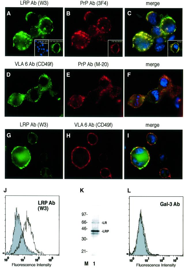
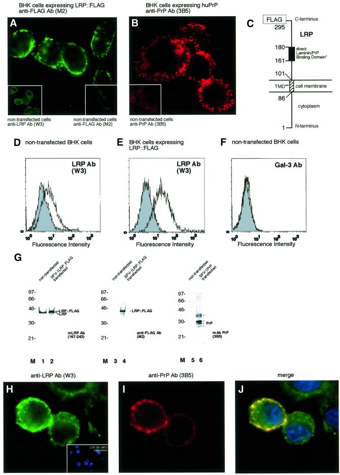
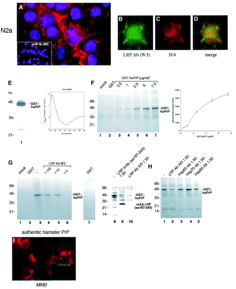
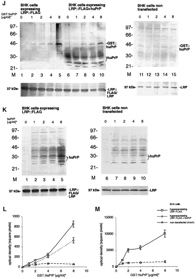
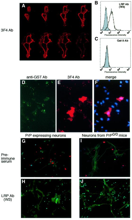
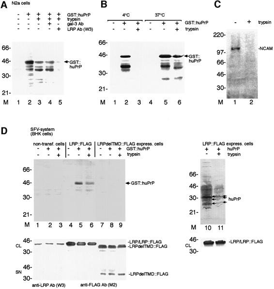
Recently, we identified the 37-kDa laminin receptor precursor (LRP) as an interactor for the prion protein (PrP). Here, we show the presence of the 37-kDa LRP and its mature 67-kDa form termed high-affinity laminin receptor (LR) in plasma membrane fractions of N2a cells, whereas only the 37-kDa LRP was detected in baby hamster kidney (BHK) cells. PrP co-localizes with LRP/LR on the surface of N2a cells and Semliki Forest virus (SFV) RNA transfected BHK cells. Cell-binding assays reveal the LRP/LR-dependent binding of cellular PrP by neuronal and non-neuronal cells. Hyperexpression of LRP on the surface of BHK cells results in the binding of exogenous PrP. Cell binding is similar in PrP(+/+) and PrP(0/0) primary neurons, demonstrating that PrP does not act as a co-receptor of LRP/LR. LRP/LR-dependent internalization of PrP is blocked at 4 degrees C. Secretion of an LRP mutant lacking the transmembrane domain (aa 86-101) from BHK cells abolishes PrP binding and internalization. Our results show that LRP/LR acts as the receptor for cellular PrP on the surface of mammalian cells.


Figures

None
Fig. 1. Plasma membrane-associated LRP/LR and PrP co-localize on the surface of neuroblastoma cells. Non-permeabilized N2a [MHM2] cells were incubated with the pAb LRP W3 [sec. Ab fluorescein isothiocyanate (FITC)] (A), pAb LRP W3 saturated with rec. GST::LRP [sec. Ab carbocyanine (Cy2), 4′-6-diamidine-2-phenylindole (DAPI)] (A, left inset) or the mAb LRP (aa 285–295 of LRP, sec. Ab Cy2) (A, right inset) and the mAb PrP 3F4 [sec. Ab indocarbocyanine (Cy3)] (B), or the pAb PrP M-20 (B, inset). Merge of (A) and (B) DAPI staining (C) (magnification ×630). N2a [MHM2] cells were incubated with the mAb VLA6 CD49f (sec. Ab Cy2) (D) and the pAb M-20 (sec. Ab Cy3) CD49f (E). Merge of (D) and (E) DAPI staining (F). N2a [MHM2] cells were incubated with the pAb LRP W3 (sec. Ab Cy2) (G) and the mAb VLA6 (sec. Ab Cy3) (H). Merge of (G) and (H) DAPI staining (I). (J) Non-permeabilized N2a cells were analysed by FACscans. Filled profile, isotype control. non-filled profile, pAb LRP W3. Fluorescence intensity (abscissa) is plotted against relative cell numbers (ordinate). (K) Purified plasma membranes from N2a cells were analysed by western blotting employing a mAb LRP (directed against aa 167–243) (lane 1). Molecular weight markers are indicated. (L) Non-permeabilized N2a cells were analysed by FACscans. Filled profile, isotype control, non-filled profile, anti-gal-3 antibody. Fluorescence intensity (abscissa) is plotted against relative cell numbers (ordinate).
None
Fig. 2. Orientation, localization of LRP::FLAG and PrP and co-localization of both proteins in BHK cells transfected with rec. SFV RNAs. (A) Immunolocalization of LRP::FLAG to the cell membrane of non-permeabilized BHK cells transfected with rec. SFV LRP-FLAG RNA. Subcellular location was determined by IF using the mAb FLAG M2 (sec. Ab FITC). (Insets) Untransfected BHK cells incubated with the pAb LRP W3 (left), mAb FLAG M2 (right). (B) Immunolocalization of human PrPc to the cell membrane of non-permeabilized BHK cells transfected with rec. SFV-huPrP1–253 RNA. Subcellular location was determined by IF using the mAb PrP 3B5 (sec. Ab Texas Red). Inset: untransfected BHK cells (Ab 3B5). (C) Orientation of LRP on the cell surface. Orientation and localization of LRP on the cell surface is confirmed in (A). *The direct PrP-binding domain suggested by (Rieger et al., 1997) and mapped in detail by (Hundt et al., 2001) is identical with the laminin-binding domain (Castronovo et al., 1991). **The transmembrane domain (TMD) was first suggested by (Castronovo et al., 1991). Secretion of an LRP mutant lacking the transmembrane domain (LRPdelTMD) to the extracellular space of BHK cells (Figure 5D) confirmed that the TMD indeed stretches from aa 86 to 101 of LRP. FACscans of non-permeabilized non-transfected (D) and SFV LRP-FLAG RNA transfected BHK cells (E). Filled profile, isotype control; non-filled profile, pAb LRP W3. (F) FACscans of non-permeabilized non-transfected BHK cells. Filled profile, isotype control; non-filled profile, pAb gal-3. Fluorescence intensity (abscissa) plotted against relative cell numbers (ordinate). (G) Western blot analysis of plasma membrane fractions from non-transfected and rec. SFV transfected BHK cells. Purified plasma membranes from non-transfected cells (lanes 1, 3 and 5) and cells transfected with SFV LRP-FLAG RNA (lanes 2 and 4) or SFV-huPrP1–253 RNA (lane 6) were analysed by western blotting using mAb LRP (aa 167–243) (lanes 1 and 2), mAb FLAG M2 (lanes 3 and 4) or mAb 3B5 (lanes 5 and 6). (H–J) IF analysis of non-permeabilized BHK cells co-transfected with rec. SFV RNAs encoding for LRP::FLAG and human PrP. Immunostaining was performed using (H) the pAb LRP W3 non-sarurated and (H, inset) saturated with rec. GST::LRP (sec. Ab Cy2, DAPI staining) and (I) mAb 3B5 (sec. Ab Cy3). (J) Merge of (H) and (I) DAPI staining (magnification A, B, H–J, ×630).
None
Fig. 3. LRP/LR-dependent binding of PrP by neuronal and BHK cells transfected with rec. SFV RNAs. (A) N2a cells (pre-incubated with pre-immune serum) were incubated with GST::huPrP23–230 (6 µg/ml). Binding of the rec. protein was assessed by IF using mAb GST (sec. Ab Cy3). (Inset) Pre-incubation of cells with pAb LRP W3 (dilution 1:50). (B–D) Co-localization of exogeneous GST::PrP23–230 with endogeneous LRP/LR on non-permeabilized N2a cells. Cells were incubated with GST::huPrP23–230 (4 µg/ml). Endogeneous LRP was detected by IF using the pAb LRP W3 (sec. Ab Cy2, B), exogeneous GST::huPrP was detected by mAb 3F4 (sec. Ab Cy3, C). Merge (D) of (B) and (C) (magnification A–D ×630). (E) Analysis of GST::huPrP23–230 by SDS–PAGE and FAR-UV CD spectroscopy. One microgram of GST::huPrP23–230 (lane 1) was analysed on a 12% SDS–PA-gel stained with silver. FAR-UV CD spectrum (right panel) of GST::huPrP23–230 in 10 mM sodium phosphate buffer, pH 7.4. (F) Western blot analysis of the binding assay illustrated in (A–D). Binding of GST::huPrP to N2a cells: 500 ng/ml (lane 3), 1 µg/ml (lane 4), 2.5 µg/ml (lane 5), 5 µg/ml (lane 6) and 7.5 µg/ml (lane 7) of GST::huPrP23–230, 7.5 µg/ml GST (lane 2) and no protein (lane 1) were incubated with N2a cells. Total cell extracts were loaded. Protein detection by mAb 3F4. The binding curve (right panel) of GST::huPrP23–230 to N2a cells was obtained by densitometric quantification (square pixels) of the western blot signals for GST::huPrP23–230 plotted against the dose of rec. PrP (µg/ml). kD = 1 × 10⁻⁷ mol/l (calculation described in Supplementary data). (G) pAb LRP W3 and mAb LRP (aa 167–243) displacement of the GST::huPrP-binding to N2a cells. Cells were incubated in the absence of protein (lane 1), with 7.5 µg/ml GST (lanes 2 and 7), 3 µg/ml GST::huPrP23–230 (lanes 3 and 8), 3 µg/ml GST::huPrP23–230 after pre-incubation with pAb LRP W3 at 1:100 (lane 4), 1:10 (lane 5), 1:5 (lane 6), anti-LRP mAb (aa 167–243) at 1:50 (lane 9) and pAb LRP W3 at 1:50 (lane 10). Proteins were detected by mAb 3 F4 (lanes 1–6 and lanes 8–10) or the pAb GST (lane 7). (H) GST::huPrP displacement on N2a cells with antibodies directed against molecular chaperones. Cells were incubated with 3 µg/ml of GST::huPrP23–230 without antibodies (lane 1) and with pAb LRP W3 (lane 2), antibodies directed against Hsp60 (lane 3), Hsp70 (lane 4) and Hsp90 (lane 5). Antibody dilution: 1:50. Blots were developed with the mAb 3F4. (I) LRP-dependent binding of authentic PrP isolated from hamster brains on MNB cells. MNB cells were incubated with 2 µg/ml of purified PrPc from hamster brain. Immunostaining was performed with the mAb 3F4 (sec. Ab Texas Red). Inset: MNB cells saturated with the pAb LRP W3 (dilution 1:50) prior to PrP treatment (magnification ×630). (J–M) Increased PrP-binding by rec. SFV RNA transfected BHK cells overexpressing LRP at the cell surface. BHK cells (J) were either transfected with SFV LRP-FLAG RNA (lanes 1–5), SFV LRP-FLAG RNA plus SFV-huPrP1–253 RNA (lanes 6–10) or non-transfected (lanes 11–15). Amounts of 0 µg/ml (lanes 1, 6 and 11), 1 µg/ml (lanes 2, 7 and 12), 2 µg/ml (lanes 3, 8 and 13), 4 µg/ml (lanes 4, 9 and 14) and 8 µg/ml (lanes 5, 10 and 15) of GST::huPrP23–230 were added to the cells. Total cell extracts were analysed by western blotting employing the mAb 3F4 (J, upper panels) or the pAb LRP W3 (J, lower panels). Please note that endogenously expressed huPrP appeared as non-, mono- and di-glycosylated isoforms (J). BHK cells (K) were transfected with SFV LRP-FLAG RNA (lanes 1–5) or non-transfected (lanes 6–10). Amounts of 0 µg/ml (lanes 1 and 6), 1 µg/ml (lanes 2 and 7), 2 µg/ml (lanes 3 and 8), 4 µg/ml (lanes 4 and 9) and 8 µg/ml of huPrP23–230 (SFV system) (lanes 5 and 10) were added to the cells. Total cell extracts were analysed by western blotting with the mAb 3F4 (K, upper panels) and the pAb LRP W3 (K, lower panels). Please note that externally added rec. non-tagged human PrP used for binding studies appeared as non-, mono- and di-glycosylated isoforms (K). Binding curves were obtained by quantitating the western blot signals for GST::huPrP in (J) (L) and for huPrP in (K) (M) by densitometry (square pixels). For binding studies the cells were incubated for 18 h with GST::huPrP before staining with the indicated individual antibody was performed. *GST::huPrP and huPrP concentrations represent the concentration of added recombinant protein in the cell media. Values (F, right panel, L and M) were calculated by optical scanning methods (see Supplementary data).
None
Fig. 4. Endogenous PrP does not act as a co-receptor of LRP/LR for the binding of exogeneous PrP. (A–F) Unaltered GST::huPrP23–230-binding by HeLa cells overexpressing human PrP at the cell surface. (A) Confocal z series of HeLa cells transiently transfected with cDNA encoding for human PrP1–253. Transfected HeLa cells were analysed employing the mAb 3F4 (sec. Ab Texas Red). Confocal scanning was performed from the cell surface (top panel left) towards the interior of the cell (bottom panel, right) (magnification ×630). Non-permeabilized HeLa cells were analysed by FACscans. Filled profile, isotype control (B and C), non-filled profile, pAb LRP W3 (B), pAb-gal-3 (C). Fluorescence intensity (abscissa) is plotted against relative cell numbers (ordinate). (D–F) Binding of GST::huPrP23–230 by HeLa cells transfected with pCR3-uni™-huPrP1–253. (D) Cells were analysed by IF with pAb GST (sec. Ab FITC), (E) Immunostaining mAb 3F4 (sec. Ab Texas Red). (F) Triple labelling with PrP and GST antibodies, DAPI staining. pCR3-uni™-huPrP1-253 transfected cells are red-coloured (magnification ×400). (G–J) LRP-dependent binding of GST::huPrP23–230 by primary culture of neurons isolated from PrP wild-type and PrP0/0 mice. Primary cultures of neurons from wild-type mice (G and H) or PrP0/0 mice (I and J) were incubated with GST::huPrP23–230 (4 µg/ml) after pre-incubation with either pre-immune serum (G and I) or pAb LRP W3 (dilution 1:50) (H and J). Immunostaining was performed with mAb 3F4, DAPI staining and neuron staining with MAP-2 antibody (sec. Ab FITC) (magnification ×400).
None
Fig. 5. LRP/LR-dependent binding and internalization of GST::huPrP by N2a cells. An LRP mutant lacking the transmembrane domain (LRPdelTMD) totally abolishes PrP binding and internalization on BHK cells. (A) Internalization of GST::huPrP by N2a cells. N2a cells not pre-incubated with antibodies (lane 1–3), pre-incubated with pAb gal-3 (dilution 1:5; lane 4), pAb LRP W3 (dilution 1:5; lane 5) were incubated with 8 µg/ml of GST::huPrP (lanes 2–5). Non-treated cells (lanes 1 and 2) and trypsin-treated cells (lanes 3–5) were analysed by western blotting employing mAb 3F4. (B) Temperature-dependent internalization of PrP by N2a cells. Cells were incubated with 8 µg/ml of GST::huPrP (lanes 2, 3, 5 and 6) at 4°C (lanes 1–3) and 37°C (lanes 4–6). Non-treated cells (lanes 1, 2, 4 and 5) and trypsin-treated cells (lanes 3 and 6) were analysed by western blotting employing mAb 3F4. (C) Total cell extracts from trypsin-treated N2a cells (lane 2) or non-treated cells (lane 1) were analysed by western blotting employing pAb N-CAM directed against the neuron-specific cell adhesion molecule (N-CAM). (D) Binding and internalization of GST::huPrP by BHK cells hyperexpressing full-length LRP::FLAG or an LRP mutant lacking the transmembrane domain (aa 86–101) termed LRPdelTMD::FLAG. BHK cells either non-transfected (lanes 1–3), hyperexpressing LRP::FLAG (lanes 4–6, 10 and 11) or LRPdelTMD::FLAG (lanes 7–9) by the SFV system were incubated with either 5 µg/ml of GST::huPrP (lanes 1–9) or 5 µg/ml huPrP (generated in the SFV system, lanes 10 and 11). Total cell extracts from non trypsin-treated (lanes 1, 2, 4, 5, 7, 8 and 10) and trypsin-treated cells (lanes 3, 6, 9 and 11) were analysed by western blotting employing the mAb 3F4 (upper panels), pAb LRP W3 (middle/lower panels, lanes 1–3, 10 and 11) or the mAb FLAG M2 (middle/lower panel, lanes 4–9). CL, crude lysate; SN, supernatant.

References

    1. Ardini E., Pesole,G., Tagliabue,E., Magnifico,A., Castronovo,V., Sobel,M.E., Colnaghi,M.I. and Menard,S. (1998) The 67-kDa laminin receptor originated from a ribosomal protein that acquired a dual function during evolution. Mol. Biol. Evol., 15, 1017–1025. - PubMed
    1. Auth D. and Brawerman,G. (1992) A 33-kDa polypeptide with homology to the laminin receptor: component of translation machinery. Proc. Natl Acad. Sci. USA, 89, 4368–4372. - PMC - PubMed
    1. Brown D.R. et al. (1997) The cellular prion protein binds copper in vivo. Nature, 390, 684–687. - PubMed
    1. Bueler H., Aguzzi,A., Sailer,A., Greiner,R.A., Autenried,P., Aguet,M. and Weissmann,C. (1993) Mice devoid of PrP are resistant to scrapie. Cell, 73, 1339–1347. - PubMed
    1. Buto S. et al. (1998) Formation of the 67-kDa laminin receptor by acylation of the precursor. J. Cell. Biochem., 69, 244–251. - PubMed

Publication types

MeSH terms