. 2001 Nov 1;20(21):5940-9.
doi: 10.1093/emboj/20.21.5940.

Cystinosin, the protein defective in cystinosis, is a H(+)-driven lysosomal cystine transporter

Affiliations

Cystinosin, the protein defective in cystinosis, is a H(+)-driven lysosomal cystine transporter

V Kalatzis et al. EMBO J. .

Abstract

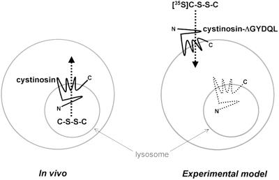
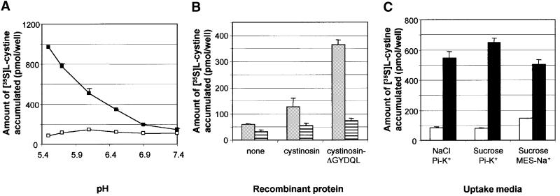
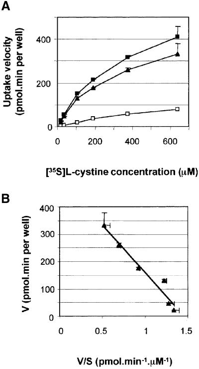
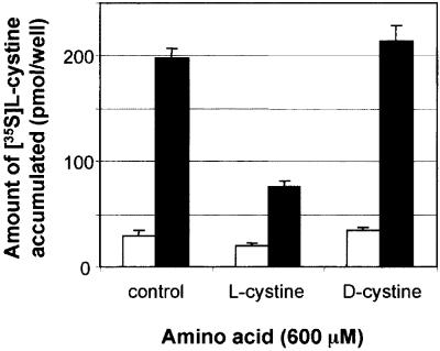
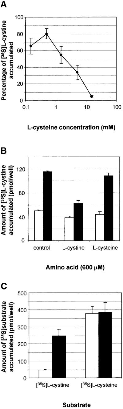
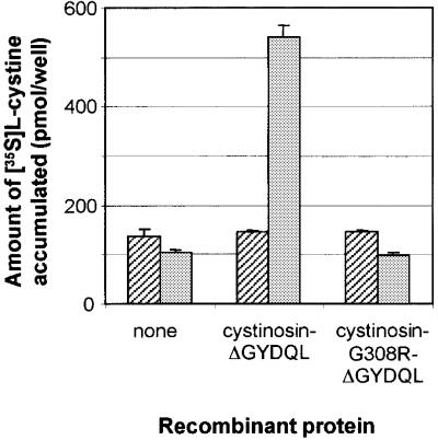
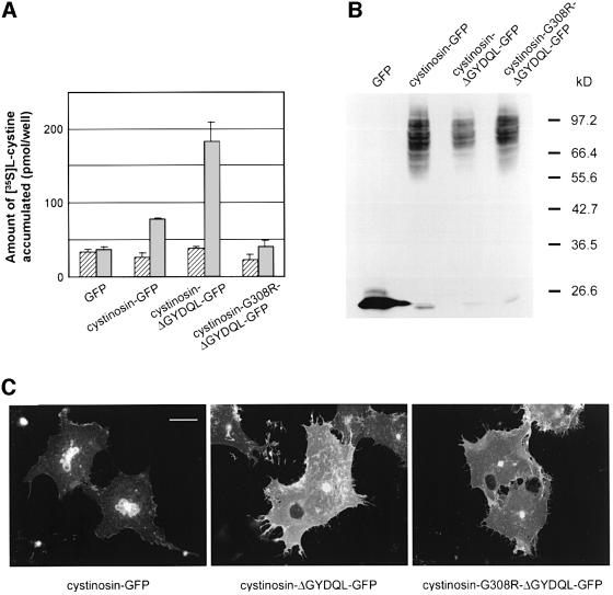
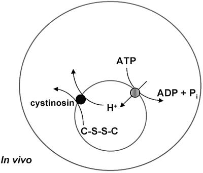
Cystinosis is an inherited lysosomal storage disease characterized by defective transport of cystine out of lysosomes. However, the causative gene, CTNS, encodes a seven transmembrane domain lysosomal protein, cystinosin, unrelated to known transporters. To investigate the molecular function of cystinosin, the protein was redirected from lysosomes to the plasma membrane by deletion of its C-terminal GYDQL sorting motif (cystinosin-DeltaGYDQL), thereby exposing the intralysosomal side of cystinosin to the extracellular medium. COS cells expressing cystinosin-DeltaGYDQL selectively take up L-cystine from the extracellular medium at acidic pH. Disruption of the transmembrane pH gradient or incubation of the cells at neutral pH strongly inhibits the uptake. Cystinosin-DeltaGYDQL is directly involved in the observed cystine transport, since this activity is highly reduced when the GYDQL motif is restored and is abolished upon introduction of a point mutation inducing early-onset cystinosis. We conclude that cystinosin represents a novel H(+)-driven transporter that is responsible for cystine export from lysosomes, and propose that cystinosin homologues, such as mammalian SL15/Lec35 and Saccharomyces cerevisiae ERS1, may perform similar transport processes at other cellular membranes.


Figures

None
Fig. 1. Rationale used for the cystine transport assay of cystinosin. In vivo, wild-type cystinosin is localized at the lysosomal membrane (smaller grey circle) and is thought to transport cystine (C-S-S-C) from the lysosomal lumen to the cytosol. In our experimental model, we have deleted the C-terminal lysosomal targeting signal from cystinosin (cystinosin-ΔGYDQL). This results in a partial redirection of this protein to the plasma membrane (larger grey circle) in transfected COS cells. In this model, the extracellular medium is topologically equivalent to the lysosomal lumen, and cystinosin-ΔGYDQL would thus act to transport [35S]cystine from the extracellular medium into the cytosol.
None
Fig. 2. Cystine uptake ability of cystinosin-ΔGYDQL-expressing cells. (A) Assay of transfected cells for [35S]cystine uptake in a neutral (pH 7.4, hatched bars) or acidic (pH 5.6, grey bars) extracellular medium. At neutral pH, cells expressing cystinosin-ΔGYDQL show a modest increase in the amount of accumulated [35S]cystine as compared with mock-transfected cells or wild-type cystinosin-expressing cells. At acidic pH, a dramatic increase in accumulated [35S]cystine is observed in cystinosin-ΔGYDQL-expressing cells but not in mock-transfected cells. A small amount of [35S]cystine is also taken up by wild-type cystinosin-expressing cells. Error bars correspond to the SEM for all figures. (B) Cystinosin-ΔGYDQL-mediated [35S]cystine uptake (black squares) remained linear for 10 min. [35S]cystine uptake mediated by mock-transfected cells is indicated by white squares. (C) Amount of accumulated [35S]cystine remaining after a 3 and a 6 min incubation with 20 µM digitonin treatment of mock-transfected (white squares) or cystinosin-ΔGYDQL-expressing (black squares) cells.
None
Fig. 3. Effect of a transmembrane pH gradient on cystine uptake. (A) [35S]cystine uptake by mock-transfected (white squares) and cystinosin-ΔGYDQL (black squares) expressing cells was performed in standard uptake buffer (see Materials and methods) adjusted to a pH ranging from 5.6 to 7.4 with 20 mM potassium phosphate. Cystinosin-mediated uptake increased with decreasing pH. (B) Amount of [35S]cystine accumulated by mock-transfected, cystinosin-expressing and cystinosin-ΔGYDQL-expressing cells in an acidic extracellular medium (grey boxes). The addition of 5 µM nigericin to the uptake media (striped boxes) abolished [35S]cystine uptake by cystinosin and cystinosin-ΔGYDQL, demonstrating the dependence of cystine transport on a proton gradient. (C) Effect of the presence of extracellular Na+ and K+ (uptake media containing NaCl as the major osmolyte, buffered with potassium phosphate K+-Pi), solely K+ (sucrose, Pi-K+) and solely Na+ (sucrose, MES-Na+) on [35S]cystine uptake by mock-transfected (white bars) and cystinosin-ΔGYDQL-expressing (black bars) cells. These changes do not significantly alter the amount of [35S]cystine taken up by cystinosin-ΔGYDQL, demonstrating that cystine transport does not require other ions.
None
Fig. 4. Saturation kinetics of cystinosin-mediated cystine uptake. (A) Plot of the velocity (V) of [35S]cystine uptake versus increasing substrate concentration (S) for mock-transfected (white squares) and cystinosin-ΔGYDQL-expressing (black squares) cells over an 8 min uptake period (linear phase). A deduction of background levels demonstrates that cystine uptake by cystinosin-ΔGYDQL is saturable (black triangles). (B) A linear Eadie–Hostee plot of the cystinosin-ΔGYDQL-dependent data demonstrates that cystine transport follows Michaelis–Menten kinetics. KM = 350 µM and Vmax = 507 pmol/min per well for this experiment.
None
Fig. 5. Stereoselectivity of cystine uptake. Assay of [35S]cystine uptake by mock-transfected (white bars) or cystinosin-ΔGYDQL (black bars) expressing cells in the absence (control) or presence of 600 µM l- or d-cystine. l-cystine inhibits [35S]cystine uptake by cystinosin-ΔGYDQL by >60%, whereas its stereoisomer d-cystine has no significant effect.
None
Fig. 6. Cysteine uptake ability of cystinosin-ΔGYDQL-expressing cells. (A) [35S]cystine accumulated by cystinosin-ΔGYDQL in the presence of increasing concentrations of l-cysteine (logarithmic scale) is expressed as a percentage of uptake in the absence of cysteine. Half-inhibition of [35S]cystine uptake was obtained for a cysteine concentration of 1.5 mM, a value ∼5-fold higher than the cystine concentration that half-saturates cystinosin (278 ± 49 µM). (B) At equal concentrations, l-cystine inhibits [35S]cystine uptake by cystinosin-ΔGYDQL (black bars), whereas l-cysteine has no effect. [35S]cystine uptake by mock-transfected cells is shown as white bars. (C) At equal substrate occupancy (i.e. in the presence of a 5-fold higher concentration of [35S]cysteine as opposed to [35S]cystine), cystinosin does not translocate cysteine significantly. Bars as for (B).
None
Fig. 7. Effect of the cystinotic point mutation G308R on cystine uptake. Amount of [35S]cystine accumulated by mock-transfected, cystinosin-ΔGYDQL-expressing and cystinosin-G308R-ΔGYDQL-expressing cells in a neutral (hatched bars) and acidic (grey bars) uptake medium. The introduction of the G308R point mutation associated with infantile cystinosis abolishes cystine transport by cystinosin-ΔGYDQL.
None
Fig. 8. Effect of G308R on the amount of recombinant protein produced or its subcellular localization. (A) Amount of [35S]cystine accumulated by cells expressing GFP or the fusion proteins cystinosin–GFP, cystinosin-ΔGYDQL–GFP and cystinosin-G308R-ΔGYDQL–GFP in a neutral (hatched bars) and acidic (grey bars) uptake medium. (B) Western blot analysis of the same lot of transfected cells using an anti-GFP monoclonal antibody demonstrates that cystinosin-G308R-ΔGYDQL–GFP is not produced at a lower level than cystinosin–GFP or cystinosin-ΔGYDQL–GFP. (C) Immunofluorescence studies on the same lot of transfected cells demonstrate that cystinosin-ΔGYDQL–GFP and cystinosin-G308R-ΔGYDQL–GFP have the same subcellular localization pattern, and that both of these fusion proteins are present at a much higher level at the plasma membrane than cystinosin–GFP. Scale bar 40 µm for all panels.
None
Fig. 9. Chemiosmotic coupling between cystinosin and the lysosomal H+-ATPase. The interior of the lysosome (small grey circle) is acidified by a H+-ATPase present in its membrane. The efflux of cystine (C-S-S-C) from the lysosome by the lysosomal cystine transporter, cystinosin, is coupled to an efflux of H+. Thus, the influx of H+ by the lysosomal H+-ATPase drives cystinosin-mediated cystine transport to the cystosol.

References

    1. Anand M., Rush,J.S., Ray,S., Doucey,M.A., Weik,J., Ware,F.E., Hofsteenge,J., Waechter,C.J. and Lehrman,M.A. (2001) Requirement of the Lec35 gene for all known classes of monosaccharide-P-dolichol-dependent glycosyltransferase reactions in mammals. Mol. Biol. Cell, 12, 487–501. - PMC - PubMed
    1. Anikster Y., Shotelersuk,V. and Gahl,W.A. (1999) CTNS mutations in patients with cystinosis. Hum. Mutat., 14, 454–458. - PubMed
    1. Attard M., Jean,G., Forestier,L., Cherqui,S., van’t Hoff,W., Broyer,M., Antignac,C. and Town,M. (1999) Severity of phenotype in cystinosis varies with mutations in the CTNS gene: predicted effect on the model of cystinosin. Hum. Mol. Genet., 8, 2507–2514. - PubMed
    1. Chairoungdua A. et al. (1999) Identification of an amino acid transporter associated with the cystinuria-related type II membrane glycoprotein. J. Biol. Chem., 274, 28845–28848. - PubMed
    1. Cherqui S., Kalatzis,V., Trugnan,G. and Antignac,C. (2001) The targeting of cystinosin to the lysosomal membrane requires a tyrosine-based signal and a novel sorting motif. J. Biol. Chem., 276, 13314–13321. - PubMed

Publication types

MeSH terms