. 2001 Nov 1;20(21):5971-81.
doi: 10.1093/emboj/20.21.5971.

The pre-autophagosomal structure organized by concerted functions of APG genes is essential for autophagosome formation

Affiliations

The pre-autophagosomal structure organized by concerted functions of APG genes is essential for autophagosome formation

K Suzuki et al. EMBO J. .

Abstract

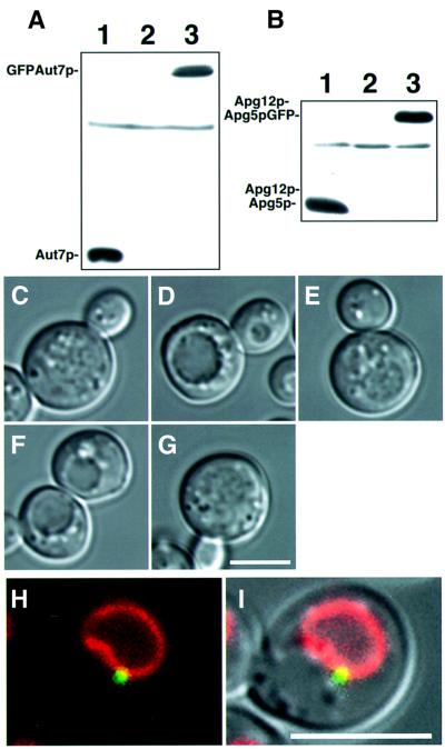
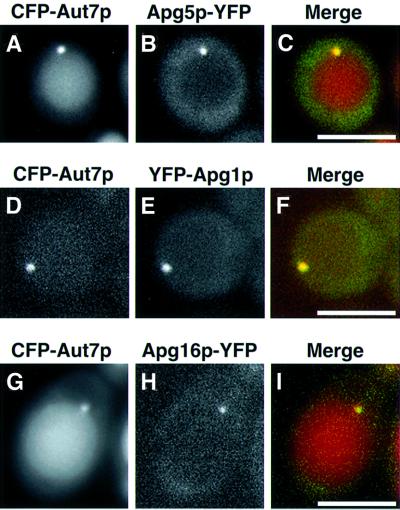
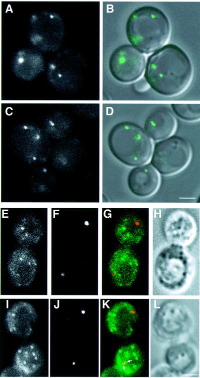
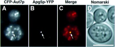
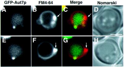
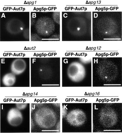
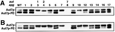
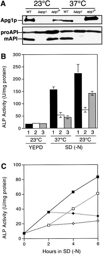
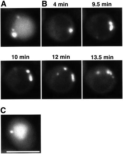
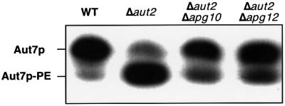
Macroautophagy is a bulk degradation process induced by starvation in eukaryotic cells. In yeast, 15 Apg proteins coordinate the formation of autophagosomes. Several key reactions performed by these proteins have been described, but a comprehensive understanding of the overall network is still lacking. Based on Apg protein localization, we have identified a novel structure that functions in autophagosome formation. This pre-autophagosomal structure, containing at least five Apg proteins, i.e. Apg1p, Apg2p, Apg5p, Aut7p/Apg8p and Apg16p, is localized in the vicinity of the vacuole. Analysis of apg mutants revealed that the formation of both a phosphatidylethanolamine-conjugated Aut7p and an Apg12p- Apg5p conjugate is essential for the localization of Aut7p to the pre-autophagosomal structure. Vps30p/Apg6p and Apg14p, components of an autophagy- specific phosphatidylinositol 3-kinase complex, Apg9p and Apg16p are all required for the localization of Apg5p and Aut7p to the structure. The Apg1p protein kinase complex functions in the late stage of autophagosome formation. Here, we present the classification of Apg proteins into three groups that reflect each step of autophagosome formation.


Figures

None
Fig. 1. Functional GFP–Aut7p and Apg5p–GFP are expressed from natural promoters at physiological levels. (A) The expression level of GFP–Aut7p under growing conditions. Cell lysates were prepared as described in Materials and methods. (1) Wild-type (KA311A), (2) Δaut7 (YYK218) and (3) Δaut7 (YYK218) expressing GFP–Aut7p. (B) The Apg12p–Apg5p conjugate was produced normally in Δapg5 cells expressing Apg5p–GFP. Cell lysates were prepared as described in Materials and methods. (1) Wild-type (KA311A), (2) Δapg5 (GYS59) and (3) Δapg5 (GYS59) expressing Apg5p–GFP. (CG) The accumulation of autophagic bodies was examined under a light microscope. Cells were incubated for 6 h in 0.17% yeast nitrogen base w/o amino acid and ammonium sulfate containing 1 mM PMSF. Nomarski images of (C) wild-type (KA311A), (D) Δaut7 (YYK218), (E) Δaut7 (YYK218) expressing GFP–Aut7p, (F) Δapg5 (GYS59) and (G) Δapg5 (GYS59) expressing Apg5p–GFP. (H–I) GFP–Aut7p visualized in Δaut7 cells (KVY5) under growing conditions. A punctate structure containing GFP–Aut7p is detected close to the vacuole, identified by FM4-64 labelling. (H) Fluorescence of GFP–Aut7p (green) and a FM4-64-labelled vacuole (red). (I) The Nomarski image is overlaid with GFP–Aut7p and FM4-64 fluorescence. The punctate structures, close to the vacuole, were detected in 43 of 206 cells (21%). Bar: 5 µm.
None
Fig. 2. Colocalization of Apg1p, Apg5p, Apg16p and Aut7p on a punctate structure close to the vacuole. (AC) Δapg5 cells (YNM119) expressing Apg5p–YFP and CFP–Aut7p were treated with rapamycin for 3 h: (A) Apg5p–YFP, (B) CFP–Aut7p, (C) merged image of Apg5p–YFP (green) and CFP–Aut7p (red). (DF) Δapg1 cells (NNY20) expressing CFP–Aut7p and YFP–Apg1p were treated with rapamycin for 30 min: (D) CFP–Aut7p, (E) YFP–Apg1p, (F) merged image of CFP–Aut7p (red) and YFP–Apg1p (green). (GI) Δapg16 cells (KVY117) expressing CFP–Aut7p and YFP–Apg16p were treated with rapamycin for 6 h: (G) CFP–Aut7p, (H) YFP–Apg16p, (I) merged image of CFP–Aut7p (red) and YFP–Apg16p (green). Bar: 5 µm.
None
Fig. 3. Localization of Aut7p and Apg9p–GFP. (A and B) Apg9p–GFP was visualized in Δapg9 cells (CTD1) under growing conditions: (A) Apg9p–GFP, (B) the Nomarski image is overlaid with the fluorescence of Apg9p–GFP. (C and D) Apg9p–GFP in Δapg9Δapg14 cells (GYS29) was identified under growing conditions: (C) Apg9p–GFP, (D) the Nomarski image is overlaid with the fluorescence of Apg9p–GFP. (EL) Δapg9 cells (CTD1) expressing Apg9p–GFP were treated with rapamycin for 2 h. Immunofluorescence microscopy was performed as described in Materials and methods. (E and I) Apg9p–GFP, (F and J) Aut7p, (G and K) merged images of Apg9p–GFP (green) and Aut7p (red) and (H and L) Nomarski images. Apg9p and Aut7p were occasionally found in close proximity (arrows in K). Bar: 2 µm.
None
Fig. 4. Localization of Aut7p and Apg5p in Δypt7 cells. Δypt7Δapg5 cells (YAK3) expressing CFP–Aut7p and Apg5p–YFP were treated with rapamycin for 5 h. (A) Autophagosomes stained with CFP–Aut7p; (B) Apg5p–YFP; (C) merged image of CFP–Aut7p (red) and Apg5p–YFP (green); (D) Nomarski image: CFP–Aut7p and Apg5p–YFP colocalized on a punctate structure (arrows). Bar: 5 µm.
None
Fig. 5. Localization of Aut7p and the class E compartment. The class E compartment of Δvps4 cells (MBY3) expressing GFP–Aut7p was labelled with FM4-64 as described in Materials and methods, following treatment with rapamycin for 2 h. (A and E) GFP–Aut7p. (B and F) The class E compartments stained with FM4-64 (arrows). (C and G) Merged images of GFP–Aut7p (green) and FM4-64 (red). (D and H) Nomarski images. Bar: 2 µm.
None
Fig. 6. Localization of GFP–Aut7p and Apg5p–GFP in the apg mutants grown in SD + CA medium. (A) Δapg1 cells (NNY20). GFP–Aut7p was detected on a punctate structure close to the vacuole in 30 of 161 cells (18%). Δapg13 (C; TFD13W2), Δapg17 (YYK111), apg2 (MT2-4-4) cells exhibited an identical phenotype. (B) Δapg1 cells (YYK36) expressing Apg5p–GFP. Punctate structures were detected in 11 of 91 cells (12%). Δapg13 (D; TFD13W2), Δapg17 (YYK111) and apg2 (MT2-4-4) cells demonstrated an identical phenotype. (E) This image of a Δaut2 cell (GYS6) expressing GFP–Aut7p is representative of the Aut7 system. The punctate structures were detected in one of 78 cells (1%). Δapg7 (GYS9) and Δaut1 (GYS5) cells exhibited the same phenotype. (F) Δaut2 cells (GYS6) expressing Apg5p–GFP. A single punctate structure was detected in 11 of 52 cells (21%). Δaut1 (GYS5) and Δaut7 (YYK218) cells demonstrated a similar phenotype. (G) This Δapg12 cell (GYS13) expressing GFP–Aut7p is representative of mutants of the Apg12 system. Punctate structures were detected in one of 84 cells (1%). Δapg10 (TFD10-L1) and Δapg5 (SKD5-1D) cells possessed an identical phenotype. (H) A Δapg5Δapg12 cell (YNM117) expressing Apg5p–GFP. A single punctate structure was detected in 16 of 104 cells (15%). Δapg7 (GYS9), Δapg10 (TFD10-L1) and Δapg5 (SKD5-1D) cells all exhibited an identical phenotype. (I) A Δapg14 cell (SKD14–1C) expressing GFP–Aut7p. None of the 51 cells displayed a punctate structure. Δvps30 cells (SKD6-1D) and Δapg9 cells (CTD1) possessed an identical phenotype. (J) A Δapg14 cell (AKY12) expressing Apg5p–GFP under growing conditions. Punctate structures were observed in one of 248 cells (<1%). Δvps30 cells (AKY74) and Δapg9 cells (CTD1) exhibited an identical phenotype. (K) A Δapg16 cell (KVY117) expressing GFP–Aut7p. No punctate structures were observed in any of the 77 cells. (L) A Δapg5Δapg16 cell (YNM126) expressing Apg5p–GFP under growing conditions. No punctate structures were observed in the 184 cells examined. Bar: 5 µm.
None
Fig. 7. Levels of Aut7p–PE present in each apg mutant. Lysates were prepared by glass bead disruption prior to SDS–PAGE as described in Materials and methods. (A) Cells in vegetative growth. (B) Cells following a 4.5 h starvation in SD (–N) medium. We examined the phenotype of wild type (SEY6210), Δapg1 (GYS102), apg2 (MT2-4-4), Δaut1 (KVY113), Δaut2 (KVY13), Δapg5 (KVY142), Δvps30 (KVY135), Δapg7 (KVY118), Δaut7 (KVY5), Δapg9 (KVY114), Δapg10 (KVY136), Δapg12 (KVY115), ΔΔapg13 (KVY116), Δapg14 (GYS115), Δapg16 (KVY117) and Δapg17 (YYK111) cells.
None
Fig. 8. apg1ts mutant possessing functional temperature sensitivity. (A) Although temperature-sensitive Apg1p was detectable, the maturation of API was completely blocked at 37°C in apg1ts cells. The API proform (proAPI) is processed to mature API (mAPI) in an Apg1p activity-dependent manner. Wild-type cells (TN125), Δapg1 cells (YYK126) and Δapg1 cells (YYK126) carrying the apg1ts plasmid were grown in YEPD medium at 23°C or 37°C overnight. Apg1p and API were detected as described in Materials and methods. (B) Δapg1 cells (YYK126) carrying (1) the APG1 plasmid, (2) the vector or (3) the apg1ts plasmid were grown in YEPD medium at 23°C. They were then transferred into SD (–N) medium at 23°C or 37°C and incubated for 6 h before ALP activity was measured. (C) Δapg1 cells (YYK126) carrying the apg1ts plasmid were grown in YEPD medium at 23°C. The cells were then transferred into SD (–N) medium and incubated at 23°C or 37°C. Closed squares, cells starved continuously at 23°C. Open squares, cells incubated for 2 h at 23°C and then transferred to 37°C. Open lozenges, cells starved continuously at 37°C. Closed lozenges, cells incubated for 2 h at 37°C and then transferred to 23°C.
None
Fig. 9. Time-lapse microscopy of apg1ts cells expressing GFP–Aut7p. Δapg1 cells (NNY20) carrying the apg1ts and GFP–Aut7p plasmids were used. (A) Cells treated with rapamycin for 3 h at 30°C. (B) Time-lapse images following temperature decrease. Cells were treated with rapamycin for 4 h at 37°C before decreasing the temperature to 30°C. (C) Cell incubated for 2 h at 30°C after temperature decrease. Bar: 5 µm.
None
Fig. 10. Amounts of Aut7p–PE in Δaut2 cells expressing Aut7FGp during vegetative growth. Lysates were prepared by glass bead disruption and subjected to SDS–PAGE as described in Materials and methods. The strains used in this experiment are as follows: wild type (SEY6210), Δaut2 (KVY13), Δaut2Δapg10 (KVY150) and Δaut2Δapg12 (KVY148).

References

    1. Abeliovich H., Darsow,T. and Emr,S.D. (1999) Cytoplasm to vacuole trafficking of aminopeptidase I requires a t-SNARE-Sec1p complex composed of Tlg2p and Vps45p. EMBO J., 18, 6005–6016. - PMC - PubMed
    1. Abeliovich H., Dunn,W.A., Jr, Kim,J. and Klionsky,D.J. (2000) Dissection of autophagosome biogenesis into distinct nucleation and expansion steps. J. Cell Biol., 151, 1025–1033. - PMC - PubMed
    1. Baba M., Takeshige,K., Baba,N. and Ohsumi,Y. (1994) Ultrastructural analysis of the autophagic process in yeast: detection of autophagosomes and their characterization. J. Cell Biol., 124, 903–913. - PMC - PubMed
    1. Babst M., Sato,T.K., Banta,L.M. and Emr,S.D. (1997) Endosomal transport function in yeast requires a novel AAA-type ATPase, Vps4p. EMBO J., 16, 1820–1831. - PMC - PubMed
    1. Cadwell R.C. and Joyce,G.F. (1995) Mutagenic PCR. In Dieffenbach,C.W. and Dveksler,G.S. (eds), PCR Primer. Cold Spring Harbor Laboratory Press, Cold Spring Harbor, NY, pp. 583–589.

Publication types

MeSH terms

Substances