. 2001 Nov 1;20(21):5999-6007.
doi: 10.1093/emboj/20.21.5999.

Cytoplasmic O-glycosylation prevents cell surface transport of E-cadherin during apoptosis

Affiliations

Cytoplasmic O-glycosylation prevents cell surface transport of E-cadherin during apoptosis

W Zhu et al. EMBO J. .

Abstract

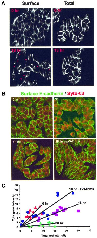
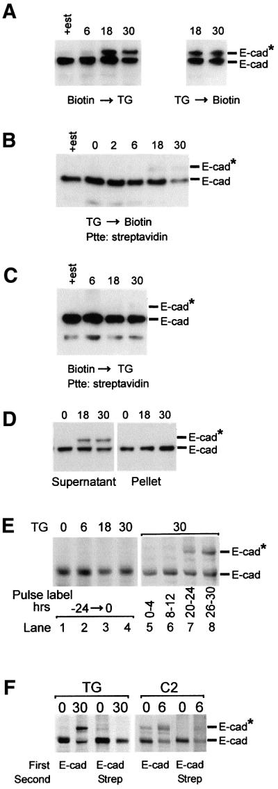
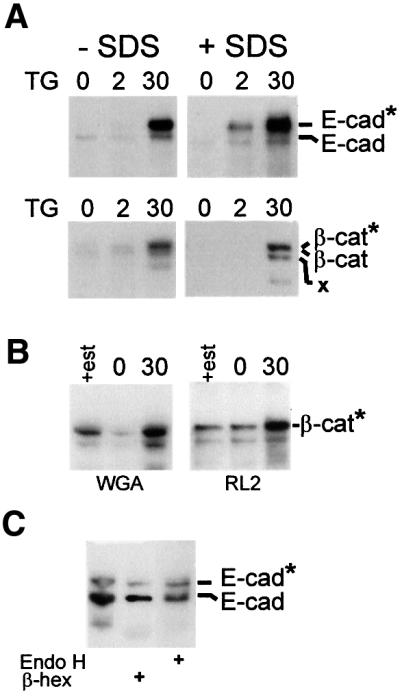
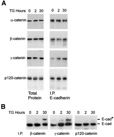
Cellular adhesion is regulated by members of the cadherin family of adhesion receptors and their cytoplasmic adaptor proteins, the catenins. Adhesion complexes are regulated by recycling from the plasma membrane and proteolysis during apoptosis. We report that in MCF-7, MDA-MB-468 and MDCK cells, induction of apoptosis by agents that cause endoplasmic reticulum (ER) stress results in O-glycosylation of both beta-catenin and the E-cadherin cytoplasmic domain. O-glycosylation of newly synthesized E-cadherin blocks cell surface transport, resulting in reduced intercellular adhesion. O-glycosylated E-cadherin still binds to beta- and gamma-catenin, but not to p120-catenin. Although O-glycosylation can be inhibited with caspase inhibitors, cleavage of caspases associated with the ER or Golgi complex does not correlate with E-cadherin O-glycosylation. However, agents that induce apoptosis via mitochondria do not lead to E-cadherin O-glycosylation, and decrease adhesion more slowly. In MCF-7 cells, this is due to degradation of E-cadherin concomitant with cleavage of caspase-7 and its substrate poly(ADP-ribose) polymerase. We conclude that cytoplasmic O-glycosylation is a novel, rapid mechanism for regulating cell surface transport exploited to down-regulate adhesion in some but not all apoptosis pathways.


Figures

None
Fig. 1. Loss of E-cadherin from the cell surface during apoptosis is blocked by the caspase inhibitor zVAD-fmk. (A) E-cadherin localized by immunofluorescence and confocal microscopy after estrogen depletion (0 h) and incubation with thapsigargin (TG) for 18 h. E-cadherin was localized using a monoclonal antibody to an extracellular epitope and FITC-labeled secondary antibodies. Staining of non-permeabilized cells identifies E-cadherin on the plasma membrane (Surface). Permeabilization of the cells with Triton X-100 prior to labeling allows visualization of all of the E-cadherin in the cell (Total). Extracellular E-cadherin staining is reduced at areas of cell contact after 18 h exposure to thapsigargin (arrowheads). (B) Non-permeabilized cells were stained with a monoclonal antibody to E-cadherin and secondary antibodies labeled with FITC (green). Cell contents were labeled using Syto-63, a dye that in fixed cells stains principally extranuclear nucleic acids. The intensity of green relative to red indicates the amount of E-cadherin on the cell surface after treatment with thapsigargin for 0, 18 and 30 h. Addition of zVAD-fmk prevents loss of E-cadherin from the cell surface. (C) Quantification of cell surface E-cadherin by image analysis. Each point represents one image similar to those seen in (B). Images were thresholded auto matically to remove background noise and the total amount of red and green staining summed. The total red intensity is directly proportional to cell number in each image; therefore, the slope of each line reflects the amount of E-cadherin externalized/cell. Lines of best fit were calculated by linear regression. After estrogen depletion, prior to thapsigargin, 0 h, red triangles; incubation in thapsigargin 18 h, purple squares; with zVAD-fmk, blue circles. Incubation in thapsigargin 30 h, green diamonds.
None
Fig. 2. Newly synthesized E-cadherin is modified and blocked for cell surface transport. (A) Biotinylation of cell surface proteins before or after adding thapsigargin does not affect the total amount or modification of E-cadherin. Cells were grown for 6 days in estrogen-depleted medium (+est indicates cells in estrogen-replete medium) and then cell surface proteins biotinylated using EZ-Link™ Biotin for 30 min prior to (left panel) or after (right panel) addition of thapsigargin. Cells were incubated in thapsigargin for the time indicated above the lanes. After solubilization using a buffer containing Triton X-100, proteins were separated by SDS–PAGE and E-cadherin was identified by immunoblotting. (B) Modified E-cadherin is not transported to the cell surface. Cells were grown in estrogen-depleted medium, and treated with thapsigargin for the time indicated above the lanes. Cell surface proteins were biotinylated and solubilized; biotinlyated proteins were isolated using streptavidin magnetic microspheres (Ptte: streptavidin), separated by SDS–PAGE and E-cadherin identified by immunoblotting. (C) Pre-existing cell surface E-cadherin is not modified during apoptosis. E-cadherin on the cell surface was biotinylated and cells treated with thapsigargin. At the times indicated, biotinylated molecules were isolated, and analyzed by SDS–PAGE and immunoblotting. (D) Modified E-cadherins are not found in the Triton X-100-insoluble cytoskeleton. Cells were grown in estrogen-depleted medium, treated with thapsigargin for the times indicated and solubilized with buffer containing 1% Triton X-100. The Triton X-100-insoluble cytoskeleton was pelleted by centrifugation and solubilized with SDS–PAGE loading buffer. Aliquots containing proteins from 40 000 cells were separated by SDS–PAGE and E-cadherin identified by immunoblotting. (E) Newly synthesized but not pre-existing E-cadherin is modified during thapsigargin-induced apoptosis. Lanes 1–4: cells were labeled continuously with [35S]methionine for 24 h and apoptosis induced with thapsigargin for the time indicated above the lanes; E-cadherin was immunoprecipitated from Triton X-100 cell lysates and analyzed by SDS–PAGE. Lanes 5–8: thapsigargin was added to the cells and then incubated for 30 h. The cells were pulse-labeled with [35S]methionine during the time period indicated below the lanes. E-cadherin was immunoprecipitated from Triton X-100 cell lysates and analyzed by SDS–PAGE (separation time 7.5 h). (F) Cell surface transport of modified E-cadherin is blocked selectively. Untreated cells (0) or cells exposed to thapsigargin (TG) or C2-ceramide (C2) for the time indicated above the lanes (h) were labeled with [35S]methionine for 4 h and cell surface proteins biotinylated. The cells were lysed in Triton X-100, and E-cadherin immunoprecipitates were analyzed by SDS–PAGE (separation time 7.5 h) and autoradiography either directly (first two lanes in each panel) or after release with SDS and re-precipitation with streptavidin (last two lanes in each panel). The migration positions of E-cadherin (E-cad) and modified E-cadherin (E-cad*) are indicated to the right of the panels. Addition of streptavidin magnetic microspheres is indicated as Streptavidin or Strep.
None
Fig. 3. Cytoplasmic O-glycosylation of E-cadherin and β-catenin during apoptosis. (A) Binding of E-cadherin and β-catenin to WGA increases during apoptosis. MCF-7 cells were incubated for the indicated time after thapsigargin, and then lysed in Triton X-100 (–SDS) or incubated in 10 mM Tris pH 7.5, 100 mM NaCl, 0.4% sodium deoxycholate, 0.3% SDS, 1% NP-40 to dissociate protein complexes. After incubation with WGA–agarose, beads were washed with the same buffer, and the precipitated proteins separated by SDS–PAGE and analyzed by immunoblotting with antibodies to E-cadherin (top panels) or β-catenin (bottom panels). The migration positions of E-cadherin and β-catenin are indicated; * designates the migration position of the glycosylated forms of the molecules. X indicates a breakdown product of β-catenin seen in some but not all experiments. (B) Thapsigargin increased binding of β-catenin to a monoclonal antibody directed towards O-GlcNAc on some proteins. Cells were treated with thapsigargin for 30 h and Triton X-100-soluble proteins were precipitated with WGA–agarose or after incubation with mAb RL2 with protein G–Sepharose. The precipitated proteins were analyzed by SDS–PAGE and immunoblotting for β-catenin. (C) The modified form of E-cadherin is digested preferentially by β-hexosaminidase. E-cadherin was precipitated from Triton X-100 cell lysates of cells treated for 30 h with thapsigargin using a monoclonal antibody; the precipitated products were incubated with either β-hexosaminidase (β-hex) or endoglycosidase H (Endo H). After incubation with these enzymes overnight, the products were analyzed by SDS–PAGE and immunoblotting. Although all three lanes come from the same experiment, contaminating protease activity in reactions containing β-hexosaminidase resulted in digestion of some of the E-cadherin and, therefore, a longer exposure of lane 2 of the blot to film was necessary to visualize the bands at approximately the same density as the proteins incubated with endoglycosidase H.
None
Fig. 4. (A) Modification of E-cadherin in response to thapsigargin is blocked by the caspase inhibitors zVAD-fmk (V), DEVD-CHO (D) and YVAD-CHO (Y). After the addition of thapsigargin, MCF-7 cells were incubated in the indicated concentration of inhibitor for 30 h, lysed in Triton X-100, analyzed by SDS–PAGE and immunoblotted for E-cadherin. Identical results were obtained using DEVD-fmk and YVAD-fmk (not shown). The migration positions of E-cadherin (E-cad) and modified E-cadherin (E-cad*) are indicated to the right of the panels. (B) E-cadherin is O-glycosylated in MCF-7, MB-MDA-468 and MDCK cells when apoptosis is induced by thapsigargin (TG) or C2-ceramide (C) but not adriamycin (A). After the addition of thapsigargin, C2-ceramide or adriamycin, cells were incubated for 30, 6 or 18 h, respectively, and then lysed in Triton X-100 and analyzed by SDS–PAGE and immunoblotting. None of the cell lines were estrogen depleted prior to drug treatment. The migration positions of E-cadherin (E-cad) and modified E-cadherin (E-cad*) are indicated to the right. (C) Degradation of E-cadherin correlates with activation of caspase-7. Cells were incubated in thapsigargin (TG), adriamycin (A) or TNF-α/cycloheximide (TNF) for the times indicated, lysed in Triton X-100 and analyzed by SDS–PAGE and immunoblotting with antibodies to E-cadherin (E-cad), PARP, caspase-7, caspase-2 or caspase-12. Migration positions for the relevant proteins and their cleavage products are illustrated to the right. A longer exposure of the Δcaspase-7 region of the blot is also provided to facilitate visualization of the primary cleavage product. The monoclonal antibody for rodent caspase-12 recognizes a single protein species in untreated MCF-7, Jurkat (J) and HL60 (H) cells. Caspase-2 and the ‘caspase-12-like’ protein are cleaved when cells are grown in estrogen-depleted medium (Δcaspase-2, Δcaspase-12, respectively).
None
Fig. 5. E-cadherin–catenin binding during apoptosis. (A) E-cadherin continues to bind to catenins during thapsigargin (TG)-induced apoptosis. After the addition of thapsigargin, MCF-7 cells were incubated for the indicated time and then lysed in Triton X-100 and analyzed either directly (Total Protein) or after immunoprecipitation with antibodies to E-cadherin (I.P. E-cadherin). Samples were separated by SDS–PAGE, transferred to a PVDF membrane and immunoblotted with the indicated antibody. (B) Glycosylated E-cadherin binds β- and γ-catenin but not p120-catenin. After the addition of thapsigargin, MCF-7 cells were incubated for the indicated time and then lysed in Triton X-100 and, after immunoprecipitation with antibodies to β-, γ- or p120-catenin, analyzed by SDS–PAGE and immunoblotting for E-cadherin. The migration positions of E-cadherin (E-cad) and modified E-cadherin (E-cad*) are indicated to the right of the panels.

References

    1. Anastasiadis P.Z., Moon,S.Y., Thoresen,M.A., Mariner,D.J., Crawford,H.C., Zheng,Y. and Reynolds,A.B. (2000) Inhibition of RhoA by p120 catenin. Nature Cell Biol., 2, 637–644. - PubMed
    1. Annis M., Zamzami,N., Zhu,W., Penn,L.Z., Kroemer,G., Leber,B. and Andrews,D.W. (2000) Endoplasmic reticulum localized Bcl-2 prevents apoptosis when redistribution of cytochrome c is a late event. Oncogene, 20, 1939–1952. - PubMed
    1. Chen Y.T., Stewart,D.B. and Nelson,W.J. (1999) Coupling assembly of the E-cadherin/β-catenin complex to efficient endoplasmic reticulum exit and basal–lateral membrane targeting of E-cadherin in polarized MDCK cells. J. Cell Biol., 144, 687–699. - PMC - PubMed
    1. Comer F.I. and Hart,G.W. (2000) O-Glycosylation of nuclear and cytosolic proteins. Dynamic interplay between O-GlcNAc and O-phosphate. J. Biol. Chem., 275, 29179–29182. - PubMed
    1. Daniel J.M. and Reynolds,A.B. (1997) Tyrosine phosphorylation and cadherin/catenin function. Bioessays, 19, 883–891. - PubMed

Publication types

MeSH terms