. 2001 Nov 1;20(21):6008-16.
doi: 10.1093/emboj/20.21.6008.

The vasorelaxant effect of H(2)S as a novel endogenous gaseous K(ATP) channel opener

Affiliations

The vasorelaxant effect of H(2)S as a novel endogenous gaseous K(ATP) channel opener

W Zhao et al. EMBO J. .

Abstract

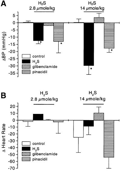
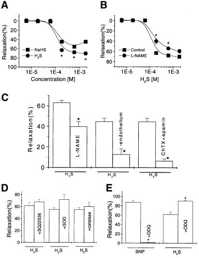
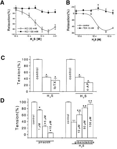
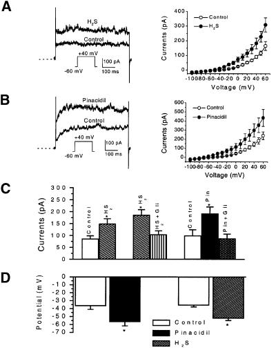
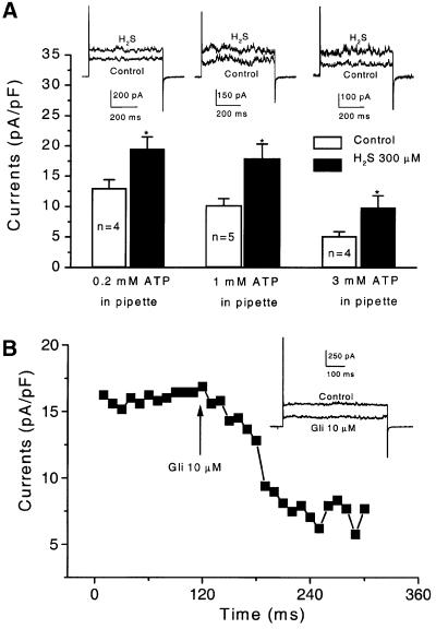
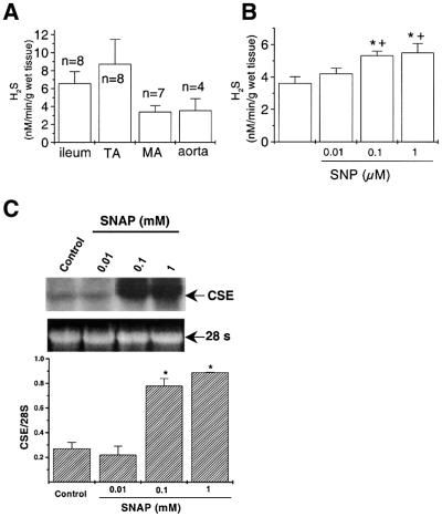
Hydrogen sulfide (H(2)S) has been traditionally viewed as a toxic gas. It is also, however, endogenously generated from cysteine metabolism. We attempted to assess the physiological role of H(2)S in the regulation of vascular contractility, the modulation of H(2)S production in vascular tissues, and the underlying mechanisms. Intravenous bolus injection of H(2)S transiently decreased blood pressure of rats by 12- 30 mmHg, which was antagonized by prior blockade of K(ATP) channels. H(2)S relaxed rat aortic tissues in vitro in a K(ATP) channel-dependent manner. In isolated vascular smooth muscle cells (SMCs), H(2)S directly increased K(ATP) channel currents and hyperpolarized membrane. The expression of H(2)S-generating enzyme was identified in vascular SMCs, but not in endothelium. The endogenous production of H(2)S from different vascular tissues was also directly measured with the abundant level in the order of tail artery, aorta and mesenteric artery. Most importantly, H(2)S production from vascular tissues was enhanced by nitric oxide. Our results demonstrate that H(2)S is an important endogenous vasoactive factor and the first identified gaseous opener of K(ATP) channels in vascular SMCs.


Figures

None
Fig. 1. The effect of H2S in vivo on mean arterial blood pressure (BP) and heart rate of rats. (A) Intravenous injection of H2S induced significant decrease in BP. This effect was mimicked by intravenous injection of pinacidil (2.8 µmol/kg) and antagonized by a prior intravenous injection of glibenclamide (2.8 µmol/kg). (B) Effect of H2S on rat heart rate. Heart rate was recorded 30 s after intravenous injection of PBS (control), H2S or pinacidil. *P < 0.05 compared with control.
None
Fig. 2. The H2S-induced relaxation of rat aortic rings and the underlying mechanisms. (A) Relaxation of the PHE-precontracted tissues by H2S in the form of either standard NaHS solution (square) or H2S gas-saturated solution (circle). (B) Inhibitory effect of l-NAME (100 µM, 20 min, circle) on the H2S-induced relaxation (control, square). (C) The effects of H2S (180 µM) on the endothelium-free or endothelium-intact aortic tissues pretreated with l-NAME or charybdotoxin (ChTX)/apamin. (D) The relaxant effect of H2S was not affected by pretreating the tissues with SQ22536, SOD or catalase, respectively. (E) The effect of ODQ treatment (10 µM for 10 min) on the relaxant effects of SNP (0.1 µM) or H2S (600 µM). n = 8 for each data point. *P < 0.05 compared with control.
None
Fig. 3. The K+ channel-mediated vascular effects of H2S. (A) The relaxant effect of H2S on the aortic tissues precontracted with 20 or 100 mM KCl. (B) Inhibitory effect of TEA on the H2S-induced vasorelaxation. The concentration-dependent vasorelaxant effects of H2S with or without TEA (10 mM) pretreatment of aortic rings were determined. (C) The H2S (600 µM)-induced vasorelaxation was not affected by pretreatment of aortic tissues with either 10 µM iberiotoxin (IbTX) or 2.5 mM 4-aminopyridine (4-AP). *P < 0.05 compared with control, n = 8. (D) The vasoactive effects of pinacidil (left) and H2S (right) on the precontracted aortic tissues. The vasorelaxant effect of H2S (600 µM) was examined in the presence of glibenclamide at different concentrations. *P < 0.05 compared with control, **P < 0.05 compared with the H2S group in the absence of glibenclamide, n = 8.
None
Fig. 4. The effect of H2S and pinacidil on KATP channel currents in rat aortic SMCs. (A) The effect of H2S on KATP channel currents. The original records from one cell (left) and the mean current–voltage relationship of six cells (right) in the absence and then presence of H2S (300 µM) are shown. (B) The effect of pinacidil (5 µM) on KATP channel currents. The original records from one cell (left) and the mean current–voltage relationship of five cells (right) in the absence and then presence of pinacidil are shown. Dashed line indicates zero current level. (C) The antagonistic effect of glibenclamide (Gli, 5 µM) on the effect of H2S or pinacidil (Pin) on KATP channel currents (holding potential, –60 mV; test potential, +40 mV). (D) The membrane hyperpolarization induced by H2S (300 µM) or pinacidil (5 µM), respectively. *P < 0.05 compared with control; n = 3–7 for each group.
None
Fig. 5. The modulation of KATP channel currents by H2S with different intracellular ATP concentrations and by glibenclamide (Gli) in rat aortic SMCs. (A) The ATP-dependence of the H2S (300 µM)-induced increases in KATP channel currents. Holding potential, –60 mV; test potential, +40 mV. *P < 0.05 compared with control. (B) The time course of the inhibition of KATP currents by glibenclamide. Aortic SMCs were dialysed with 0.2 mM ATP and 1 mM GDP. The voltage pulses of +30 mV were applied from a holding potential of –60 mV. Inset shows the representative of original current traces before and after the application of 10 µM glibenclamide.
None
Fig. 6. Differential expression of CSE in rat vascular tissues. (A) RT–PCR analysis of the expression of CSE (234 bp) in rat liver, mesenteric artery, tail artery, pulmonary artery and aorta. (B) Quantitative comparison of CSE mRNA levels in rat tail artery, mesenteric artery, pulmonary artery and aorta with RPA. This is representative of three experiments. *P < 0.05 compared with mesenteric artery; **P < 0.01 compared with tail artery, mesenteric artery and aorta. A, artery. (C) The transcriptional expressions of CSE and β-actin in cultured SMCs and EC (endothelial cell) detected by RT–PCR. (DIn situ hybridization showing the location of CSE mRNA in rat aorta wall by antisense probe on the left and sense probe as control on the right.
None
Fig. 7. Regulation of the endogenous H2S production in different rat tissues. (A) Accumulated endogenous H2S levels in rat tail artery (TA), mesenteric artery (MA), aorta and ileum. (B) H2S production rate of aorta tissues was stimulated by SNP in a concentration-dependent manner. *P < 0.05 compared with control, n = 3. (C) The SNAP-induced concentration-dependent upregulation of CSE transcriptional expression in cultured aortic SMCs, determined using northern blotting. The 28S ribosome RNA was assayed as the housekeeping control. n = 3, *P < 0.05 compared with control.

Similar articles

Cited by

References

    1. Abe K. and Kimura,H. (1996) The possible role of hydrogen sulfide as an endogenous neuromodulator. J. Neurosci., 16, 1066–1071. - PMC - PubMed
    1. Beech D.J., Zhang,H., Nakao,K. and Bolton,T.B. (1993) K channel activation by nucleotide diphosphates and its inhibition by glibenclamide in vascular smooth muscle cells. Br. J. Pharmacol., 110, 573–582. - PMC - PubMed
    1. Broillet M.C. (1999) S-nitrosylation of proteins. Cell Mol. Life Sci., 55, 1036–1042. - PMC - PubMed
    1. Doughty J.M., Plane,F. and Langton,P.D. (1999) Charybdotoxin and apamin block EDHF in rat mesenteric artery if selectively applied to the endothelium. Am. J. Physiol., 276, H1107–H1112. - PubMed
    1. Hattori Y., Kawasaki,H., Fukao,M. and Kanno,M. (1995) Phorbol esters elicit Ca2+-dependent delayed contractions in diabetic rat aorta. Eur. J. Pharmacol., 279, 51–58. - PubMed

Publication types

MeSH terms