. 2001 Nov 1;20(21):6037-49.
doi: 10.1093/emboj/20.21.6037.

Two mechanisms activate PTPalpha during mitosis

Affiliations

Two mechanisms activate PTPalpha during mitosis

X M Zheng et al. EMBO J. .

Abstract

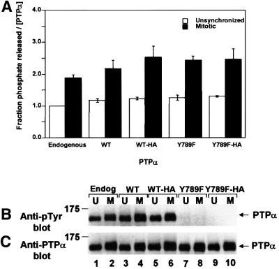
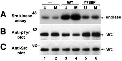
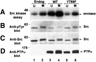
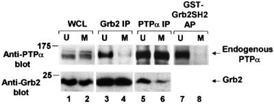
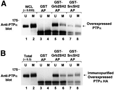
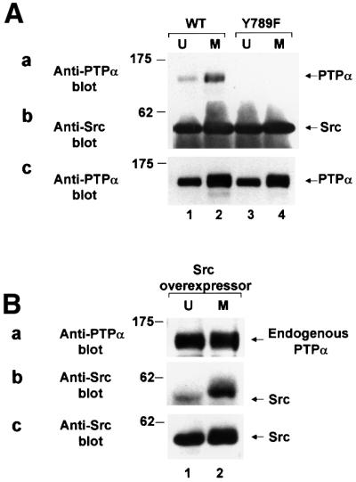
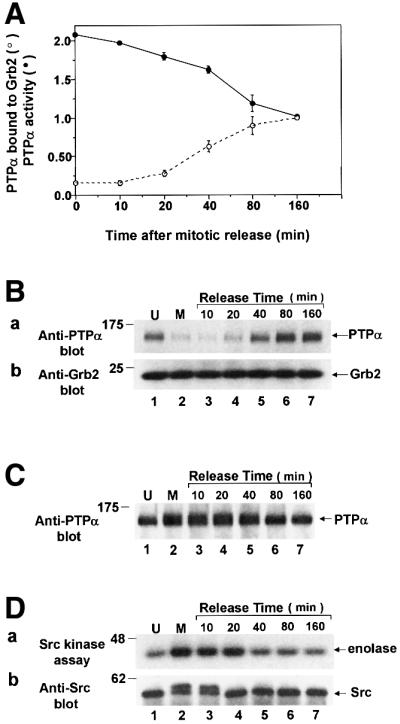
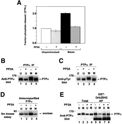
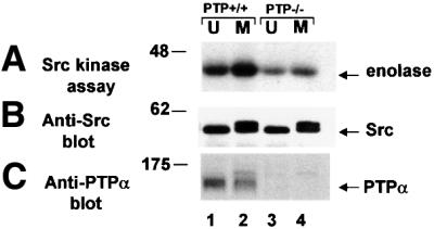
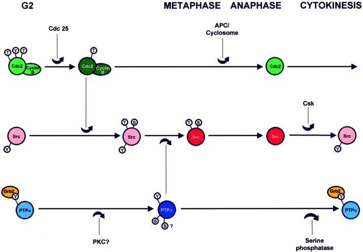
We show that, dependent on serine hyperphosphorylation, protein tyrosine phosphatase alpha (PTPalpha) is activated by two different mechanisms during mitosis: its specific activity increases and its inhibitory binding to Grb2 decreases. The latter effect probably abates Grb2 inhibition of the phosphotyrosine displacement process that is required specifically for Src dephosphorylation and causes a mitotic increase in transient PTPalpha-Src binding. Thus, part of the increased protein tyrosine phosphatase activity may be specific for Src family members. These effects cease along with Src activation when cells exit mitosis. Src is not activated in mitosis in PTPalpha-knockout cells, indicating a unique mitotic role for this phosphatase. The activation of PTPalpha, combined with the effects of mitotic Cdc2-mediated phosphorylations of Src, quantitatively accounts for the mitotic activation of Src, indicating that PTPalpha is the membrane-bound, serine phosphorylation-activated, protein tyrosine phosphatase that activates Src during mitosis.


Figures

None
Fig. 1. Phosphatase activity of PTPα from unsynchronized and mitotic cells. Endogenous mouse PTPα (Endog) and overexpressed human PTPα (WT), PTPα-HA (WT-HA), PTPα(Y789F) (Y789F) and PTPα(Y789F)-HA (Y789F-HA) were immunoprecipitated with anti-PTPα(D2) antibody from lysates from non-overexpressor or overexpressor NIH 3T3-derived cells that were either unsynchronized (U) or arrested in mitosis (M). (The lysates were adjusted to contain approximately equal amounts of PTPα.) Aliquots of immunoprecipitates were incubated with [32P]pTyr-containing MBP in phosphatase buffer for 15 min at 30°C or subjected to anti-pTyr or-PTPα immunoblots. (A) Amount of 32P released per molecule of PTPα after 15 min incubation, normalized to the amount released by overexpressed PTPα from unsynchronized cells. Error bars indicate the SEM. (B) Anti-pTyr immunoblot of the immunoprecipitated PTPα. (C) Anti-PTPα immunoblot of the immunoprecipitated PTPα. SDS–PAGE was in 10% gels. The positions of molecular weight standards are indicated in kDa.
None
Fig. 2. Tyrosine-dephosphorylation and activation of Src in vitro by PTPα from unsynchronized and mitotic cells. Src that had been immunoprecipitated from chicken-Src overexpressing NIH 3T3-derived cells was incubated in phosphatase buffer with PTPα-HA (lanes 3 and 4) and PTPα(Y789F)-HA (lanes 5 and 6) that had been immuno purified from unsynchronized or mitotic PTPα-overexpressor cells using anti-HA antibody or with mock-immunopurified protein from control cells that did not express any HA-tagged protein (–, lanes 1 and 2). The partially dephosphorylated Src immunoprecipitates were washed to remove PTPα and then incubated with enolase and [γ-32P]ATP in kinase buffer. (A) Autoradiograph of [32P]enolase after the Src kinase assay. (B) Anti-pTyr immunoblot of the Src immuno precipitates after PTPα treatment. (C) Anti-Src immunoblot of the immunoprecipitates. SDS–PAGE was in 10% gels. The positions of molecular weight standards are indicated in kDa.
None
Fig. 3. Src kinase activity and Tyr527 phosphorylation in unsynchronized and mitotic normal and PTPα-overexpressor cells. Endogenous Src was immunoprecipitated from unsynchronized (U) or mitotic (M) non-overexpressor cells (lanes 1–2), or cells overexpressing wt PTPα-HA (lanes 3 and 4) or PTPα(Y789F)-HA (lanes 5 and 6). The immunoprecipitates were split into aliquots and used in the assays shown in A–D. (A) The in vitro kinase activity of the Src immunoprecipitate was measured by incubating it with enolase substrate and [γ-32P]ATP in phosphorylation buffer, separating the reaction products by electrophoresis and autoradiography. (B) Anti-phosphotyrosine immunoblot of the immunoprecipitate. (C) Anti-Src immunoblot of the immunoprecipitate. (D) Anti-PTPα immunoblot of cell lysates containing 10 µg total cell protein. SDS–PAGE was in 9% gels. The positions of molecular weight standards are indicated in kDa.
None
Fig. 4. Decreased in vivo binding of Grb2 to PTPα during mitosis. The association of endogenous PTPα with Grb2 in unsynchronized and mitotic cells was measured by complementary co-immunoprecipitation experiments and by Grb2 SH2 domain affinity-precipitation experiments. Immunoprecipitates made with either anti-Grb2 (lanes 3 and 4) or -PTPα (lanes 5 and 6) antibody and lysates from unsynchronized (U) or mitotic (M) non-overexpressor cells were analyzed by 10% SDS–PAGE and immunoblotted with anti-PTPα or -Grb2 antibody as indicated. Lanes 7 and 8: PTPα was affinity-precipitated from lysates from unsynchronized or mitotic cells using GST–Grb2 SH2 domain fusion protein bound to Sepharose beads and immunoblotted with anti-PTPα antibody only. Direct immunoblots of the whole cell lysates used in lanes 5 and 6 (lanes 1 and 2, bottom panel) or lanes 7 and 8 (lanes 1 and 2, top panel) are also shown. To optimize detection, the different experiments used lysates containing differing amounts of total cell protein: lanes 1 and 2, top, 0.05 mg; lanes 1 and 2 bottom, 0.025 mg; lanes 3 and 4, 0.4 mg; lanes 5 and 6, 1.5 mg; lanes 7 and 8, 1 mg. The positions of molecular weight standards are indicated in kDa.
None
Fig. 5. In vitro binding of unsynchronized and mitotic PTPα to the Grb2 and Src SH2 domains. (A) Lysates from unsynchronized (U, odd lanes) or mitotic (M, even lanes) PTPα-HA overexpressor cells were affinity-precipitated by incubation with GST (lanes 3 and 4), a GST–Grb2 SH2 domain fusion protein (lanes 5 and 6) or a GST–Src SH2 domain fusion protein (lanes 7 and 8) bound to Sepharose beads. The washed beads were then analyzed by 9% SDS–PAGE and anti-PTPα immunoblotting. For comparison, lanes 1 and 2 (WCL) contained 0.033 times the amount of complete whole cell lysate. (B) PTPα-HA was immunopurified from unsynchronized or mitotic overexpressor cells and affinity-precipitated by GST or the GST–SH2 domain fusion proteins used above, analyzed by 9% SDS–PAGE and immunoblotted with anti-PTPα antibody (lanes 3–8). For comparison, lanes 1 and 2 (Total) contain 0.3 times the amount of immunopurified PTPα used in the affinity precipitations. The positions of molecular weight standards are indicated in kDa.
None
Fig. 6. Increased co-association in vivo of Src and PTPα during mitosis. (A) Immunoprecipitates made with anti-Src antibody from lysates (containing 1.5 mg total cell protein) from unsynchronized (U, odd lanes) or mitotic (M, even lanes) cells overexpressing wt PTPα-HA (lanes 1–2) or PTPα(Y789F)-HA (lanes 3–4) were analyzed by 9% SDS–PAGE and immunoblotted with anti-PTPα or -Src antibody. The positions of molecular weight standards are indicated in kDa. (a) Anti-PTPα immunoblot of anti-Src immunoprecipitate. (b) Anti-Src immunoblot of anti-Src immunoprecipitate. (c) Anti-PTPα immunoblot of cell lysates containing 10 µg total cell protein. (B) Immunoprecipitates made with anti-PTPα antibody from lysates (containing 1.5 mg total cell protein) from unsynchronized or mitotic Src-overexpressor cells were analyzed by 10% SDS–PAGE and immunoblotted with anti-Src or -PTPα antibody. The positions of molecular weight standards are indicated in kDa. (a) Anti-PTPα immunoblot of anti-PTPα immunoprecipitate. (b) Anti-Src immunoblot of anti-PTPα immunoprecipitate. (c) Anti-Src immunoblot of cell lysates containing 5 µg total cell protein.
None
Fig. 7. PTPα tyrosine phosphatase activity and Grb2 binding following release from mitotic arrest. Mitotic non-overexpressor cells were collected after arrest with nocodazole and were then incubated for the indicated amounts of time in normal medium without nocodazole. Cell lysates were then prepared and analyzed for PTPα activity, binding to Grb2 in vivo, electrophoretic mobility and Src activity. (A) The ability of immunoprecipitated endogenous PTPα to dephosphorylate [32P]pTyr-containing MBP was measured as described in Figure 1. The amount of 32P released per molecule of PTPα normalized to the amount released by overexpressed PTPα after 160 min is shown (filled circles). The binding of PTPα to Grb2 was measured in experiments like the one shown in Figure 7B. The amount of PTPα bound normalized to the amount bound after 160 min release is shown (open circles). Error bars indicate the SEM (two experiments). (B) Grb2 was immunoprecipitated from the cell lysates and the amount of co-immunoprecipitated endogenous PTPα was revealed by anti-PTPα immunoblotting (panel a). Immunoblotting of the lower portion of the blot with anti-Grb2 antibody showed that there was no change in the level of Grb2 throughout the release period (panel b). Experimental procedures were as in Figure 4. (C) Anti-PTPα immunoblot of immunoprecipitated endogenous PTPα. (D) Endogenous Src was immunoprecipitated from the cell lysates and its ability to phosphorylate enolase was measured as described in Figure 3A. Aliquots of each immunoprecipitate were immunoblotted with anti-Src antibody (panel b). The positions of molecular weight standards are indicated in kDa.
None
Fig. 8. Effect of serine-dephosphorylation on phosphatase activity and Grb2 binding of PTPα from unsynchronized and mitotic cells. Wild-type PTPα-HA was immunoprecipitated with anti-HA antibody from unsynchronized (U) or mitotic (M) overexpressor cells, either treated (+) or not treated (–) with serine/threonine phosphatase PP2A, and then assayed. (A) The ability of the immunoprecipitates to dephosphorylate [32P]pTyr-containing MBP was assayed as in Figure 1A. Error bars indicate the SEM. (B) Anti-PTPα immunoblot in 10% SDS–PAGE of the immunoprecipitates. (C) Anti-pTyr immunoblot in 10% SDS–PAGE of the immunoprecipitates. The slightly decreased level of tyrosine phosphorylation in unsynchronized untreated cells was not routinely reproducible. (D) The treated and untreated PTPα was eluted and assayed for its ability to dephosphorylate and active immunoprecipitated overexpressed Src as in Figure 2. (E) The treated and untreated PTPα was eluted and affinity-precipitated by GST–Grb2 SH2 domain fusion protein, analyzed by 9% SDS–PAGE and immunoblotted with anti-PTPα antibody (lanes 5–8) as in Figure 5B. For comparison, lanes 1–4 (Total) each contain 40% of the amount of eluant which was affinity-precipitated. The positions of molecular weight standards are indicated in kDa.
None
Fig. 9. Effect of PTPα knockout on Src kinase activity in unsynchronized and mitotic cells. Src was immunoprecipitated from unsynchronized (U) or mitotic (M) cells and (A) assayed for in vitro kinase activity by incubating it with enolase substrate and [γ-32P]ATP in phosphorylation buffer, separating the reaction products by 9% SDS–PAGE and autoradiography. (B) Anti-Src immunoblot of the immunoprecipitates. (C) Anti-PTPα immunoblot of cell lysates containing 50 µg total cell protein. Lanes: 1 and 2, mouse E3 PTPα-knockout cells into which PTPα gene had been reintroduced; lanes 3 and 4, E3 PTPα-knockout cells (Su et al., 1999). The positions of molecular weight markers are indicated in kDa.
None
Fig. 10. Temporal model for activation of PTPα and Src during mitosis. Hypothetical time lines for phosphorylation and activation of the Cdc2–Cyclin B complex, Src and PTPα are shown which consolidate the multiple known functional and temporal relationships: (1) Cdc 25 activates the Cdc2-Cyclin B complex by dephosphorylating Thr14 and Tyr15 in Cdc2 (Kishimoto and Okumura, 1997); (2) the complex initiates nuclear envelope breakdown (Kishimoto and Okumura, 1997) and (3) phosphorylates mammalian Src at Thr36 and Ser74 (Shenoy et al., 1989; Lin, 1994); (4) these phosphorylations reduce Src SH2-pTyr527 affinity (Stover et al, 1994), thus (5) making pTyr527 more accessible for dephosphorylation (Bagrodia et al., 1991, 1994; Stover et al., 1994); (6) PTPα is activated by Ser hyperphosphorylation (this study) and (7) dephosphorylates pTyr527 by a phosphotyrosine displacement mechanism involving PTPα pTyr789 (Zheng et al., 2000); (8) both PTPα and Src are activated during metaphase (this study and Chackalaparampil and Shalloway, 1988); (9) APC/cyclosome deactivates the Cdc2–Cyclin B complex leading to anaphase (Page and Hieter, 1999); (10) the mitotic Ser/Thr Src phosphorylations are removed and Src is subsequently deactivated by rephosphorylation at Tyr527 at about the time of cytokinesis (Chackalaparampil and Shalloway, 1988), (11) probably by Csk (Sabe et al., 1994); (12) the mitotic modifications of PTPα (i.e. serine decreased Grb2 binding and enhanced specific activity) are removed during an extended period surrounding cytokinesis (this stydy). Dark colors indicate the functionally activated state. Small circles surrounding S, T or Y indicate phosphorylation of the respective amino acid types: in Cdc2, the inhibitory sites, Thr14 and Tyr15, and the stimulatory site Thr161; in mammalian Src, the mitosis-specific targets of Cdc2 phosphorylation, Thr36 and Ser74, and Tyr527; and in PTPα, Tyr789 and at least one serine site. The relative timing of all events is established except for that of activation of PTPα relative to activation of the Cdc2–Cyclin B complex, which is unknown. The absolute times of the phosphorylation of Src by Cdc2–Cyclin B and its dephosphorylation by PTPα are not known, except that they must occur sometime within an interval beginning shortly before nuclear envelope breakdown and ending at metaphase.

Similar articles

Cited by

References

    1. Arnott C.H., Sale,E.M., Miller,J. and Sale,G.J. (1999) Use of an antisense strategy to dissect the signaling role of protein-tyrosine phosphatase α. J. Biol. Chem., 274, 26105–26112. - PubMed
    1. Bagrodia S.,I. Chackalaparampil,T.E. Kmiecik and D. Shalloway (1991) Altered tyrosine 527 phosphorylation and mitotic activation of p60c–src. Nature, 349, 172–175. - PubMed
    1. Bagrodia S., Laudano,A.P. and Shalloway,D. (1994) Accessibility of the c-src SH2-domain for binding is increased during mitosis. J. Biol. Chem., 269, 10247–10251. - PubMed
    1. Bagrodia S., Taylor,S.J. and Shalloway,D. (1993) Myristylation is required for Tyr-527 dephosphorylation and activation of pp60c–src in mitosis. Mol. Cell. Biol., 13, 1464–1470. - PMC - PubMed
    1. Bhandari B., Lim,K.L. and Pallen,C.J. (1998) Physical and functional interactions between receptor-like protein-tyrosine phosphatase α and p59fyn. J. Biol. Chem., 173, 8691–8698. - PubMed

Publication types

MeSH terms