

Identification of protein kinases dysregulated in CD4(+) T cells in pathogenic versus apathogenic simian immunodeficiency virus infection
- PMID: 11689610
- PMCID: PMC114715
- DOI: 10.1128/JVI.75.23.11298-11306.2001
Identification of protein kinases dysregulated in CD4(+) T cells in pathogenic versus apathogenic simian immunodeficiency virus infection
Abstract
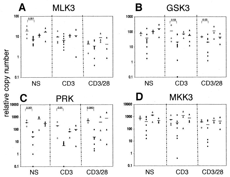
Human immunodeficiency virus infection in humans and simian immunodeficiency virus (SIV) infection in rhesus macaques (RM) leads to a generalized loss of immune responses involving perturbations in T-cell receptor (TCR) signaling. In contrast, naturally SIV-infected sooty mangabeys (SM) remain asymptomatic and retain immune responses despite relatively high viral loads. However, SIV infection in both RM and SM led to similar decreases in TCR-induced Lck phosphorylation. In this study, a protein tyrosine kinase (PTK) differential display method was utilized to characterize the effects of in vivo SIV infection on key signaling molecules of the CD4(+) T-cell signaling pathways. The CD4(+) T cells from SIV-infected RM, but not SIV-infected SM, showed chronic downregulation of baseline expression of MLK3, PRK, and GSK3, and symptomatically SIV-infected RM showed similar downregulation of MKK3. In vitro TCR stimulation with or without CD28 costimulation of CD4(+) T cells did not lead to the enhancement of gene transcription of these PTKs. While the CD4(+) T cells from SIV-infected RM showed a significant increase of the baseline and anti-TCR-mediated ROR2 transcription, SIV infection in SM led to substantially decreased anti-TCR-stimulated ROR2 transcription. TCR stimulation of CD4(+) T cells from SIV-infected RM (but not SIV-infected SM) led to the repression of CaMKKbeta and the induction of gene transcription of MLK2. Studies of the function of these molecules in T-cell signaling may lead to the identification of potential targets for specific intervention, leading to the restoration of T-cell responses.
Figures


References
-
- Anderson K A, Ribar T J, Illario M, Means A R. Defective survival and activation of thymocytes in transgenic mice expressing a catalytically inactive form of Ca2+/calmodulin-dependent protein kinase IV. Mol Endocrinol. 1997;11:725–737. - PubMed
-
- Borgatti P, Zauli G, Cantley L C, Capitani S. Extracellular HIV-1 Tat protein induces a rapid and selective activation of protein kinase C (PKC)-alpha, -epsilon, and -zeta isoforms in PC12 cells. Biochem Biophys Res Commun. 1998;242:332–337. - PubMed
-
- Borgatti P, Zauli G, Colamussi M L, Gibellini D, Previati M, Cantley L L, Capitani S. Extracellular HIV-1 Tat protein activates phosphatidylinositol 3- and Akt/PKB kinases in CD4+ T lymphoblastoid Jurkat cells. Eur J Immunol. 1997;27:2805–2811. - PubMed
-
- Bostik P, Brice G T, Greenberg K P, Mayne A E, Villinger F, Lewis M G, Ansari A A. Inverse correlation of telomerase activity/proliferation of CD4+ T lymphocytes and disease progression in simian immunodeficiency virus-infected nonhuman primates. J Acquir Immune Defic Syndr. 2000;24:89–99. - PubMed
-
- Bostik P, Mayne A E, Villinger F, Greenberg K P, Powell J D, Ansari A A. Relative resistance in the development of T cell anergy in CD4+ T cells from simian immunodeficiency virus disease-resistant sooty mangabeys. J Immunol. 2001;166:506–516. - PubMed
Publication types
MeSH terms
Substances
Grants and funding
LinkOut - more resources
Full Text Sources
Research Materials
Miscellaneous
