. 2001 Dec;75(23):11354-64.
doi: 10.1128/JVI.75.23.11354-11364.2001.

Critical roles of nuclear receptor response elements in replication of hepatitis B virus

Affiliations

Critical roles of nuclear receptor response elements in replication of hepatitis B virus

X Yu et al. J Virol. 2001 Dec.

Abstract

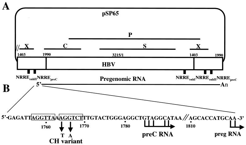
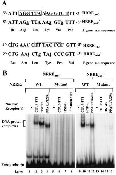
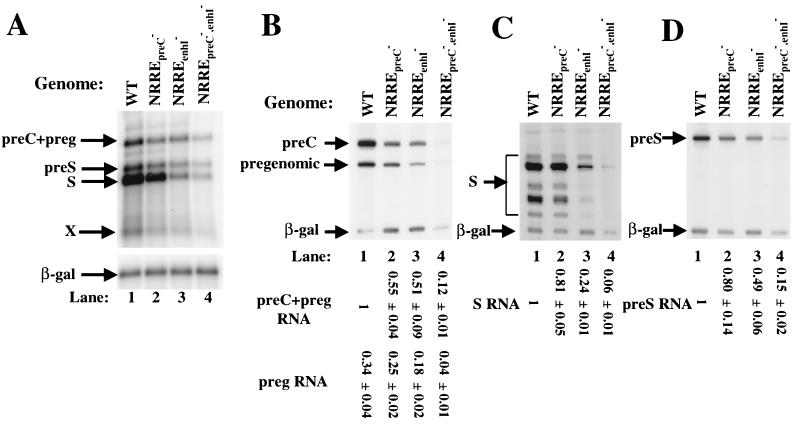
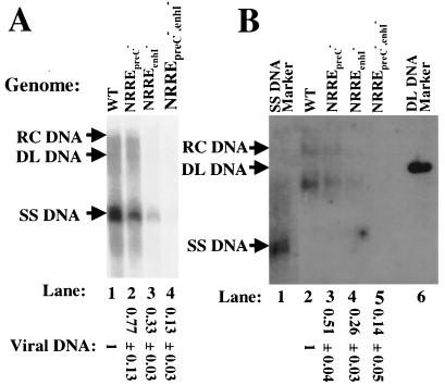
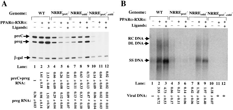
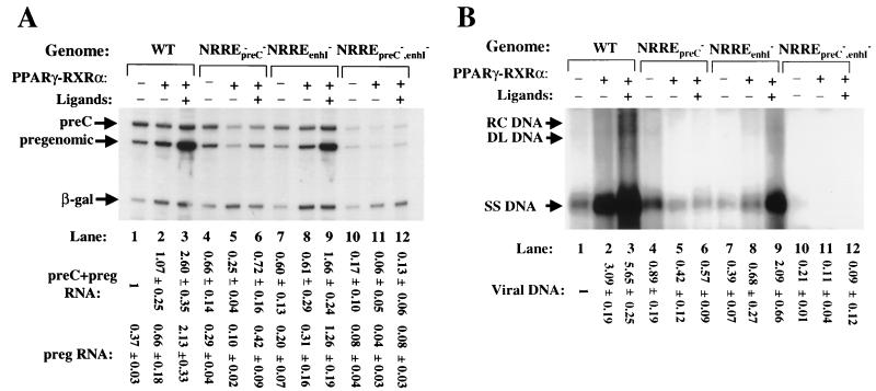
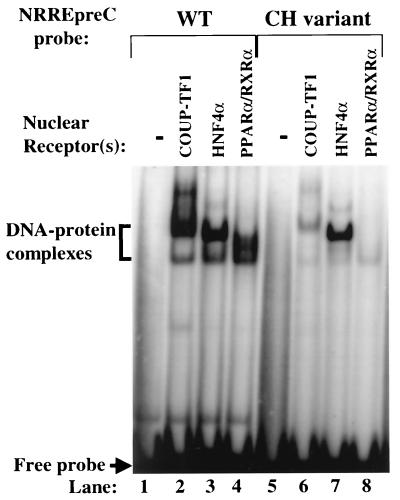
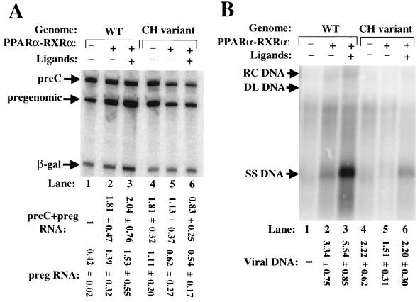
Functional analysis of the roles of the nuclear receptor response elements (NRREs) in the transcription and replication of hepatitis B virus (HBV) in the context of its whole genome has been hampered by the extensive overlapping of the NRREs with the regions encoding viral proteins. We introduced point mutations that inactivate the NRREs individually without altering the open reading frames of viral proteins. These mutations in the context of a plasmid containing 1.2 copies of the HBV genome were transiently transfected into the human hepatoma cell line Huh7. Inactivation of the NRRE in either the preC promoter (NRRE(preC)) or enhancer I (NRRE(enhI)) led to moderate reductions in synthesis of viral RNAs. Concurrent inactivation of both NRREs led to 7- to 8-fold reductions in synthesis of the preC, pregenomic, and preS RNAs and a 15-fold reduction in synthesis of the S RNA. The accumulation of viral DNA in the cytoplasmic nucleocapsids and virion particles in the culture medium was also reduced seven- to eightfold. These results suggest that these NRREs are critical for the efficient propagation of HBV in hepatocytes. In cotransfection experiments we also found that overexpression of PPARalpha-RXRalpha in the presence of their respective ligands led to a fourfold increase in pregenomic RNA synthesis and a four- to fivefold increase in viral DNA synthesis, while it had little or no effect on synthesis of the other viral RNAs. Similar effects were observed with overexpression of PPARgamma-RXRalpha in the presence of their respective ligands. This activation was dependent on NRRE(preC), because the increase in synthesis of viral RNA and DNA was not observed when this site was mutated. Likewise, no activation of synthesis of pregenomic RNA and viral DNA by PPARalpha-RXRalpha was observed in a naturally occurring NRRE(preC)(-) mutant of HBV. Our results suggest that interactions between nuclear receptors and NRREs present in the HBV genome may play critical roles in regulating its transcription and replication during HBV infection of hepatocytes.


Figures

FIG. 1
FIG. 1
(A) Schematic diagram of plasmid containing 1.2 copies of the HBV genome. A 3.8-kb, terminally redundant variant of the HBV genome, indicated by the large open rectangle, was inserted into plasmid pSP65. The locations of the P, S, C, and X open reading frames are indicated by solid lines. The locations of the NRREs in enhancer I, enhancer II, and the preC promoter are indicated by small solid rectangles. Shown at the bottom is the structure of the pregenomic RNA synthesized from this plasmid. (B) Nucleotide sequence of the region of the HBV genome surrounding the NRREpreC. The half-site sequences in the NRREpreC are boxed. The sequence changes in a naturally occurring variant of HBV (CH variant) are shown below the boxed sequences. Right-pointing arrows indicate locations of transcription initiation sites used in the synthesis of preC and pregenomic RNA (preg RNA).
FIG. 2
FIG. 2
Inactivation of the NRREs in the preC promoter and enhancer I. (A) Sequences of the point mutations introduced into NRREpreC and NRREenhI such that translation of the open reading frames of the X and P genes remains unaffected. The imperfect direct repeats of the NR half-site sequence are boxed. The base pair changes introduced by mutagenesis are shown as underlined lowercase letters. (B) EMSAs used to determine the binding of NRs to wild-type (WT) and mutant NRREs. Radiolabeled double-stranded oligonucleotides containing the wild-type and mutant NRRE sequences were used as probes. DNA-protein complexes are indicated by the bracket; free probes are indicated by the arrow.
FIG. 3
FIG. 3
Reduced viral RNA synthesis in NRRE mutants of HBV. (A) Autoradiogram of Northern blot analysis of the viral RNAs accumulated in Huh7 cells transfected with wild-type (WT) and NRRE mutant plasmids. One-half of the poly(A)-selected RNA from a 60-mm dish of cells was loaded in each lane and analyzed as described in Materials and Methods. (B through D) Autoradiograms of 8 M urea–8% polyacrylamide gels showing the results of primer extension assays used to quantify the preC and pregenomic RNAs (B), S RNAs (C), and preS RNA (D). The positions of the viral and β-Gal RNAs are indicated by arrows. preg, pregenomic. One-sixth of the RNA from a 60-mm dish of cells was used in each reaction along with radiolabeled primers specific for the viral and β-Gal RNAs. Numbers at the bottom give the amount of viral RNA in each lane relative to that synthesized from the wild-type plasmid. These numbers were determined with a PhosphorImager, normalized to β-Gal, and represent the means ± standard errors of the data obtained from three experiments similar to the one for which results are shown. The amount of preC RNA can be calculated by subtracting the pregenomic RNA from the preC-plus-pregenomic RNA in each lane.
FIG. 4
FIG. 4
Reduced viral DNA synthesis in NRRE mutants of HBV. (A) Autoradiogram of Southern blot analysis of viral DNA isolated from nucleocapsids present in the cytoplasm of transfected Huh7 cells. Cells were harvested 48 h posttransfection, and nucleocapsid viral DNA was prepared. One-third of the DNA from a 60-mm dish of cells was loaded in each lane. The total viral DNA from relaxed circular (RC) to single-stranded (SS) DNA in each lane was quantitated with a PhosphorImager. Positions of viral DNAs are indicated by arrows. WT, wild type; DL DNA, duplex linear DNA. The smears between the specific indicated DNA structures are the result of HBV DNAs with incomplete strands. Numbers at the bottom are means ± standard errors of data relative to the wild type obtained in three experiments similar to the one for which results are shown. (B) Autoradiogram of Southern blot analysis of extracellular viral DNA. Culture medium was harvested for HBV virions 5 days posttransfection. One-half of the viral DNA from the medium of a 60-mm dish of cells was loaded in each lane. Lanes 1 and 6, molecular markers for SS and DL DNAs, respectively. The DNA band below the DL DNA in lanes 2 through 4 is likely DL or RC DNA with an incomplete plus strand. Data were quantified as for panel A.
FIG. 5
FIG. 5
PPARα-RXRα activates synthesis of RNA and DNA of HBV. After incubation with the indicated DNA in a calcium phosphate precipitate for 8 h, Huh7 cells were washed and incubated in medium containing 1 mM clofibric acid (Sigma), a ligand for PPARα, and 1 μM 9-cis-retinoic acid (Sigma), a ligand for RXRα, for 40 h before they were harvested for RNA and DNA. (A) Autoradiogram showing the primer extension reactions of preC and pregenomic (preg) RNAs. One-sixth of the RNA from a 60-mm dish of cells was used in each primer extension reaction. WT, wild type. (B) Autoradiogram of Southern blot analysis of cytoplasmic viral DNA. One-third of the DNA from a 60-mm dish of cells was loaded in each lane. Quantitations were performed as described in the legends to Fig. 3 and 4. Numbers at the bottom are means ± standard errors of data obtained from three experiments similar to the one for which results are shown. RC DNA, relaxed circular DNA; DL DNA, duplex linear DNA; SS DNA, single-stranded DNA.
FIG. 6
FIG. 6
PPARγ-RXRα activates synthesis of viral RNA and DNA in Huh7 cells. The amounts of RNA and DNA used in each assay were the same as those stated for Fig. 5. Transfection of Huh7 cells was carried out as described for Fig. 5 except that a PPARγ-specific ligand, 15-deoxy-Δ12,14-prostaglandin J2, was used (8 μM) (Cayman Chemical). (A) Autoradiogram showing primer extension reactions used to quantify preC and pregenomic (preg) RNAs. (B) Autoradiogram of a Southern blot analysis of cytoplasmic viral DNA. Quantitations were performed as described in the legends to Fig. 3 and 4. Numbers at the bottom are means ± standard errors of data obtained from three experiments similar to the one for which results are shown. RC DNA, relaxed circular DNA; DL DNA, duplex linear DNA; SS DNA, single-stranded DNA.
FIG. 7
FIG. 7
Effects of base pair changes in the NRREpreC of the CH variant of HBV on its binding to NRs. Shown here are results of EMSAs performed with the indicated NRs and radiolabeled, double-stranded oligonucleotides containing the wild-type (WT) and CH variant NRREpreC sequences as probes. Bracket, DNA-protein complexes; arrow, free probe.
FIG. 8
FIG. 8
Sequence changes in the NRREpreC of the CH variant of HBV abolish activation of synthesis of viral RNA and DNA by PPARα-RXRα. (A) Autoradiogram of primer extension reactions used to quantify preC and pregenomic (preg) RNAs synthesized from the wild-type (WT) and CH variant HBV plasmids. Transient transfections of Huh7 cells and the concentrations of ligands for PPARα and RXRα added to the medium were as described for Fig. 5. (B) Autoradiogram of Southern blot analysis of cytoplasmic viral DNA present in the same cells. Numbers at the bottom are means ± standard errors of data quantified from three experiments similar to the one for which results are shown. RC DNA, relaxed circular DNA; DL DNA, duplex linear DNA; SS DNA, single-stranded DNA.

Similar articles

Cited by

References

    1. Antonucci T K, Rutter W J. Hepatitis B virus (HBV) promoters are regulated by the HBV enhancer in a tissue-specific manner. J Virol. 1989;63:579–583. - PMC - PubMed
    1. Baumert T F, Marrone A, Vergalla J, Liang T J. Naturally occurring mutations define a novel function of the hepatitis B virus core promoter in core protein expression. J Virol. 1998;72:6785–6795. - PMC - PubMed
    1. Ben-Levy R, Faktor O, Berger I, Shaul Y. Cellular factors that interact with the hepatitis B virus enhancer. Mol Cell Biol. 1989;9:1804–1809. - PMC - PubMed
    1. Bock C-T, Malek N P, Tillmann H L, Manns M P, Trautwein C. The enhancer I core region contributes to the replication level of hepatitis B virus in vivo and in vitro. J Virol. 2000;74:2193–2202. - PMC - PubMed
    1. Brown T. Southern blotting. In: Ausubel F M, Brent R, Kingston R E, Moore D D, Seidman J G, Smith J A, Struhl K, editors. Current protocols in molecular biology. New York, N.Y: John Wiley and Sons, Inc.; 1993. p. 2.9.1.

Publication types

LinkOut - more resources