. 2001 Dec;75(23):11437-48.
doi: 10.1128/JVI.75.23.11437-11448.2001.

Vaccinia virus infection disarms the mitochondrion-mediated pathway of the apoptotic cascade by modulating the permeability transition pore

Affiliations

Vaccinia virus infection disarms the mitochondrion-mediated pathway of the apoptotic cascade by modulating the permeability transition pore

S T Wasilenko et al. J Virol. 2001 Dec.

Abstract

Many viruses have evolved strategies that target crucial components within the apoptotic cascade. One of the best studied is the caspase 8 inhibitor, crmA/Spi-2, encoded by members of the poxvirus family. Since many proapoptotic stimuli induce apoptosis through a mitochondrion-dependent, caspase 8-independent pathway, we hypothesized that vaccinia virus would encode a mechanism to directly modulate the mitochondrial apoptotic pathway. In support of this, we observed that Jurkat cells, which undergo Fas-mediated apoptosis exclusively through the mitochondrial route, were resistant to Fas-induced death following infection with a crmA/Spi-2-deficient strain of vaccinia virus. In addition, vaccinia virus-infected cells subjected to the proapoptotic stimulus staurosporine exhibited decreased levels of both cytochrome c released from the mitochondria and caspase 3 activation. In all cases we found that the loss of the mitochondrial membrane potential, which occurs as a result of opening the multimeric permeability transition pore complex, was prevented in vaccinia virus-infected cells. Moreover, vaccinia virus infection specifically inhibited opening of the permeability transition pore following treatment with the permeability transition pore ligand atractyloside and t-butylhydroperoxide. These studies indicate that vaccinia virus infection directly impacts the mitochondrial apoptotic cascade by influencing the permeability transition pore.


Figures

FIG. 1
FIG. 1
Vaccinia virus strain Copenhagen protects cells from anti-Fas-mediated death. Jurkat cells were either mock infected or infected with either vaccinia virus strain Copenhagen or vaccinia virus strain Western Reserve at an MOI of 10. Following 5 h of infection, cells were treated with 250 ng of anti-Fas antibody/ml to induce apoptosis, and cell death was monitored 8 h later by 51Cr release. As controls, Jurkat cells that overexpress SPI-2 and Bcl-2 were also treated with anti-Fas, and Jurkat cells were pretreated for 30 min with 100 μM zVAD.fmk prior to addition of anti-Fas. Standard deviations were generated from three replicates.
FIG. 2
FIG. 2
Vaccinia virus strain Copenhagen infection inhibits activation of caspase 3. Jurkat cells were either mock infected or infected with vaccinia virus strain Copenhagen at an MOI of 10 and treated with 250 ng of anti-Fas antibody/ml for 2, 4, 6, or 8 h. At the times indicated, cells were permeabilized with digitonin, and caspase 3 processing was monitored by Western blot analysis. (A) Mock-infected Jurkat cells; (B) Jurkat cells infected with vaccinia virus strain Copenhagen.
FIG. 3
FIG. 3
Vaccinia virus strain Copenhagen inhibits cytochrome c translocation. Jurkat cells were either mock infected or infected with virus. Following infection, cells were treated with 250 ng of anti-Fas/ml for 2, 4, 6, or 8 h to induce cytochrome c translocation. At the times indicated, cells were permeabilized with digitonin and fractionated into the mitochondrion-containing membranous fraction and the soluble cytoplasmic fraction, and cytochrome c was assessed by Western blot analysis. (A) Mock-infected Jurkat cells; (B) mock infected Jurkat cells that overexpress Bcl-2; (C) Jurkat cells infected with vaccinia virus strain Copenhagen.
FIG. 4
FIG. 4
Vaccinia virus infection protects against granzyme B-mediated cytochrome c release from isolated mitochondria by a mechanism downstream of Bid activation. Mitochondria were isolated from mock-infected Jurkat cells, Jurkat cells overexpressing Bcl-2, and Jurkat cells infected with vaccinia virus strain Copenhagen. Purified mitochondria were incubated for 60 min at 37°C with 2, 5, or 10 ng of recombinant Bid either in the presence or in the absence of granzyme B. Following treatment, samples were fractionated into mitochondria-containing and soluble fractions and the proteins were resolved by SDS-PAGE. (A through C)Western blot analysis of Bid cleavage in mitochondria isolated either from mock-infected cells (A), from cells overexpressing Bcl-2 (B), or from vaccinia virus strain Copenhagen-infected cells (C). (D through F) Western blot analysis of cytochrome c translocation from purified mitochondria to supernatant fractions in mitochondria isolated either from mock-infected cells (D), from mock-infected cells overexpressing Bcl-2 (E), or from vaccinia virus strain Copenhagen-infected cells (F).
FIG. 5
FIG. 5
DNA fragmentation is blocked by vaccinia virus strain Copenhagen infection. Jurkat cells were either mock infected or infected with vaccinia virus strain Copenhagen. Following infection, cells were treated with 2.5 μM staurosporine for 2 h, and DNA fragmentation was assessed by TUNEL as outlined in Materials and Methods. (a) Untreated Jurkat cells; (b) Jurkat cells treated with staurosporine; (c) Jurkat cells treated with staurosporine in the presence of 100 μM zVAD.fmk; (d) Jurkat cells overexpressing Bcl-2; (e) Jurkat cells overexpressing Bcl-2 treated with staurosporine; (f) Jurkat cells infected with vaccinia virus strain Copenhagen; (g) Jurkat cells infected with vaccinia virus and treated with staurosporine.
FIG. 6
FIG. 6
Staurosporine-induced caspase 3 activation is inhibited by vaccinia virus strain Copenhagen infection. Jurkat cells were either mock infected or infected with virus; following infection, they were treated with 5 μM staurosporine for 0, 2, 4, or 6 h. At the times indicated, cells were permeabilized with digitonin and the proteins were resolved by SDS-PAGE. The activation of caspase 3 was monitored by Western blot analysis. (A) Mock-infected Jurkat cells; (B) mock-infected Jurkat cells that overexpress Bcl-2; (C) Jurkat cells infected with vaccinia virus strain Copenhagen.
FIG. 7
FIG. 7
Staurosporine-induced cytochrome c release is inhibited by vaccinia virus strain Copenhagen infection. Jurkat cells were either mock infected or infected with virus and were treated with 5 μM staurosporine for 0, 2, 4, or 6 h. At the times indicated, cells were fractionated into mitochondria-containing membrane fractions and cytoplasmic fractions by the addition of digitonin. The translocation of cytochrome c was monitored by Western blot analysis. (A) Mock-infected Jurkat cells; (B) mock-infected Jurkat cells that overexpress Bcl-2; (C) Jurkat cells infected with vaccinia virus strain Copenhagen.
FIG. 8
FIG. 8
Vaccinia virus strain Copenhagen infection inhibits staurosporine-induced disruption of the mitochondrial membrane potential. Jurkat cells were either mock infected or infected with vaccinia virus and treated with 1 μM staurosporine for 1 h, and the mitochondrial membrane potential was determined using TMRE fluorescence. (a) Untreated Jurkat cells; (b) Jurkat cells treated with staurosporine; (c) Jurkat cells treated with the membrane uncoupler ClCCP; (d) Jurkat cells treated with staurosporine in the presence of 100 μM zVAD.fmk; (e) untreated Jurkat cells overexpressing Bcl-2; (f) Jurkat cells overexpressing Bcl-2 treated with staurosporine; (g) untreated Jurkat cells infected with vaccina virus strain Copenhagen; (h) vaccinia virus-infected cells treated with staurosporine; (i) Jurkat cells infected with vaccinia virus strain Copenhagen in the presence of 40 μg of araC/ml; (j) Jurkat cells infected with vaccinia virus strain Copenhagen in the presence of araC and staurosporine; (k) untreated cells infected with UV-inactivated vaccinia virus strain Copenhagen; (l) Jurkat cells infected with UV-inactivated vaccinia virus and treated with staurosporine.
FIG. 9
FIG. 9
Vaccinia virus strain Copenhagen inhibits opening of the PT pore. (A through C) Atractyloside-induced cytochrome c release is inhibited by vaccinia virus strain Copenhagen infection. Mitochondria were purified and incubated at 37°C with 5, 10, or 15 mM atractyloside (Atrac.) for 40 min. Following treatment, samples were fractionated into mitochondria-containing and soluble fractions, and translocation of cytochrome c was analyzed by Western blot analysis. (A) Mitochondria isolated from mock-infected Jurkat cells; (B) mitochondria isolated from mock-infected Jurkat cells overexpressing Bcl-2; (C) mitochondria isolated from vaccinia virus strain Copenhagen-infected Jurkat cells. (D) Vaccinia virus inhibits t-butylhydroperoxide-induced disruption of the mitochondrial membrane potential. Jurkat cells were either mock infected or infected with vaccinia virus and treated with 300 μM t-butylhydroperoxide for 2 h. The mitochondrial membrane potential was determined using TMRE fluorescence in the presence and absence of 100 μM zVAD.fmk. Standard deviations were generated from three independent experiments.
FIG. 10
FIG. 10
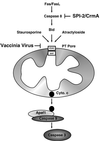
Model of vaccinia virus-mediated apoptosis inhibition. Fas initiated apoptosis occurs via activation of caspase 8 and by the subsequent proteolytic cleavage of Bid. Once cleaved, Bid translocates to the mitochondria, resulting in a loss of the inner-mitochondrial-membrane permeability transition and the release of cytochrome c. The release of cytochrome c (Cyto. c) results in activation of caspase 9 via interaction with the adapter molecule Apaf1 and the subsequent activation of caspase 3. Additionally, loss of the inner-membrane permeability transition can be triggered by staurosoporine and atractyloside. Vaccinia virus infection manipulates this pathway at two separate points. First, the vaccinia virus-encoded serine protease inhibitor SPI-2/crmA inhibits the activity of caspase 8. Second, vaccinia virus infection also inhibits apoptosis by modulating the PT pore, thereby preventing the loss of cytochrome c and activation of caspase 9.

Similar articles

Cited by

References

    1. Afonso C L, Neilan J G, Kutish G F, Rock D L. An African swine fever virus Bcl-2 homolog, 5-HL, suppresses apoptotic cell death. J Virol. 1996;70:4858–4863. - PMC - PubMed
    1. Barry M, McFadden G. Apoptosis regulators from DNA viruses. Curr Opin Immunol. 1998;10:422–430. - PubMed
    1. Barry M, Heibein J A, Pinkoski M J, Lee S F, Moyer R W, Green D R, Bleackley R C. Granzyme B short-circuits the need for caspase 8 activity during granule-mediated cytotoxic T-lymphocyte killing by directly cleaving Bid. Mol Cell Biol. 2000;20:3781–3794. - PMC - PubMed
    1. Barry M, Heibein J, Pinkoski M, Bleackley R C. Quantitative measurement of apoptosis induced by cytotoxic T lymphocytes. Methods Enzymol. 2000;322:40–46. - PubMed
    1. Bertin J, Armstrong R C, Ottilie S, Martin D A, Wang Y, Banks S, Wang G H, Senkevich T G, Alnemri E M, Moss B, Lenardo M J, Tomaselli K J, Cohen J I. Death effector domain-containing herpesvirus and poxvirus proteins inhibit both Fas- and TNFR1-induced apoptosis. Proc Natl Acad Sci USA. 1997;94:1772–1776. - PMC - PubMed

Publication types