

In vivo inhibition of anti-hepatitis B virus core antigen (HBcAg) immunoglobulin G production by HBcAg-specific CD4(+) Th1-type T-cell clones in a hu-PBL-NOD/SCID mouse model
- PMID: 11689626
- PMCID: PMC114731
- DOI: 10.1128/JVI.75.23.11449-11456.2001
In vivo inhibition of anti-hepatitis B virus core antigen (HBcAg) immunoglobulin G production by HBcAg-specific CD4(+) Th1-type T-cell clones in a hu-PBL-NOD/SCID mouse model
Abstract
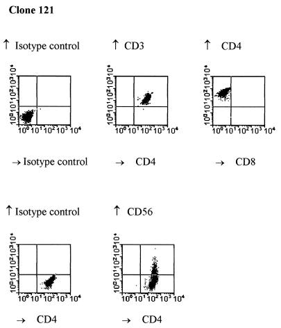
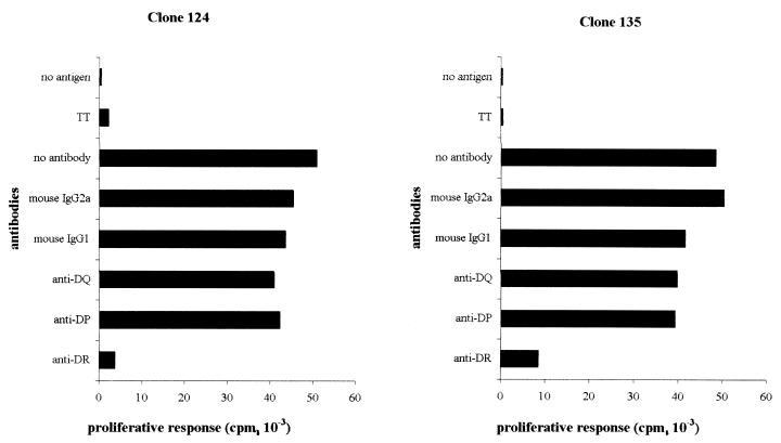
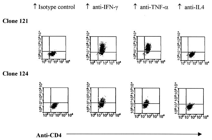
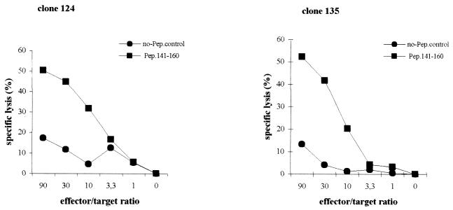
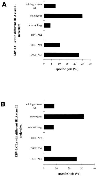
Hepatitis B virus (HBV) core antigen (HBcAg)-specific CD4(+) T-cell responses are believed to play an important role in the control of human HBV infection. In the present study, HBcAg-specific, HLA-DR13*-restricted CD4(+) Th1-type T-cell clones were generated which secreted both gamma interferon and tumor necrosis factor alpha after in vitro antigen stimulation. These HBcAg-specific CD4(+) Th1-type T cells were able to lyse HBc peptide-loaded Epstein-Barr virus-transformed lymphoblastoid target cells in vitro. To examine whether these HLA-DR13*-restricted human CD4(+) Th1 T cells also display the same cytotoxic effects in vivo, we transferred peripheral blood leukocytes (PBL) derived from HBV-infected donors or an HBV-naïve donor sharing the DR13*, together with the HBcAg-specific CD4(+) Th1-type T cells and HBcAg, directly into the spleen of optimally conditioned Nod/LtSz-Prkdc(scid)/Prkdc(scid) (NOD/SCID) mice. The production of both secondary anti-HBc-immunoglobulin G (anti-HBc-IgG) and primary HBcAg-binding IgM in hu-PBL-NOD/SCID mice was drastically inhibited by HBcAg-specific CD4(+) Th1-type T cells. No inhibition was observed when CD4(+) Th1 cells and donor PBL did not share an HLA-DR13. These results suggest that HBcAg-specific CD4(+) Th1 T cells may be able to lyse HBcAg-binding, or -specific, B cells that have taken up and presented HBcAg in a class II-restricted manner. Thus, HBcAg-specific CD4(+) Th1-type T cells can modulate the function and exert a regulatory role in deleting HBcAg-binding, or -specific, human B cells in vivo, which may be of importance in controlling the infection.
Figures
Similar articles
-
Hepatitis B virus core antigen binds and activates naive human B cells in vivo: studies with a human PBL-NOD/SCID mouse model.J Virol. 2001 Jul;75(14):6359-66. doi: 10.1128/JVI.75.14.6359-6366.2001. J Virol. 2001. PMID: 11413302 Free PMC article.
-
Plasmacytoid dendritic cells induce efficient stimulation of antiviral immunity in the context of chronic hepatitis B virus infection.Hepatology. 2012 Nov;56(5):1706-18. doi: 10.1002/hep.25879. Epub 2012 Aug 27. Hepatology. 2012. PMID: 22707082
-
Molecular basis for the interaction of the hepatitis B virus core antigen with the surface immunoglobulin receptor on naive B cells.J Virol. 2001 Jul;75(14):6367-74. doi: 10.1128/JVI.75.14.6367-6374.2001. J Virol. 2001. PMID: 11413303 Free PMC article.
-
HBcAg-specific CD4+CD25+ regulatory T cells modulate immune tolerance and acute exacerbation on the natural history of chronic hepatitis B virus infection.J Biomed Sci. 2007 Jan;14(1):43-57. doi: 10.1007/s11373-006-9129-z. Epub 2006 Nov 16. J Biomed Sci. 2007. PMID: 17109186 Clinical Trial.
-
Hybrid hepatitis B virus core antigen as a vaccine carrier moiety: I. presentation of foreign epitopes.J Biotechnol. 1996 Jan 26;44(1-3):91-6. doi: 10.1016/0168-1656(95)00118-2. J Biotechnol. 1996. PMID: 8717391 Review.
Cited by
-
Association between HLA class II gene and susceptibility or resistance to chronic hepatitis B.World J Gastroenterol. 2003 Oct;9(10):2221-5. doi: 10.3748/wjg.v9.i10.2221. World J Gastroenterol. 2003. PMID: 14562382 Free PMC article.
-
Fighting Viral Infections and Virus-Driven Tumors with Cytotoxic CD4+ T Cells.Front Immunol. 2017 Feb 27;8:197. doi: 10.3389/fimmu.2017.00197. eCollection 2017. Front Immunol. 2017. PMID: 28289418 Free PMC article. Review.
-
A Systematic Review of T Cell Epitopes Defined from the Proteome of Hepatitis B Virus.Vaccines (Basel). 2022 Feb 8;10(2):257. doi: 10.3390/vaccines10020257. Vaccines (Basel). 2022. PMID: 35214714 Free PMC article. Review.
-
Negative HBcAg in immunohistochemistry assay of liver biopsy is a predictive factor for the treatment of patients with nucleos(t)ide analogue therapy.J Cell Mol Med. 2018 Mar;22(3):1675-1683. doi: 10.1111/jcmm.13444. Epub 2017 Nov 29. J Cell Mol Med. 2018. PMID: 29193766 Free PMC article.
-
Induction of humoral and cell-mediated immune responses by hepatitis B virus epitope displayed on the virus-like particles of prawn nodavirus.Appl Environ Microbiol. 2015 Feb;81(3):882-9. doi: 10.1128/AEM.03695-14. Epub 2014 Nov 21. Appl Environ Microbiol. 2015. PMID: 25416760 Free PMC article.
References
-
- Ahn S H, Han K H, Park J Y, Lee C K, Kang S W, Chon C Y, Kim Y S, Park K, Kim D K, Moon Y M. Association between hepatitis B virus infection and HLA-DR type in Korea. Hepatology. 2000;31:1371–1373. - PubMed
-
- Ando K, Hiroishi K, Kaneko T, Moriyama T, Muto Y, Kayagaki N, Yagita H, Okumura K, Imawari M. Perforin, Fas/Fas ligand, and TNF-alpha pathways as specific and bystander killing mechanisms of hepatitis C virus-specific human CTL. J Immunol. 1997;158:5283–5291. - PubMed
-
- Barnaba V, Franco A, Alberti A, Benvenuto R, Balsano F. Selective killing of hepatitis B envelope antigen-specific B cells by class I-restricted, exogenous antigen-specific T lymphocytes. Nature. 1990;345:258–260. - PubMed
-
- Barnaba V, Franco A, Paroli M, Benvenuto R, De Petrillo G, Burgio V L, Santilio I, Balsano C, Bonavita M S, Cappelli G. Selective expansion of cytotoxic T lymphocytes with a CD4+CD56+ surface phenotype and a T helper type 1 profile of cytokine secretion in the liver of patients chronically infected with Hepatitis B virus. J Immunol. 1994;152:3074–3087. - PubMed
Publication types
MeSH terms
Substances
LinkOut - more resources
Full Text Sources
Other Literature Sources
Molecular Biology Databases
Research Materials
