. 2001 Dec;75(23):11565-72.
doi: 10.1128/JVI.75.23.11565-11572.2001.

A short N-proximal region in the large envelope protein harbors a determinant that contributes to the species specificity of human hepatitis B virus

Affiliations

A short N-proximal region in the large envelope protein harbors a determinant that contributes to the species specificity of human hepatitis B virus

P Chouteau et al. J Virol. 2001 Dec.

Abstract

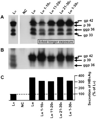
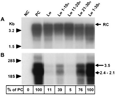
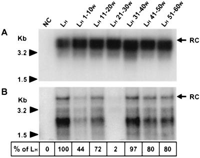
Infection by hepatitis B virus (HBV) is mainly restricted to humans. This species specificity is likely determined at the early phase of the viral life cycle. Since the envelope proteins are the first viral factors to interact with the cell, they represent attractive candidates for controlling the HBV host range. To investigate this assumption, we took advantage of the recent discovery of a second virus belonging to the primate Orthohepadnavirus genus, the woolly monkey HBV (WMHBV). A recombinant plasmid was constructed for the expression of all WMHBV envelope proteins. In additional constructs, N-terminal sequences of the WMHBV large envelope protein were substituted for their homologous HBV counterparts. All wild-type and chimeric WMHBV surface proteins were properly synthesized by transfected human hepatoma cells, and they were competent to replace the original HBV proteins for the production of complete viral particles. The resulting pseudotyped virions were evaluated for their infectious capacity on human hepatocytes in primary culture. Virions pseudotyped with wild-type WMHBV envelope proteins showed a significant loss of infectivity. By contrast, infectivity was completely restored when the first 30 residues of the large protein originated from HBV. Analysis of smaller substitutions within this domain limited the most important region to a stretch of only nine amino acids. Reciprocally, replacement of this motif by WMHBV residues in the context of the HBV L protein significantly reduced infectivity of HBV. Hence this short region of the L protein contributes to the host range of HBV.


Figures

FIG. 1
FIG. 1
Organization of the HBV gene coding for the envelope proteins. The L, M, and S proteins are encoded by a single ORF. Triangles and vertical bar, positions of the three different in-frame initiation sites and the common stop codon, respectively, which divide the gene into the pre-S1, pre-S2, and S domains (boxes). Sizes in amino acids of the regions are specified at the bottom.
FIG. 2
FIG. 2
Expression and secretion of the WT and chimeric WMHBV envelope proteins by a human hepatoma cell line. HepG2 cells were transfected with 20 μg of vectors designed to express viral surface proteins: LH, expression plasmid driving the synthesis of the WT HBV proteins; NC, plasmid without any HBV insert (negative control); LW and LW x-yH, expression plasmids driving the translation of WT and chimeric WMHBV proteins, respectively. Analyses were performed 7 dpt. Proteins were extracted from cells (A) or were precipitated from the culture medium (B) and studied by Western blotting. The primary monoclonal antibody was directed against the pre-S2H region. gp 42, p 39, ggp 36, and gp 33, migration positions of the glycosylated and unglycosylated LH and LW proteins and the diglycosylated and glycosylated MH and MW proteins, respectively. In panel A, the exposure time of the autoradiogram was 5-fold longer for samples NC, LW, and LW x-yH than for the control, LH. (C) Assessment of the secreted HBsAg by radioimmunoassay. Data are percentages of the positive-control (LH) value.
FIG. 3
FIG. 3
Production and infectivity of pseudotyped viruses. (A) HepG2 cells were either transfected with a plasmid (pHBV L- env-) containing a WT replication-competent HBV genome (PC for positive control) or cotransfected with an envelope protein-defective genome complemented with vectors expressing different envelope proteins: NC, plasmid without any HBV insert (negative control); LW and LW x-yH, expression plasmids driving the translation of WT and chimeric WMHBV proteins, respectively. Complete viral particles were immunoprecipitated with a polyclonal anti-HBsAg antibody from HepG2 cell supernatants collected between 4 and 7 dpt. DNA was extracted from the immunoprecipitates and analyzed by Southern blotting. Molecular size markers are on the left, and the position of the relaxed circular DNA (RC) is shown on the right. (B) Adult human hepatocytes in primary culture were incubated with concentrated supernatants obtained from HepG2 cells transfected or cotransfected as described for panel A. Total RNAs of inoculated cells were extracted 7 dpi and analyzed by Northern blotting. For each sample, 10 μg of total RNA was analyzed. The migration positions of 18S and 28S rRNAs and viral RNAs are indicated on the left and right, respectively. Shown at the bottom are the percentages of detected signal intensities relative to that revealed for PC.
FIG. 4
FIG. 4
Production and infectivity of reciprocal mutants. (A) HepG2 cells were cotransfected with the L-defective HBV genome (pHBV L-) complemented with vectors expressing different envelope proteins: NC, plasmid without any HBV insert (negative control); LH and LH x-yW, expression plasmids driving the translation of WT and mutant HBV surface proteins, respectively. Complete viral particles were immunoprecipitated with a polyclonal anti-HBsAg antibody from HepG2 cell supernatants collected between 4 and 7 dpt. DNA was extracted from the immunoprecipitates and analyzed by Southern blotting. (B) Adult human hepatocytes in primary culture were incubated with concentrated supernatants obtained from HepG2 cells cotransfected as described for panel A. Intracellular viral DNA was selectively extracted from hepatocytes collected 15 dpi and analyzed by Southern blotting. The positions of the relaxed circular DNA (RC) are shown on the right, and molecular size markers are on the left. Shown at the bottom are the percentages of detected signal intensities relative to that revealed for LH.
FIG. 5
FIG. 5
Comparison of WMHBV and HBV sequences in the N terminus pre-S1 domain. The WMHBV amino acid sequence is compared with the homologous sequence of HBV subtype ayw 3 used in this work. Six other human isolates, representing genotypes A to F, were also aligned. The genotype and the GenBank accession number of each sequence are in parentheses. The amino acid sequence from Met 1 to Thr 40 of pre-S1H (subtype ayw 3) is entirely shown in one-letter code. Dashes and lowercase letters, residues identical or similar to those in the corresponding positions of subtype ayw 3, respectively. Grey backgrounds indicate the two domains involved in the determination of HBV species specificity. Black box and open box, WMHBV-specific amino acids located at positions where the corresponding residues are conserved or variable among HBV strains, respectively; dotted-line box, residues different from those present in the ayw 3 subtype HBV but found in some other HBV isolates.

References

    1. Aden D P, Fogel A, Plotkin S, Damjanov I, Knowles B B. Controlled synthesis of HBsAg in a differentiated human liver carcinoma-derived cell line. Nature (London) 1979;282:615–616. - PubMed
    1. Bruss V. A short linear sequence in the pre-S domain of the large hepatitis B virus envelope protein required for virion formation. J Virol. 1997;71:9350–9357. - PMC - PubMed
    1. Bruss V, Ganem D. The role of envelope proteins in hepatitis B virus assembly. Proc Natl Acad Sci USA. 1991;88:1059–1063. - PMC - PubMed
    1. Bruss V, Hagelsten J, Gerhardt E, Galle P R. Myristylation of the large surface protein is required for hepatitis B virus in vitro infectivity. Virology. 1996;218:396–399. - PubMed
    1. Bruss V, Lu X Y, Thomssen R, Gerlich W H. Post-translational alterations in transmembrane topology of the hepatitis B virus large envelope protein. EMBO J. 1994;13:2273–2279. - PMC - PubMed

Publication types

Substances

LinkOut - more resources