. 2001 Dec;75(23):11709-19.
doi: 10.1128/JVI.75.23.11709-11719.2001.

Identification, phylogeny, and evolution of retroviral elements based on their envelope genes

Affiliations

Identification, phylogeny, and evolution of retroviral elements based on their envelope genes

L Bénit et al. J Virol. 2001 Dec.

Abstract

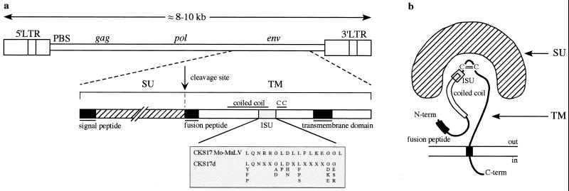
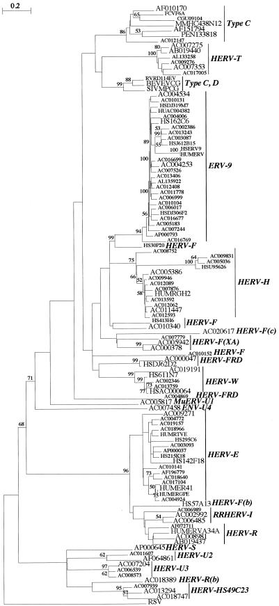
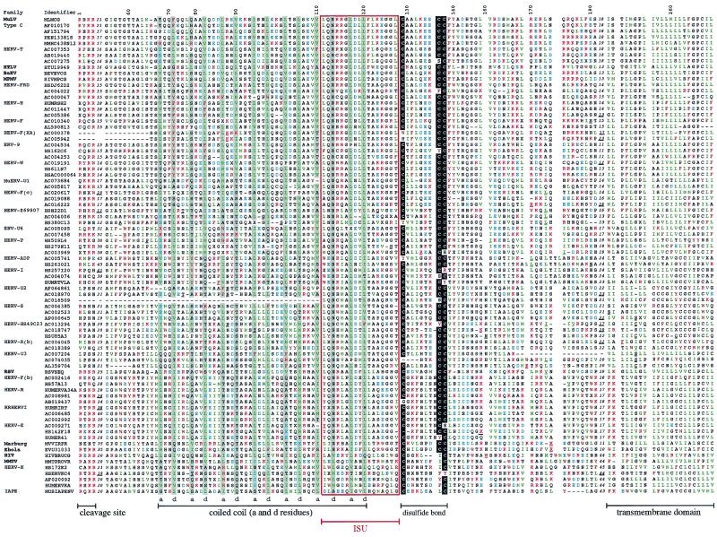
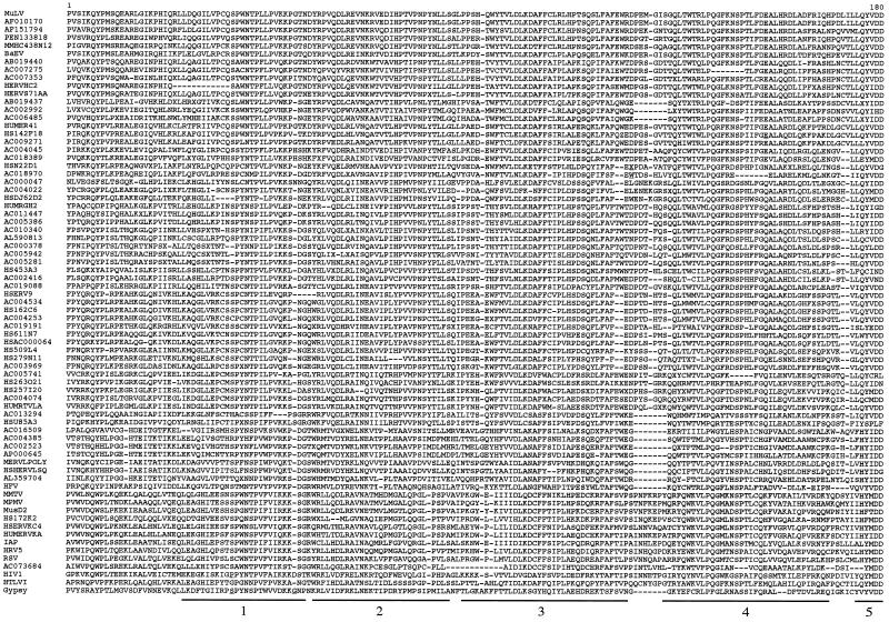
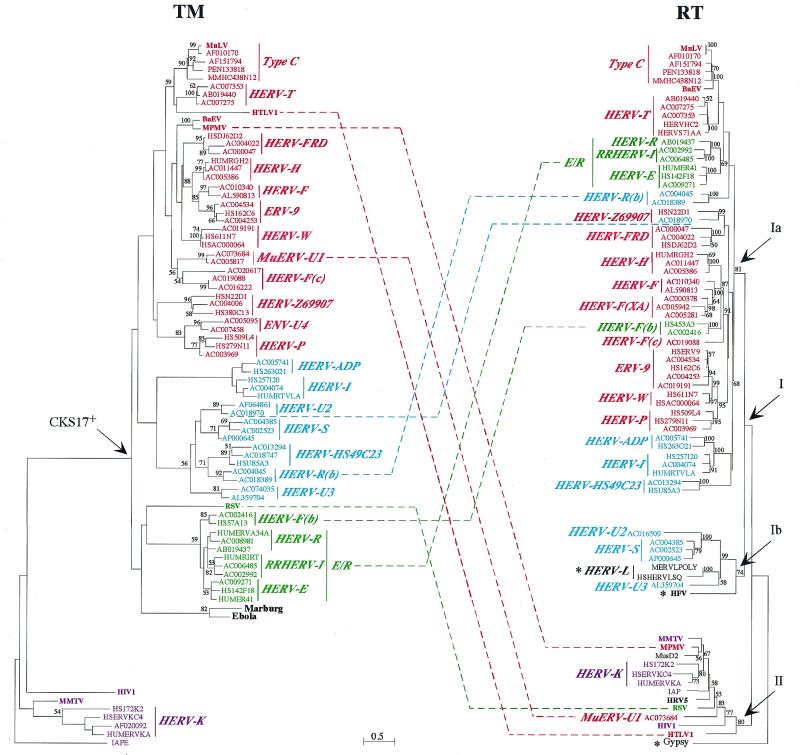
Phylogenetic analyses of retroviral elements, including endogenous retroviruses, have relied essentially on the retroviral pol gene expressing the highly conserved reverse transcriptase. This enzyme is essential for the life cycle of all retroid elements, but other genes are also endowed with conserved essential functions. Among them, the transmembrane (TM) subunit of the envelope gene is involved in virus entry through membrane fusion. It has also been reported to contain a domain, named the immunosuppressive domain, that has immunosuppressive properties most probably essential for virus spread within the host. This domain is conserved among a large series of retroviral elements, and we have therefore attempted to generate phylogenetic links between retroviral elements identified from databases following tentative alignments of the immunosuppressive domain and adjacent sequences. This allowed us to unravel a conserved organization among TM domains, also found in the Ebola and Marburg filoviruses, and to identify a large number of human endogenous retroviruses (HERVs) from sequence databases. The latter elements are part of previously identified families of HERVs, and some of them define new families. A general phylogenetic analysis based on the TM proteins of retroelements, and including those with no clearly identified immunosuppressive domain, could then be derived and compared with pol-based phylogenetic trees, providing a comprehensive survey of retroelements and definitive evidence for recombination events in the generation of both the endogenous and the present-day infectious retroviruses.


Figures

FIG. 1
FIG. 1
Schematic representation of the proviral form of a retrovirus and its env gene products. (a) Genomic proviral structure and delineation of the TM subunit encoded by the env gene. The Mo-MuLV immunosuppressive domain (ISU) is shown, as is the degenerate CKS17d motif used for the initial screening of the databases (see text and Table 1; an extended optimized universal CKS17u motif [see Materials and Methods] which recognizes at least one member from each retroelement family of the CKS17-positive group in Fig. 3 and 5 was also devised). (b) Schematic structure of the env products, adapted from reference . CC, disulfide bond.
FIG. 2
FIG. 2
CKS17 nucleotide tree. A phylogenetic tree of a set of 110 nucleotide sequences positive for the CKS17d motif is shown, with the RSV sequence as an outgroup. This tree was determined by the neighbor-joining method with branch lengths proportional to the degrees of divergence between the sequences. Percent bootstrap values obtained from 100 replicates are indicated on the branches only when they are >50%. The names of the sequences correspond to the GenBank identifiers. The sequences retained for the TM alignment are indicated by larger characters. The HERV families or retrovirus types are indicated.
FIG. 3
FIG. 3
TM protein sequence alignment of ERVs and exogenous retroviruses (extracellular and TM domains). Sequences not selected from the CKS17 search and added in the alignment (see text) were found by BLAST searches or were derived from the literature (45). Sequence names correspond to the GenBank identifiers. The HERV or MuERV families are indicated on the left, as are the virus names. The order is the same as that of the TM tree (see Fig. 5, left) from top to bottom. Variable regions are indicated with the number of omitted residues, underlined positions correspond to insertions or frameshifts, and dashes correspond to deletions. The numbers above the alignment are relative to the full-length alignment (including insertions), which is 269 positions long. The region aligned to establish the initial CKS17 nucleotide phylogeny (Fig. 2) corresponds to positions 54 to 181. The immunosuppressive domain is boxed in red. The cysteine residues potentially involved in a short internal disulfide bond are highlighted in black, and the a and d positions in the heptad repeats within the coiled coil are in grey. Basic residues are in red, acidic residues are in blue, aromatic residues are in brown, and hydrophobic (aliphatic) residues are in green.
FIG. 4
FIG. 4
Partial RT protein sequence alignment. Sequence names correspond to the GenBank identifiers for ERVs, while common names are used for infectious retroviruses (with their GenBank identifiers given in Fig. 3 and 5 and their legends). The order of the sequences is the same as that for the RT tree (Fig. 4, right). Underlined positions correspond to insertions, and dashes correspond to deletions. The first five common peptide domains among the seven domains described by Xiong and Eickbush (51) are delineated below the sequences (the fifth is partial).
FIG. 5
FIG. 5
TM and RT protein phylogenetic trees. Both trees were determined by the neighbor-joining method with horizontal branch lengths proportional to the degree of divergence between the sequences (common scale for both trees). Vertical bars are only for presentation, with a few of them lengthened to highlight the major groups of sequences. The TM tree (left) is presented with the IAPE sequence (a murine retrotransposon envelope) as an outgroup, and the RT tree (right) is presented with Gypsy (a Drosophila melanogaster retrotransposon) as an outgroup. Percent bootstrap values obtained from 100 replicates are indicated on the branches only when they are >50%. Asterisks in the RT tree are for families devoid of the TM region (HERV-L family) or for which the TM could not be aligned (HFV and Gypsy). Major chimerisms are indicated by dotted lines between sequences from both trees. All identifiers and ERV families names are the same as in the TM alignment (Fig. 3). The few RT-only sequences are as follow: MERVLPOLY and HSHERVLSQ, the murine and human prototypic ERV-L sequences, respectively; HFV (HSPGAGPOL); MusD2 (AF246633) (27), a new mouse ERV devoid of the env region; HRV5 (HRU46939); IAP (AC006584); and Gypsy (AF033821). Within the HERV-T family, HERVHC2 and HUMS71AA (in red) are two known HERV sequences, but they carry internal deletions in the PBS (among others) which had precluded the definite naming of the family. For the sequences exhibiting large deletions in the TM (Fig. 3) and thus not included in the TM tree, a phylogeny calculated on reduced TM alignments led to the expected conclusions that the HERV-F(XA) sequences (AC000378 and AC005942) branch with the HERV-F family, the AC016509 sequence branches with the HERV-U2 family, and the AC007204 sequence branches with the HERV-U3 family.

Similar articles

Cited by

References

    1. Altschul S F, Madden T L, Schaffer A A, Zhang J, Zhang Z, Miller W, Lipman D J. Gapped BLAST and PSI-BLAST: a new generation of protein database search programs. Nucleic Acids Res. 1997;25:3389–3402. - PMC - PubMed
    1. An D S, Xie Y M, Chen I S Y. Envelope gene of the human endogenous retrovirus HERV-W encodes a functional retrovirus envelope. J Virol. 2001;75:3488–3489. - PMC - PubMed
    1. Anderson M M, Lauring A S, Burns C C, Overbaugh J. Identification of a cellular cofactor required for infection by feline leukemia virus. Science. 2000;287:1828–1830. - PubMed
    1. Bénit L, Lallemand J B, Casella J F, Philippe H, Heidmann T. ERV-L elements: a family of endogenous retrovirus-like elements active throughout the evolution of mammals. J Virol. 1999;73:3301–3308. - PMC - PubMed
    1. Best S, Le Tissier P, Towers G, Stoye J P. Positional cloning of the mouse retrovirus restriction gene Fv1. Nature. 1996;382:826–829. - PubMed

Publication types

Substances

LinkOut - more resources