

Molecular genetic analysis of revertants from a poliovirus mutant that is specifically adapted to the mouse spinal cord
- PMID: 11689657
- PMCID: PMC114762
- DOI: 10.1128/JVI.75.23.11766-11772.2001
Molecular genetic analysis of revertants from a poliovirus mutant that is specifically adapted to the mouse spinal cord
Abstract
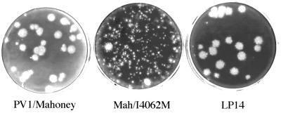
SA virus, a mutant of the Mahoney strain of type 1 poliovirus (PV1/Mahoney), replicates specifically in the spinal cords of mice and causes paralysis, although the PV1/Mahoney strain does not show any mouse neurovirulence (Q. Jia, S. Ohka, K. Iwasaki, K. Tohyama, and A. Nomoto, J. Virol. 73:6041-6047, 1999). The key mutation site for the mouse neurovirulence of SA was mapped to nucleotide (nt) 928 of the genome (A to G), resulting in the amino acid substitution of Met for Ile at residue 62 within the capsid protein VP4 (VP4062). A small-plaque phenotype of SA appears to be indicative of its mouse-neurovirulent phenotype. To identify additional amino acid residues involved in the host range determination of PV, a total of 14 large-plaque (LP) variants were isolated from a single point mutant, Mah/I4062M, that showed the SA phenotype. All the LP variants no longer showed any mouse neurovirulence when delivered via an intraspinal inoculation route. Of these, 11 isolates had a back mutation at nt 928 (G to A) that restored the nucleotide of the PV1/Mahoney type. The reversions of the remaining three isolates (LP8, LP9, and LP14) were mediated by a second site mutation. Molecular genetic analysis involving recombinants between Mah/I4062M and the LP variants revealed that the mere substitution of an amino acid residue at position 107 in VP1 (Val to Leu) (LP9), position 33 in VP2 (Val to Ile) (LP14), or position 231 in VP3 (Ile to Thr) (LP8) was sufficient to restore the PV1/Mahoney phenotype. These amino acid residues are located either on the surface or inside of the virus particle. Our results indicate that the mouse neurovirulence of PV is determined by the virion surface structure, which is formed by all four capsid proteins.
Figures
Similar articles
-
Isolation and molecular characterization of a poliovirus type 1 mutant that replicates in the spinal cords of mice.J Virol. 1999 Jul;73(7):6041-7. doi: 10.1128/JVI.73.7.6041-6047.1999. J Virol. 1999. PMID: 10364356 Free PMC article.
-
Molecular characterization of mouse-virulent poliovirus type 1 Mahoney mutants: involvement of residues of polypeptides VP1 and VP2 located on the inner surface of the capsid protein shell.J Virol. 1993 Jul;67(7):3808-17. doi: 10.1128/JVI.67.7.3808-3817.1993. J Virol. 1993. PMID: 8389907 Free PMC article.
-
Mouse neurovirulence determinants of poliovirus type 1 strain LS-a map to the coding regions of capsid protein VP1 and proteinase 2Apro.J Virol. 1994 Nov;68(11):7507-15. doi: 10.1128/JVI.68.11.7507-7515.1994. J Virol. 1994. PMID: 7933134 Free PMC article.
-
Mutation of a single conserved nucleotide between the cloverleaf and internal ribosome entry site attenuates poliovirus neurovirulence.J Virol. 2005 Nov;79(22):14235-43. doi: 10.1128/JVI.79.22.14235-14243.2005. J Virol. 2005. PMID: 16254358 Free PMC article.
-
[Molecular genetic analysis of poliovirus infection].Yakugaku Zasshi. 1989 Sep;109(9):622-35. doi: 10.1248/yakushi1947.109.9_622. Yakugaku Zasshi. 1989. PMID: 2481725 Review. Japanese.
References
-
- Couderc T, Martin A, Wychowski C, Girard M, Horaud F, Crainic R. Analysis of neutralization-escape mutants selected from a mouse virulent type 1/type 2 chimeric poliovirus: identification of a type 1 poliovirus with antigenic site 1 deleted. J Gen Virol. 1991;72:973–977. - PubMed
Publication types
MeSH terms
Substances
Grants and funding
LinkOut - more resources
Full Text Sources
Research Materials
Miscellaneous
