. 2001 Dec;75(23):11791-802.
doi: 10.1128/JVI.75.23.11791-11802.2001.

Interaction of zyxin, a focal adhesion protein, with the e6 protein from human papillomavirus type 6 results in its nuclear translocation

Affiliations

Interaction of zyxin, a focal adhesion protein, with the e6 protein from human papillomavirus type 6 results in its nuclear translocation

Y Y Degenhardt et al. J Virol. 2001 Dec.

Abstract

Zyxin, a focal adhesion molecule, interacts specifically with the E6 protein from human papillomavirus (HPV) type 6 in a yeast two-hybrid screen of a cDNA library prepared from human keratinocytes. Zyxin does not interact significantly with E6 proteins from HPV types 11, 16, or 18. The interaction was confirmed by in vitro and in vivo analyses and it requires the LIM domains (Lin-11, Isl-1, and Mec-3 [G. Freyd, S. K. Kim, and H. R. Horvitz, Nature 344:876-879, 1990]) found at the carboxyl terminus of zyxin. Cotransfection of E6 from HPV ((6)E6) and zyxin results in the accumulation of zyxin in the nucleus where it can function as a transcriptional activator. (6)E6 can also mobilize endogenous zyxin to the nucleus.


Figures

FIG. 1
FIG. 1
Zyxin binding to GST-E6 proteins. In vitro-translated and 35S-labeled zyxin was allowed to interact with identical amounts of purified GST-6E6, GST-18E6, or GST alone. After binding at 50 mM [K+], the beads were washed extensively with buffers containing 50, 150, or 250 mM salt. Bound proteins were eluted and separated by SDS-PAGE. Zyxin was visualized by autoradiography of the dried gel. The numbers above each lane are the millimolar salt concentrations used for washing the beads.
FIG. 2
FIG. 2
Coimmunoprecipitation of 6E6 with zyxin. 293T cells were transfected with DNA encoding zyxin-Flag, Gps2-Flag, PKC-Flag, or the Flag vector alone. Thirty-six hours after transfection, cell lysates were prepared and mixed with in vitro-translated and 35S-labeled 6E6 or 18E6 (A). Flag-tagged proteins were then immunoprecipitated with an anti-Flag antibody and the coprecipitated E6 proteins were visualized with a phosphorimager after separation by SDS-PAGE. The Flag proteins retained on the Sepharose beads after immunoprecipitation were detected by Western analysis with an anti-Flag antibody after boiling the beads in SDS loading buffer and separating the bound proteins by SDS-PAGE (B). Cos7 cells were transfected with DNA encoding Flag-tagged zyxin or Gps2 alone, E6-Myc constructs alone, or the combination of a Flag-tagged protein construct and an E6 construct. Thirty-six hours after transfection, cells were labeled with 35S-translabel for 3 h, and the cells were harvested and lysed. Transfected cell lysates with equal amounts of total protein were reacted with an M2 anti-Flag affinity gel. E6 proteins that were spun out with the gel were visualized by autoradiography following SDS-PAGE separation of the bound proteins. The bands corresponding to zyxin, Gps2, and the E6 proteins are marked with arrows (C). (The protein levels of E6 in each of the transfected cell lysates were detected by Western analysis using an anti-Myc antibody after SDS-PAGE and are shown in the lower part of this panel.) This experiment was repeated without labeling the cells. After separating the proteins that eluted off of the gel by SDS-PAGE, the E6 proteins and the Flag-tagged proteins were detected by Western analysis using an anti-Myc antibody (upper panel) and an anti-Flag antibody (lower panel), respectively (D).
FIG. 3
FIG. 3
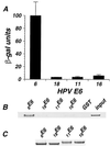
Interaction between E6 proteins and zyxin. (A) Plasmid DNAs encoding Gal4-BD fusions with 6E6, 11E6, 16E6, or 18E6 were cotransformed with a construct expressing a zyxin–Gal4-AD fusion into yeast strain YGH1. Liquid β-Gal assays were performed on the cotransformed colonies as described in Materials and Methods. The numbers for the β-Gal units are the averages of three separate experiments. (B) In vitro-translated 35S-labeled zyxin was allowed to bind to the same amounts of purified GST-E6 fusion proteins or GST alone as described in Materials and Methods. The bound zyxin protein was visualized by autoradiography after SDS-PAGE analysis. One-twentieth of the zyxin input was also electrophoresed on the gel. (C) Coomassie brilliant blue staining of the gel after SDS-PAGE was used to detect the amount of each GST fusion protein used for this assay.
FIG. 4
FIG. 4
Specificity of zyxin interactions with 6E6. (A) The amino acid sequences of 6E6 and 11E6 are aligned for comparison and only the amino acids in 11E6 that differ are shown. The vertical line denotes the junction between the sequences from 11E6 and the sequences from 6E6 in the chimeric protein 11/6E6. (B) Schematic representations of E6 proteins and their abilities to interact with zyxin in the yeast two-hybrid system or in the far Western analysis are shown. +, zyxin interaction is seen; −, no interaction; ND, not tested. (C) Equal amounts of GST fusion proteins with 6E6, 11/6E6, or 11E6 were tested for binding to in vitro-translated 35S-zyxin in a far Western analysis as described in Materials and Methods, and the resulting autoradiograph is shown in the upper panel. The GST-E6 fusion proteins used for this assay were detected by Coomassie brilliant blue staining and are shown in the lower panel.
FIG. 5
FIG. 5
Schematic diagram of zyxin. The boundaries of zyxin and the various zyxin deletion mutants constructed for this study are shown, with 6(3) being the shortest clone that interacted with 6E6 in the two-hybrid library screen. The FPPPP homology, the 5′ proline-rich domain, and the three LIM domains at the C terminus are identified.
FIG. 6
FIG. 6
Identification of 6E6 interactive domains in zyxin. (A) Yeast strain YGH1 was cotransformed with DNA constructs encoding Gal4-AD fusions to zyxin deletions and 6E6-Gal4-BD (+E6) or Gal4-BD vector (−E6). Data for the LIM proteins LIM-1+2+3 (1+2+3), LIM-2+3 (2+3), LIM-3 (3), LIM-1+2 (1+2), and LIM-1 (1) are shown. Liquid β-Gal assays were performed on the cotransformants as described in Materials and Methods. The values are the average β-Gal units from three separate experiments. (B) Yeast cotransformants were grown in Leu Trp liquid medium until the OD600 reached 0.4 to 0.6. Cells were pelleted and boiled in SDS loading buffer. Equal amounts of total cell extracts were subjected to SDS-PAGE. The expression of the Gal4-AD fusion proteins was detected by Western analysis using an anti-HA antibody. (C) Binding between zyxin deletion proteins and GST-6E6. In vitro-translated 35S-labeled full-length zyxin (FL) or zyxin deletion mutant proteins Zy-5′ and LIM-1+2+3 were allowed to interact with the same amount of GST-6E6 fusion protein or GST alone as described in Materials and Methods. The bound proteins were separated by SDS-PAGE and visualized by autoradiography. One-twentieth of the input of each 35S-labeled protein was loaded on the same gel to quantify relative binding.
FIG. 7
FIG. 7
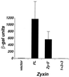
Analysis of the transcriptional activator activity of zyxin in yeast. Yeast strain YGH1 was transformed with DNAs expressing the Gal4-BD fused to full-length zyxin (FL), zyxin amino acids 1 to 382 (Zy-5′), and LIM-1+2+3 (1+2+3). The nomenclature used to describe the proteins is described in the legend for Fig. 5. Liquid β-Gal assays were performed on the yeast transformants and β-Gal units are plotted on the y axis.
FIG. 8
FIG. 8
Cellular localization of zyxin. One-half of a microgram of FL-M-GBD or Zy-5′-M-GBD DNA was cotransfected into Cos7 cells with 1.5 μg of a construct expressing 6E6-Myc (B, D) or the Myc vector (A, C). Thirty-six hours after transfection, cells were fixed and zyxin proteins were detected with a polyclonal rabbit anti-zyxin antibody and visualized after interaction with a rhodamine-conjugated anti-rabbit serum (A to D), while E6 proteins were detected with a mouse monoclonal anti-Myc antibody followed by fluorescein isothiocyanate-labeled anti-mouse antibody (E to H).
FIG. 9
FIG. 9
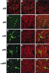
Distribution of endogenous zyxin in Mewo cells cotransfected with E6 proteins. Mewo cells were transfected with DNA constructs expressing 6E6-Myc (A to F), 11E6-Myc (G to I), 18E6-Myc (J to L), or 11/6E6-Myc (M to O). Thirty-six hours after transfection, cells were fixed and the distribution of zyxin (A, B, C, E, H, K, and N) and E6 (A, B, C, D, G, J, and M) in the cells was detected by confocal microscopy as described in Materials and Methods. Panels A, B, and C are the same image photographed at three different depths of field to demonstrate that the E6 signal is nuclearly located. Panels F, I, L, and O are the merged images of panels D and E, G and H, J and K, and M and N, respectively.

Similar articles

Cited by

References

    1. Androphy E J, Hubbert N L, Schiller J T, Lowy D R. Identification of the HPV-16 E6 protein from transformed mouse cells and human cervical carcinoma cell lines. EMBO J. 1987;6:989–992. - PMC - PubMed
    1. Arber S, Hunter J J, Ross J, Jr, Hongo M, Sansig G, Borg J, Perriard J C, Chien K R, Caroni P. MLP-deficient mice exhibit a disruption of cardiac cytoarchitectural organization, dilated cardiomyopathy, and heart failure. Cell. 1997;88:393–403. - PubMed
    1. Bach I. The LIM domain: regulation by association. Mech Dev. 2000;91:5–17. - PubMed
    1. Bacharach E, Goff S P. Binding of the human immunodeficiency virus type 1 Gag protein to the viral RNA encapsidation signal in the yeast three-hybrid system. J Virol. 1998;72:6944–6949. - PMC - PubMed
    1. Barbosa M S, Lowy D R, Schiller J T. Papillomavirus polypeptides E6 and E7 are zinc-binding proteins. J Virol. 1989;63:1404–1407. - PMC - PubMed

MeSH terms

LinkOut - more resources