. 2001 Dec;75(23):11834-50.
doi: 10.1128/JVI.75.23.11834-11850.2001.

Differential infection of polarized epithelial cell lines by sialic acid-dependent and sialic acid-independent rotavirus strains

Affiliations

Differential infection of polarized epithelial cell lines by sialic acid-dependent and sialic acid-independent rotavirus strains

M Ciarlet et al. J Virol. 2001 Dec.

Abstract

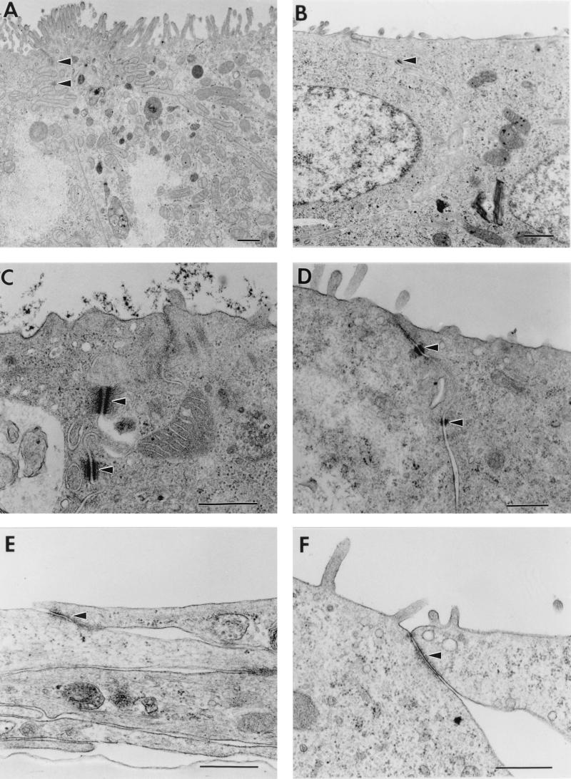
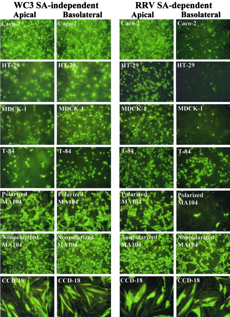
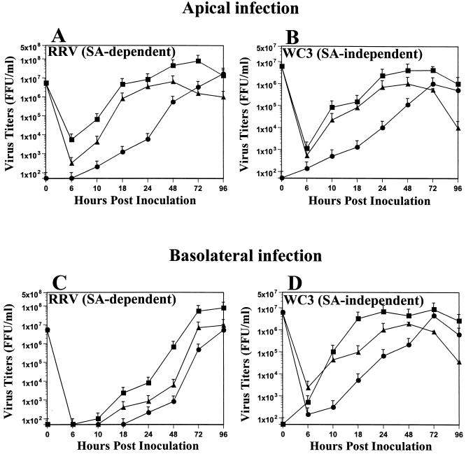
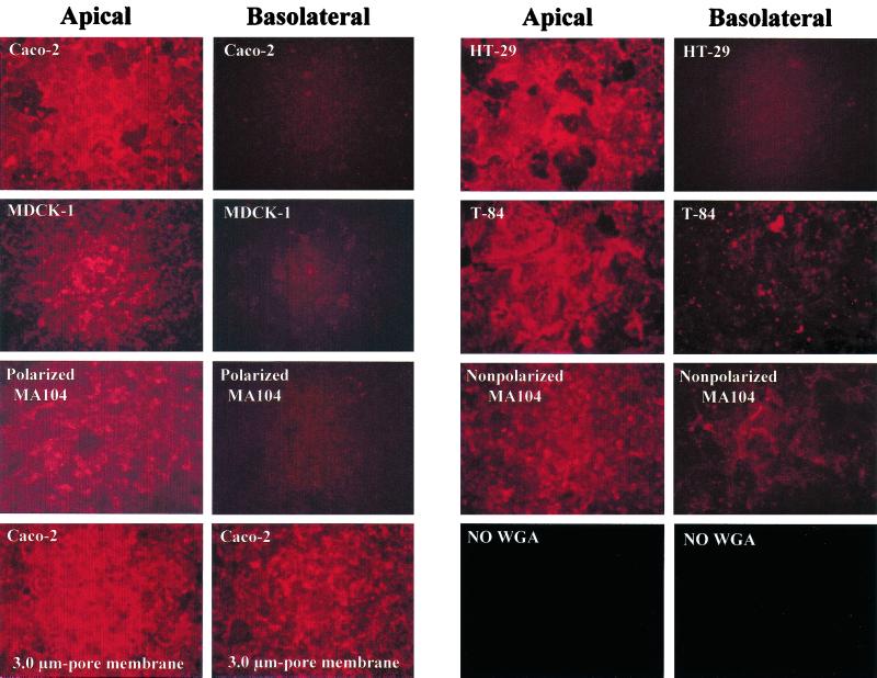
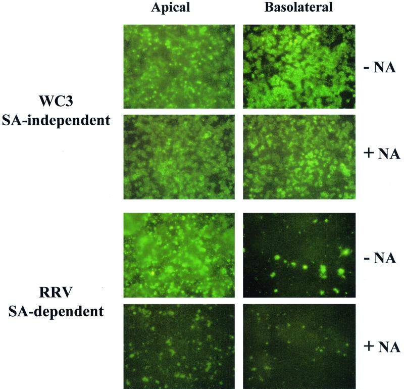
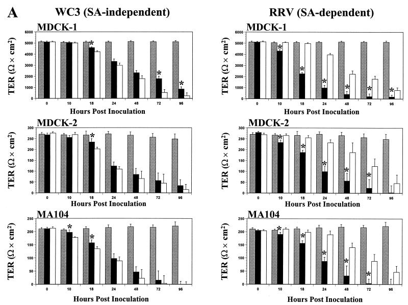
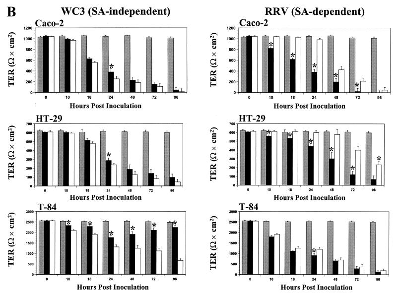
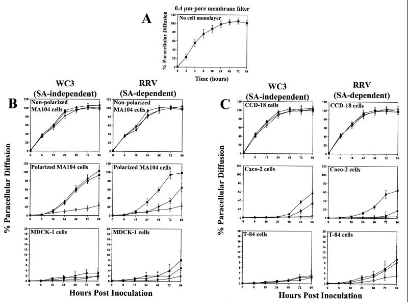
Infection of epithelial cells by some animal rotaviruses, but not human or most animal rotaviruses, requires the presence of N-acetylneuraminic (sialic) acid (SA) on the cell surface for efficient infectivity. To further understand how rotaviruses enter susceptible cells, six different polarized epithelial cell lines, grown on permeable filter membrane supports containing 0.4-microm pores, were infected apically or basolaterally with SA-independent or SA-dependent rotaviruses. SA-independent rotaviruses applied apically or basolaterally were capable of efficiently infecting both sides of the epithelium of all six polarized cell lines tested, while SA-dependent rotaviruses only infected efficiently through the apical surface of five of the polarized cell lines tested. Regardless of the route of virus entry, SA-dependent and SA-independent rotaviruses were released almost exclusively from the apical domain of the plasma membrane of polarized cells before monolayer disruption or cell lysis. The transepithelial electrical resistance (TER) of cells decreased at the same time, irrespective of whether infection with SA-independent rotaviruses occurred apically or basolaterally. The TER of cells infected apically with SA-dependent rotaviruses decreased earlier than that of cells infected basolaterally. Rotavirus infection decreased TER before the appearance of cytopathic effect and cell death and resulted in an increase in the paracellular permeability to [(3)H]inulin as a function of loss of TER. The presence of SA residues on either the apical or basolateral side was determined using a Texas Red-conjugated lectin, wheat germ agglutinin (WGA), which binds SA residues. WGA bound exclusively to SA residues on the apical surface of the cells, confirming the requirement for SA residues on the apical cell membrane for efficient infectivity of SA-dependent rotaviruses. These results indicate that the rotavirus SA-independent cellular receptor is present on both sides of the epithelium, but SA-dependent and SA-independent rotavirus strains infect polarized epithelial cells by different mechanisms, which may be relevant for pathogenesis and selection of vaccine strains. Finally, rotavirus-induced alterations of the epithelial barrier and paracellular permeability suggest that common mechanisms of pathogenesis may exist between viral and bacterial pathogens of the intestinal tract.


Figures

FIG. 1
FIG. 1
Electron micrographs of polarized intestinal Caco-2 (A), HT-29 (B), and T-84 (C) and polarized kidney MDCK-1 (D) and MA104 (E and F) cells. Cells were grown on permeable supports containing 0.4-μm pores. Intestinal Caco-2, HT-29, and T-84 cells exhibit an enterocyte-like appearance, with an apical brush border, microvilli, and typical tight junctions and desmosomes (arrows). Kidney MDCK-1 and MA104 cells show evidence of some microvilli along the apical surface typical of the polarized renal epithelium, and tight junctions and junctional complexes, respectively (arrows). Bars, 0.5 μm. Original magnifications: ×13,000 (A), ×16,000 (B), ×40,000 (C, E, and F), and ×32,000 (D).
FIG. 2
FIG. 2
Immunofluorescence analysis of the infectivity of SA-independent (WC3) and SA-dependent (RRV) rotavirus strains inoculated apically or basolaterally with 10 FFU per cell onto polarized Caco-2, HT-29, T-84, MDCK-1, and MA104 and nonpolarized MA104 and CCD-18 epithelial cells grown on permeable supports containing 0.4-μm pores. At 24 hpi, the monolayers were fixed with methanol, stained for viral antigen with a rotavirus-specific rabbit polyclonal hyperimmune serum followed by fluorescein-conjugated goat anti-rabbit Ig antibody, and examined by immunofluorescence.
FIG. 3
FIG. 3
Comparison of yields of progeny of SA-dependent strain RRV and SA-independent strain WC3 in Caco-2 cells inoculated apically or basolaterally with RRV (A and C) or WC3 (B and D) at an MOI of 10 FFU per cell. Caco-2 cell monolayers displayed a TER of >900 Ω × cm2 prior to infection. At the indicated time points, Caco-2 cells (▴) and apical (▪) and basolateral (●) supernatant culture media were collected separately, and virus titers were determined by FFA (5). Values shown are arithmetic means of at least three independent experiments. Error bars represent 1 standard error of the mean.
FIG. 4
FIG. 4
Detection of SA residues of polarized Caco-2, HT-29, T-84, MDCK-1, and MA104 and nonpolarized MA104 epithelial cells grown on permeable supports containing 0.4-μm pores, using a Texas Red-conjugated lectin, WGA. WGA (5 μg/ml) was added apically or basolaterally to cells, incubated for 20 min at 37°C, and washed before SA residue staining was visualized by fluorescence. The bottom left panels show the SA residue staining pattern in polarized Caco-2 cells grown on permeable supports containing 3.0-μm pores as the result of cells migrating through these large pores and growing on the opposite face of the membrane filter. Similar SA residue staining was detected when polarized HT-29, T-84, MDCK-1, and MA104 cells were grown on membrane filters containing 3.0-μm pores (data not shown). Background SA residue staining of Caco-2 cells grown on permeable supports containing 0.4-μm pores when no WGA was applied is also shown (bottom right panels).
FIG. 5
FIG. 5
Immunofluorescence analysis of the infectivity of SA-independent (WC3) and SA-dependent (RRV) rotavirus strains inoculated apically or basolaterally at 10 FFU per cell onto polarized Caco-2 epithelial cells grown on permeable supports containing 0.4-μm pores and treated with 40 mU of neuraminidase (Arthrobacter ureafaciens) per ml (+ NA) or mock-treated with TNC buffer (− NA) for 1 h at 37°C. At 24 hpi, the monolayers were fixed with methanol, stained for viral antigen with a rotavirus-specific rabbit polyclonal hyperimmune serum followed by fluorescein-conjugated goat anti-rabbit Ig antibody, and examined by immunofluorescence.
FIG. 6
FIG. 6
TER of renal MDCK-1, MDCK-2, and MA104 polarized intestinal Caco-2, HT-29, and T-84 (A) and (B) epithelial cell monolayers grown on permeable supports containing 0.4-μm pores and mock inoculated (shaded bars) or inoculated apically (solid bars) or basolaterally (open bars) with 10 FFU per cell of the SA-independent rotavirus WC3 or SA-dependent RRV strain. The net TER was calculated by subtracting the background (membrane filter without cells) and multiplying the resistance (Ω) by the area (0.33 cm2) of the filter. TER measurements were taken at the indicated times. Each bar shows the arithmetic mean of at least three independent experiments. Error bars represent 1 standard error of the mean. A significant (P < 0.05, Mann-Whitney U) difference in TER of cells infected apically with respect to those infected basolaterally at a given time point is indicated by an asterisk.
FIG. 6
FIG. 6
TER of renal MDCK-1, MDCK-2, and MA104 polarized intestinal Caco-2, HT-29, and T-84 (A) and (B) epithelial cell monolayers grown on permeable supports containing 0.4-μm pores and mock inoculated (shaded bars) or inoculated apically (solid bars) or basolaterally (open bars) with 10 FFU per cell of the SA-independent rotavirus WC3 or SA-dependent RRV strain. The net TER was calculated by subtracting the background (membrane filter without cells) and multiplying the resistance (Ω) by the area (0.33 cm2) of the filter. TER measurements were taken at the indicated times. Each bar shows the arithmetic mean of at least three independent experiments. Error bars represent 1 standard error of the mean. A significant (P < 0.05, Mann-Whitney U) difference in TER of cells infected apically with respect to those infected basolaterally at a given time point is indicated by an asterisk.
FIG. 7
FIG. 7
Paracellular diffusion of [3H]inulin across 0.4-μm-pore membrane filters with (A) no cells (×), (B) monolayers of nonpolarized kidney MA104 and polarized MA104 and MDCK-1 cells, and (C) monolayers of nonpolarized intestinal CCD-18 and polarized Caco-2 and T-84 epithelial cells, grown on permeable supports containing 0.4-μm pores, infected apically (▪) or basolaterally (●) at 10 FFU per cell with SA-independent rotavirus WC3 or SA-dependent RRV strain or left uninfected (○) at the indicated times. Paracellular diffusion of [3H]inulin across cell monolayers is expressed as a percentage of total [3H]inulin present in the apical chamber. Values shown are the arithmetic means of at least three replicate experiments. Error bars represent 1 standard error of the mean.

Similar articles

Cited by

References

    1. Ball J M, Tian P, Zeng C Q-Y, Morris A P, Estes M K. Age-dependent diarrhea induced by a rotaviral nonstructural glycoprotein. Science. 1996;272:101–104. - PubMed
    1. Blank C, Anderson D, Beard M, Lemon S M. Infection of polarized cultures of human intestinal epithelial cells with hepatitis A virus: vectorial release of progeny virions through apical cellular membranes. J Virol. 2000;74:6476–6484. - PMC - PubMed
    1. Canil C, Rosenshine I, Ruschkowski S, Donnenberg M, Kaper J, Finlay B B. Enteropathogenic Escherichia coli decreases the transepithelial electrical resistance of polarized epithelial monolayers. Infect Immun. 1993;61:2755–2762. - PMC - PubMed
    1. Casola A, Estes M K, Crawford S E, Ogra P L, Ernst P B, Garofalo R, Crowe S E. Rotavirus infection of cultured intestinal epithelial cells induces secretion of CXC and CC chemokines. Gastroenterology. 1998;114:947–955. - PubMed
    1. Ciarlet M, Estes M K. Human and most animal rotavirus strains do not require the presence of sialic acid on the cell surface for efficient infectivity. J Gen Virol. 1999;80:943–948. - PubMed

Publication types

Substances

LinkOut - more resources