

doi: 10.1128/JVI.75.23.11897-11901.2001.
An N-terminal domain of herpes simplex virus type Ig E is capable of forming stable complexes with gI
Affiliations
- PMID: 11689673
- PMCID: PMC114778
- DOI: 10.1128/JVI.75.23.11897-11901.2001
Item in Clipboard
An N-terminal domain of herpes simplex virus type Ig E is capable of forming stable complexes with gI
J Virol.
2001 Dec.
Abstract
Using limited proteolytic analyses, we show that gE present in soluble herpes simplex virus type 1 gE-gI complexes is cleaved into a C-terminal (CgE) and an N-terminal (NgE) domain. The domain boundary is in the vicinity of residue 188 of mature gE. NgE, but not CgE, forms a stable complex with soluble gI.
Figures
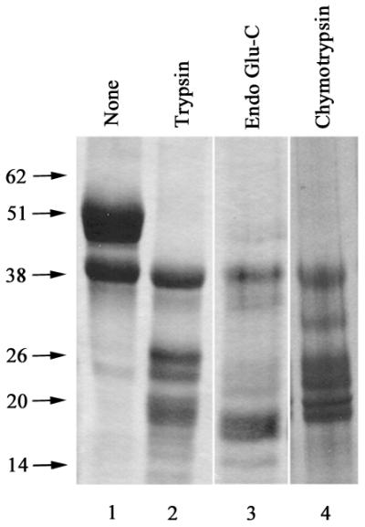
Digestions of soluble gE-gI with three different proteases show that gI is stable while gE is degraded into 20- to 25-kDa fragments. Soluble gE-gI complexes were subjected to limited proteolytic digestion with trypsin, endoproteinase Glu-C (endo Glu-C), or chymotrypsin at 4°C for 30, 30, or 120 min, respectively. Products of tryptic digestion were analyzed by SDS-PAGE. Lane 1, intact gE-gI complexes (20 μg); lane 2, trypsin-digested gE-gI (20 μg); lane 3, endoproteinase Glu-C-digested gE-gI (5 μg); lane 4, chymotrypsin digested gE-gI (5 μg).
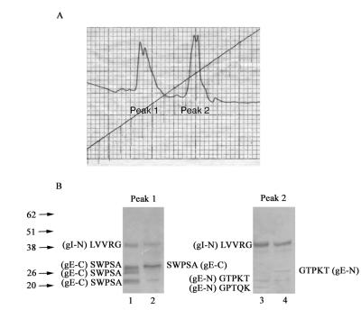
Analysis of tryptic digestion products yields information about a domain boundary in gE. (A) Soluble gE-gI complexes were digested with trypsin (substrate/enzyme ratio, 80:1 by weight) at 4°C, and digested proteins were separated on a Mono Q column. (B) Chromatographic peaks 1 and 2 from panel A were analyzed by SDS-PAGE followed by N-terminal sequencing. Lanes 1 and 2, proteins contained in peak 1, including gI (with the N-terminal sequence LVVRG) and gE fragments with SWPSA as the N-terminal sequence (initiating at residue 189 of mature gE). Lanes 3 and 4, proteins contained in peak 2, including gI (with the N-terminal sequence LVVRG) and gE fragments with the N-terminal sequence GTPKT (initiating at the N terminus of mature gE) or GPTQK (initiating at residue 23 of mature gE).
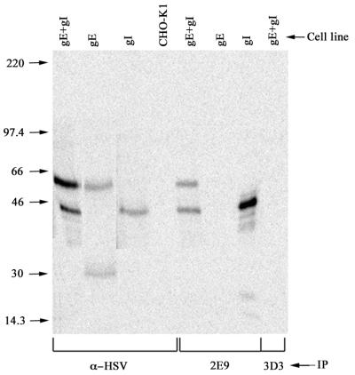
The 2E9 hybridoma line recognizes gI and gE-gI complexes. CHO cells expressing both soluble gE and gI, or either soluble gE or soluble gI alone, or untransfected CHO-K1 cells as negative controls were metabolically labeled with [35S]methionine/cysteine (ICN Biomedicals) for 5 h. Cell supernatants were immunoprecipitated with 10 μg of rabbit anti-HSV IgG to visualize the expressed HSV proteins, or with 0.05 ml of 2E9 ascites fluid, or with 0.05 ml of a control ascites fluid (generated using the 3D3 hybridoma line). The 30-kDa band observed in the second lane is a spontaneously derived proteolytic fragment of gE, often visualized in cells that express gE.
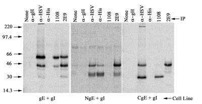
NgE but not CgE forms stable complexes with gI, as shown by coimmunoprecipitation analyses with antibodies specific for gE and gI. CHO cells expressing soluble gE and gI, or cells expressing NgE and gI, or CgE and gI, were metabolically labeled with [35S]methionine/cysteine (ICN Biomedicals) for 5 h. Cell supernatants were immunoprecipitated with the indicated antibodies, and proteins were visualized by SDS-PAGE and phosphorimaging analyses. Antibodies used were anti-gH (an irrelevant antibody), to assess nonspecific binding; anti-HSV, to visualize expressed HSV proteins; anti-His, which recognizes the hexahistidine epitope tag present on NgE; 1108, which recognizes an unknown epitope present in gE and CgE; and 2E9, which is directed against gI.
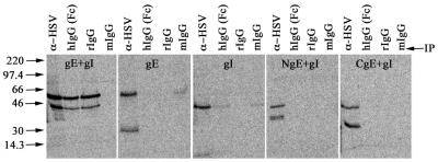
Soluble gE-gI complexes, but not the NgE-gI complexes or CgE+gI combination, function as an Fc receptor for IgG. gE-gI complexes have previously been shown to interact with the Fc domains of human and rabbit immunoglobulins but not mouse immunoglobulins. CHO cells expressing soluble gE and gI, or cells expressing either NgE plus gI, CgE plus gI, gE alone, or gI alone, were metabolically labeled with [35S]methionine/cysteine (ICN Biomedicals) for 5 h. Cell supernatants were immunoprecipitated with either anti-HSV-1, human Fc [hIgG(Fc)], rabbit IgG (rIgG) (purified from normal rabbit serum), or mouse IgG (mIgG). Immunoprecipitated proteins were visualized by SDS-PAGE followed by phosphorimaging analyses. The 30-kDa band observed in the second panel (with the gE-expressing cell line) is a spontaneously derived proteolytic fragment of gE, often visualized in cells that express gE.
Similar articles
-
UL13 protein kinase of herpes simplex virus 1 complexes with glycoprotein E and mediates the phosphorylation of the viral Fc receptor: glycoproteins E and I.Virology. 1998 Feb 1;241(1):37-48. doi: 10.1006/viro.1997.8963. Virology. 1998. PMID: 9454715
-
The extracellular domain of herpes simplex virus gE is indispensable for efficient cell-to-cell spread: evidence for gE/gI receptors.J Virol. 2005 Sep;79(18):11990-2001. doi: 10.1128/JVI.79.18.11990-12001.2005. J Virol. 2005. PMID: 16140775 Free PMC article.
-
Herpes simplex virus gE/gI must accumulate in the trans-Golgi network at early times and then redistribute to cell junctions to promote cell-cell spread.J Virol. 2006 Apr;80(7):3167-79. doi: 10.1128/JVI.80.7.3167-3179.2006. J Virol. 2006. PMID: 16537585 Free PMC article.
-
Characterization of domains of herpes simplex virus type 1 glycoprotein E involved in Fc binding activity for immunoglobulin G aggregates.J Virol. 1994 Apr;68(4):2478-85. doi: 10.1128/JVI.68.4.2478-2485.1994. J Virol. 1994. PMID: 7511171 Free PMC article.
-
Different receptors binding to distinct interfaces on herpes simplex virus gD can trigger events leading to cell fusion and viral entry.Virology. 2006 Jan 5;344(1):17-24. doi: 10.1016/j.virol.2005.09.016. Virology. 2006. PMID: 16364731 Review.
Cited by
-
Deletion of the first cysteine-rich region of the varicella-zoster virus glycoprotein E ectodomain abolishes the gE and gI interaction and differentially affects cell-cell spread and viral entry.J Virol. 2009 Jan;83(1):228-40. doi: 10.1128/JVI.00913-08. Epub 2008 Oct 22. J Virol. 2009. PMID: 18945783 Free PMC article.
-
Immunization strategies to block the herpes simplex virus type 1 immunoglobulin G Fc receptor.J Virol. 2004 Mar;78(5):2562-71. doi: 10.1128/jvi.78.5.2562-2571.2004. J Virol. 2004. PMID: 14963159 Free PMC article.
-
Responses of herpes simplex virus type 1-infected cells to the presence of extracellular antibodies: gE-dependent glycoprotein capping and enhancement in cell-to-cell spread.J Virol. 2003 Jan;77(1):701-8. doi: 10.1128/jvi.77.1.701-708.2003. J Virol. 2003. PMID: 12477873 Free PMC article.
-
Discovery and Characterization of an Aberrant Small Form of Glycoprotein I of Herpes Simplex Virus Type I in Cell Culture.Microbiol Spectr. 2022 Apr 27;10(2):e0265921. doi: 10.1128/spectrum.02659-21. Epub 2022 Mar 29. Microbiol Spectr. 2022. PMID: 35348373 Free PMC article.
-
Crystal structure of the HSV-1 Fc receptor bound to Fc reveals a mechanism for antibody bipolar bridging.PLoS Biol. 2006 Jun;4(6):e148. doi: 10.1371/journal.pbio.0040148. Epub 2006 May 2. PLoS Biol. 2006. PMID: 16646632 Free PMC article.
References
-
- Basu S, Dubin G, Basu M, Nguyen V, Friedman H M. Characterization of regions of herpes simplex virus type 1 glycoprotein E involved in binding the Fc domain of monomeric IgG and in forming a complex with glycoprotein I. J Immunol. 1995;154:260–267. - PubMed
-
- Basu S, Dubin G, Nagashunmugam T, Basu M, Goldstein L T, Wang L, Weeks B, Friedman H. Mapping regions of herpes simplex virus type 1 glycoprotein I required for formation of the viral Fc receptor for monomeric IgG. J Immunol. 1997;158:209–215. - PubMed
-
- Chapman T L, Heikema A P, Bjorkman P J. The inhibitory receptor LIR-1 uses a common binding interaction to recognize class I MHC molecules and the viral homolog UL18. Immunity. 1999;11:603–614. - PubMed
-
- Chapman T L, You I, Joseph I, Bjorkman P J, Morrison S L, Raghavan M. Characterization of the interaction between the herpes simplex virus type 1 Fc receptor and immunoglobulin G. J Biol Chem. 1999;274:6911–6919. - PubMed
Publication types
MeSH terms
Substances
Grants and funding
LinkOut - more resources
Full Text Sources
Other Literature Sources
