. 2001 Dec;21(23):7883-91.
doi: 10.1128/MCB.21.23.7883-7891.2001.

Repression of Ets-2-induced transactivation of the tau interferon promoter by Oct-4

Affiliations

Repression of Ets-2-induced transactivation of the tau interferon promoter by Oct-4

T Ezashi et al. Mol Cell Biol. 2001 Dec.

Abstract

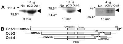
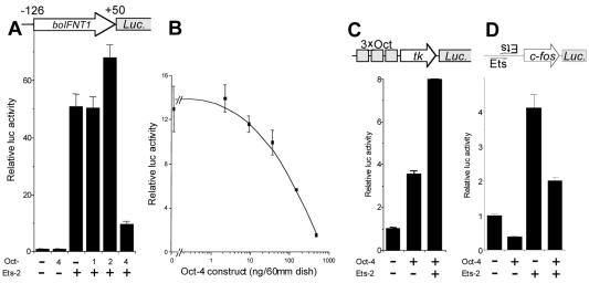
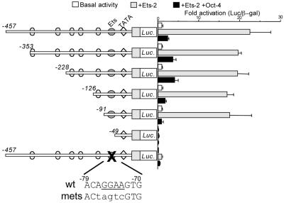
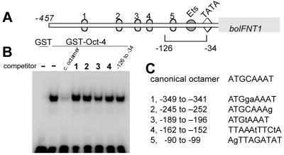
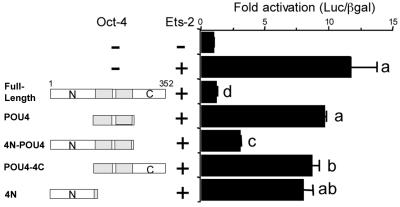
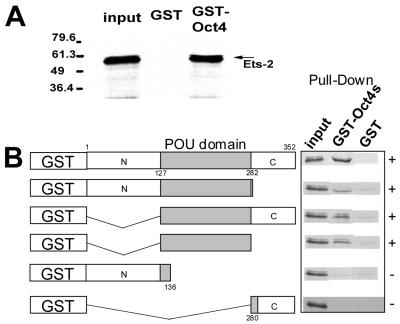
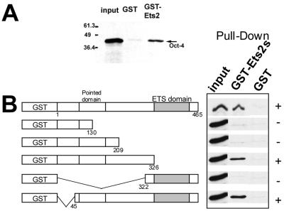
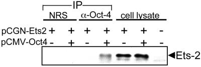
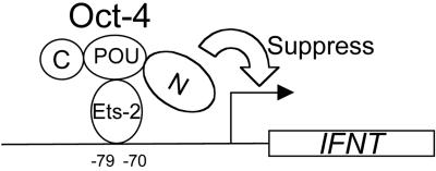
Oct-4 is a POU family transcription factor associated with potentially totipotent cells. Genes expressed in the trophectoderm but not in embryos prior to blastocyst formation may be targets for silencing by Oct-4. Here, we have tested this hypothesis with the tau interferon genes (IFNT genes), which are expressed exclusively in the trophectoderm of bovine embryos. IFNT promoters contain an Ets-2 enhancer, located at -79 to -70, and are up-regulated about 20-fold by the overexpression of Ets-2 in human JAr choriocarcinoma cells, which are permissive for IFNT expression. This enhancement was reversed in a dose-dependent manner by coexpression of Oct-4 but not either Oct-1 or Oct-2. When cells were transfected with truncated bovine IFNT promoters designed to eliminate potential octamer sites sequentially, luciferase reporter expression from each construct was still silenced by Oct-4. Full repression required both the N-terminal and POU domains of Oct-4, but neither domain used alone was an effective silencer. Oct-4 and Ets-2 formed a complex in vitro in the absence of DNA through binding of the POU domain of Oct-4 to a site located between the "pointed" and DNA binding domains of Ets-2. The two transcription factors were also coimmunoprecipitated after being expressed together in JAr cells. Oct-4, therefore, silences IFNT promoters by quenching Ets-2 transactivation. The POU domain most probably binds to Ets-2 directly, while the N-terminal domain inhibits transcription. These findings provide further evidence that the developmental switch to the trophectoderm is accompanied by the loss of Oct-4 silencing of key genes.


Figures

FIG. 1
FIG. 1
Expression of Oct proteins in transfected JAr cells. (A) Western blotting of JAr cell extracts (75 μg) before and after transfection with expression plasmids for Oct-1, Oct-2, and Oct-4. The amount of plasmid used in the transfection is shown above each panel; the exposure times for detecting chemiluminescence are shown below. Arrowheads show the positions of the respective proteins. The numbers to the left of each gel blot are molecular size markers (in thousands). (B) Structures of the Oct expression constructs that were used in transfection experiments. All plasmids share the same expression system [CMV promoter-tk leader and β-globin gene poly(A) signal]. The numbers represent the lengths (in amino acid residues) of the Oct proteins.
FIG. 2
FIG. 2
Oct-4 acts solely as a repressor of the boIFNT1 promoter. (A) The −126luc construct is suppressed specifically by Oct-4 and not by other octamer proteins. JAr cells were transfected with 3 μg of −126luc, 0.2 μg of Ets-2, and/or 0.2 μg of Oct expression plasmid. Reporter activities were normalized to the activity of cotransfected plasmid pRL-CMV. (B) Dose-dependent repression of the boIFNT1 promoter by Oct-4. JAr cells were transfected with 3.2 μg of −126luc, 0.4 μg of Ets-2, and 0 to 400 ng of Oct-4 expression plasmid. Reporter activities were normalized to the activity of cotransfected plasmid pRSVLTR-βgal. Error bars show standard errors (SE). (C) Oct-4 transactivation of an artificial promoter, 3×Oct-tk. JAr cells were transfected with 3 μg of 3×Oct-tk reporter, 0.2 μg of Oct-4, and 0.2 μg of Ets-2 expression plasmid. Reporter activities were normalized to the activity of cotransfected plasmid pRL-CMV. Results are means and SE from three independent experiments. Activity is expressed as fold activation relative to basal activity. (D) Oct-4 represses E.18 reporter activity in either the absence (−) or the presence (+) of Ets-2. JAr cells were transfected with 3.2 μg of E.18-luc reporter and 0.4 μg of Ets-2 expression plasmid. Reporter activities were normalized to the activity of cotransfected plasmid pRSVLTR-βgal. Data are reported as described for panel C.
FIG. 3
FIG. 3
Oct-4 suppresses the promoter activities of boIFNT1 promoter constructs with progressive deletions removing potential Oct binding sites. The IFNT promoter deletions (from −457 to −49 bp) linked to luc reporters were transfected into human JAr cells in either the absence (Basal) or the presence of the expression vectors for human Ets-2 (+Ets-2) and Ets-2 and murine Oct-4 together (+Ets-2 +Oct-4). As a control, expression from the −457luc construct, with its Ets binding site mutated, was measured in the presence and absence of Ets-2 with and without cotransfection with Oct-4. Reporter activities are expressed relative to the activity of cotransfected plasmid pRSVLTR-βgal. Results are means and standard errors from three independent experiments. Activity is shown as fold activation relative to the basal activity for each construct, except in two instances. For the mutated −457luc construct, fold changes in expression are shown relative to the basal expression of nonmutated −457luc. Similarly, expression from −49luc that lacks the Ets-2 binding site is compared to the basal expression of −126luc. A mutation targeted to the Ets binding site is indicated at the bottom of the figure. wt, wild type; mets, mutated Ets site.
FIG. 4
FIG. 4
Failure of Oct-4 to bind the octamer-like sites in the boIFNT1 promoter efficiently. (A) Structure of the IFNT promoter and the locations of the octamer-like sites, the Ets site, and the TATA box. (B) DNA competition binding assays with a GST–Oct-4 fusion protein. Recombinant GST (negative control) or GST–Oct-4 fusion protein was incubated with a 32P-labeled, double-stranded canonical octamer (Table 1) in the presence of either no competitor DNA (−) or a 250-fold excess of unlabeled probe (c. octamer), double-stranded oligonucleotides identical in sequence to the octamer-like sites in the boIFNT1 promoter (lanes 1 to 4), and the promoter fragment from −126 to −34 (last lane). (C) Comparisons of the putative octamer sites in the boIFNT1 promoter with canonical octamer sequences. Mismatched sequences are shown with lowercase letters.
FIG. 5
FIG. 5
Domain specificity of Oct-4 in the suppression of the boIFNT1 promoter. The −1261uc reporter was transfected into JAr cells in either the absence (−) or the presence of expression vectors for Ets-2 (+) and for both Ets-2 and Oct-4 deletion constructs. The structures of the various Oct-4 deletion proteins are shown diagrammatically on the left, with shaded rectangles representing the POU domains. Reporter activity was normalized to the activity of cotransfected plasmid pRSVLTR-βgal. Results are means and standard errors. Activity is expressed as fold activation relative to basal activity. Data from four independent transfections, each run in triplicate, were log transformed to limit the heterogeneity of variance and were analyzed by least-squares analysis of variance (PC-SAS version 6.12; Statistical Analysis System Institute, Cary, N.C.). Pairwise comparisons among treatments were completed by using F test statistics (PC-SAS). Values marked with different letters (a, b, c, and d) differ significantly (P < 0.05).
FIG. 6
FIG. 6
Recombinant Ets-2 protein binds specifically to the POU domain of Oct-4 in vitro. (A) Ets-2 protein interacts with Oct-4 protein in a GST pull-down assay. E. coli-expressed GST–Oct-4 fusion protein immobilized on Sepharose beads was mixed with [35S]methionine-labeled, in vitro-translated Ets-2 protein. Protein bound specifically was analyzed by SDS–10% PAGE. The first lane shows an analysis of the input protein (10% of in vitro-translated Ets-2). The second lane shows that immobilized GST failed to bind in vitro-translated Ets-2, while the third lane shows that GST–Oct-4 bound the radioactive protein. (B) Summary of the data from the pull-down assay, in which a series of Oct-4 truncations were tested for their ability to bind 35S-labeled Ets-2. The truncated proteins (shown diagrammatically on the left) were synthesized as GST fusion proteins and coupled to Sepharose. The ability of the proteins to bind 35S-labeled Ets-2 was then assessed in a pull-down assay. The data are consistent with the conclusion that the POU domain (aa 127 to 282) is required for Oct-4 to bind Ets-2.
FIG. 7
FIG. 7
Recombinant Oct-4 binds to a central domain of Ets-2 in vitro. (A) Oct-4 protein interacts with Ets-2 protein in a GST pull-down assay. GST–Ets-2 fusion protein and GST immobilized on Sepharose beads were mixed with [35S]methionine-labeled, in vitro-translated Oct-4. Bound protein was analyzed by SDS-PAGE as described in the legend to Fig. 6. The first lane is input protein (10% of the in vitro translation mixture of Oct-4). (B) Summary of the data from the pull-down assay, in which a series of Ets-2 truncations (shown on the left) were tested for their ability to bind 35S-labeled Oct-4. The data indicate that a central domain within Ets-2 is required for binding to Oct-4.
FIG. 8
FIG. 8
Oct-4 binds Ets-2 in vivo. Whole-cell extracts were prepared from JAr cells that had been transiently transfected with 10 μg of pCGNEts-2 in either the presence or the absence of 10 μg of pCMV-Oct4. Protein G cross-linked with antiserum against Oct-4 (α-Oct-4) or nonimmune serum (NRS) was then added. Immune complexes were collected and analyzed by SDS-PAGE and Western blotting with anti-Ets-2 antiserum (first through fourth lanes). Blots show the presence of Ets-2 protein in whole-cell lysates (10 μg) of transfected cells (fifth and sixth lanes) and in nontransfected cells (seventh lane). The fourth lane shows that Ets-2 is immunoprecipitated with anti-Oct-4 antiserum only in cells coexpressing both proteins.
FIG. 9
FIG. 9
Oct-4 likely silences IFNT gene transcription through a quenching mechanism involving binding to the transactivator Ets-2.

Similar articles

Cited by

References

    1. Ambrosetti D C, Basilico C, Dailey L. Synergistic activation of the fibroblast growth factor-4 enhancer by Sox2 and Oct-3 depends on protein-protein interactions facilitated by a specific spatial arrangement of factor binding sites. Mol Cell Biol. 1997;17:6321–6329. - PMC - PubMed
    1. Bazer F W, Spencer T E, Ott T L. Interferon tau: a novel pregnancy recognition signal. Am J Reprod Immunol. 1997;37:412–420. - PubMed
    1. Botquin V, Hess H, Fuhrmann G, Anastassiadis C, Gross M K, Vriend G, Schöler H R. New POU dimer configuration mediates antagonistic control of an osteopontin preimplantation enhancer by Oct-4 and Sox-2. Genes Dev. 1998;12:2073–2090. - PMC - PubMed
    1. Brehm A, Ohbo K, Schöler H. The carboxy-terminal transactivation domain of Oct-4 acquires cell specificity through the POU domain. Mol Cell Biol. 1997;17:154–162. - PMC - PubMed
    1. Brehm A, Ohbo K, Zwerschke W, Botquin V, Jansen-Durr P, Schöler H R. Synergism with germ line transcription factor Oct-4: viral oncoproteins share the ability to mimic a stem cell-specific activity. Mol Cell Biol. 1999;19:2635–2643. - PMC - PubMed

Publication types

MeSH terms

Substances

LinkOut - more resources