. 2001 Dec;21(23):7913-22.
doi: 10.1128/MCB.21.23.7913-7922.2001.

Methylation-mediated proviral silencing is associated with MeCP2 recruitment and localized histone H3 deacetylation

Affiliations

Methylation-mediated proviral silencing is associated with MeCP2 recruitment and localized histone H3 deacetylation

M C Lorincz et al. Mol Cell Biol. 2001 Dec.

Abstract

The majority of 5-methylcytosine in mammalian DNA resides in endogenous transposable elements and is associated with the transcriptional silencing of these parasitic elements. Methylation also plays an important role in the silencing of exogenous retroviruses. One of the difficulties inherent in the study of proviral silencing is that the sites in which proviruses randomly integrate influence the probability of de novo methylation and expression. In order to compare methylated and unmethylated proviruses at the same genomic site, we used a recombinase-based targeting approach to introduce an in vitro methylated or unmethylated Moloney murine leukemia-based provirus in MEL cells. The methylated and unmethylated states are maintained in vivo, with the exception of the initially methylated proviral enhancer, which becomes demethylated in vivo. Although the enhancer is unmethylated and remodeled, the methylated provirus is transcriptionally silent. To further analyze the repressed state, histone acetylation status was determined by chromatin immunoprecipitation (ChIP) analyses, which revealed that localized histone H3 but not histone H4 hyperacetylation is inversely correlated with proviral methylation density. Since members of the methyl-CpG binding domain (MBD) family of proteins recruit histone deacetylase activity, these proteins may play a role in proviral repression. Interestingly, only MBD3 and MeCP2 are expressed in MEL cells. ChIPs with antibodies specific for these proteins revealed that only MeCP2 associates with the provirus in a methylation-dependent manner. Taken together, our results suggest that MeCP2 recruitment to a methylated provirus is sufficient for transcriptional silencing, despite the presence of a remodeled enhancer.


Figures
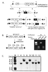
FIG. 1
FIG. 1
Principle and application of Cre-mediated targeting of methylated and unmethylated proviruses at a defined genomic site. (A) A cell line containing a stably integrated L1-HYTK-1L gene flanked by inverted loxP sites (black triangles) is transfected with the proviral construct L1-MFGhGFP-1L (also flanked by inverted loxP sites) together with a Cre recombinase expression plasmid. Recombination between the loxP sites in the two constructs results in exchange of the cassettes and loss of the TK-negative selectable marker. Alternatively, recombination between the inverted loxP sites on the same DNA molecule occurs, resulting in the inversion of the intervening DNA. Cells that have undergone the latter recombination event still express the HYTK gene and thus can be selected against with ganciclovir, allowing the isolation of cells that have undergone the targeting reaction. Note that selection is not dependent on the expression of the introduced cassette. hGFP, humanized GFP. (B) The L1-MFGhGFP-1L construct, containing the MFGhGFP retroviral vector, was methylated with SssI methylase and digested with the methylation-sensitive restriction (Restric) enzyme HpaII and its insensitive isoschizomer MspI to establish that the reaction was carried to completion. Methylated DNA was introduced into RL5 or RL6 L1-HYTK-1L MEL cells, and ganciclovir-resistant clones were generated by limiting dilution. Clones with an unmethylated L1-MFGhGFP-1L cassette were also generated. (C) Genomic DNA was isolated from RL5 clones after ganciclovir selection and digested with BamHI, which cuts only at the 3′ end of the GFP gene and thus generates junction fragments of different sizes depending on the orientation of the insertion. Southern blot analysis using a GFP probe to confirm the fidelity of targeting reveals that the majority of clones contain the provirus preferentially integrated in one of the two possible orientations. Marker lanes (M) are included on each gel.
FIG. 2
FIG. 2
Methylation of the MFGhGFP provirus is necessary and sufficient for proviral silencing. (A) Representative in vitro methylated (M8) and unmethylated (U12) L1-MFGhGFP-1L clones (black histograms) in the same genomic orientation and control RL5 cells (gray histograms) were analyzed by flow cytometry at day 46 posttransfection. Greater than 97% of clone U12 cells show GFP expression, while greater than 99% of M8 cells show low to undetectable levels of expression. (B) The lack of GFP expression was confirmed by Western blot analysis using an anti-GFP antibody. This reagent also recognizes a nonspecific protein (N), which serves as an internal control for protein loading. (C) To confirm that repression acted at the level of transcription, Northern blotting was performed using equal amounts of total RNA isolated 74 days after electroporation. As expected, the unmethylated clone expresses both spliced (S) and unspliced (U) isoforms, while the methylated clone and RL5 MEL parent cell line show no detectable signal.
FIG. 3
FIG. 3
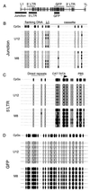
Detailed methylation mapping of methylated and unmethylated proviral clones. Genomic DNA isolated 56 days after electroporation was bisulfite converted, and the regions of interest were PCR amplified, subcloned, and sequenced (see Materials and Methods). (A) The regions amplified, including the junction region, the 5′ LTR, and the GFP gene (thick black lines), are shown relative to the L1-MFGhGFP-1L map. (B to D) Open and filled ovals correspond to CpGs and methylated CpGs, respectively. For each amplified region, at least six molecules from clones M8 and U12 were sequenced. Bisulfite sequencing of the junction region (B), the 5′ LTR (C), and the GFP gene (D) reveals the absence of methylation in the initially unmethylated clone U12 and the maintenance of methylation, with the exception of the enhancer region, in the methylated clone M8. PBS, primer binding site.
FIG. 4
FIG. 4
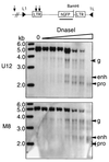
Dependence of LTR enhancer and promoter remodeling on methylation state. Nuclei were isolated from RL5 clones M8 and U12 and digested with increasing concentrations of DNase I. Subsequently, genomic DNA was isolated, digested with BamHI, and subjected to Southern hybridization with a GFP probe. Predicted HSs at the proviral enhancer (enh) and promoter (pro), as well as a genomic site (g) upstream of the introduced cassette, are labeled with arrows. A sample with no DNase I added (lane 0) is also shown. Note the absence of the promoter HS in the methylated clone. hGFP, humanized GFP.
FIG. 5
FIG. 5
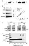
Histone acetylation is dependent on proviral methylation status. (A) A map of the L1-MFGhGFP-1L provirus is shown with the locations of the primer pairs used to amplify either the 5′ LTR or the GFP gene. SA, splice acceptor; SD, splice donor. (B) Amplification of titrated input DNA using these primer pairs and a control primer pair specific for the endogenous mouse amylase 2.1y gene is linear under the conditions used for duplex PCR (see Materials and Methods). Formaldehyde-cross-linked chromatin was purified by isopycnic centrifugation and immunoprecipitated with antibodies recognizing AcH4 or AcH3. After reversal of the cross-link, duplex PCR was performed for the input and antibody-bound chromatin fractions (equivalent to approximately 1 to 2 ng of DNA) with the amylase 2.1y primer pair in combination with either the 5′ LTR or the GFP gene primer pair in the presence of radiolabeled deoxycytidine. (C) The PCR products from the input (I) and antibody-bound DNA (H3 and H4) were resolved by electrophoresis on a nondenaturing acrylamide gel. Amy, amylase 2.1y. (D) To determine the enrichment of proviral sequences relative to the nonexpressed, hypoacetylated amylase gene, products from three independent duplex amplifications were quantified by PhosphorImager analysis. The ratio of the two PCR products was determined for the antibody-bound fraction and normalized to the ratio obtained from the input material prior to immunoprecipitation. The mean and standard error of the mean are plotted. The x axis is set at 1, which reflects no enrichment relative to the amylase 2.1y gene.
FIG. 6
FIG. 6
MeCP2 and MBD3 are expressed in MEL cells and are detectable in chromatin preparations. (A) mRNAs from MEL cells and mouse brain as a positive control were reverse transcribed, and the cDNA generated was used as a template for PCR with primers specific for MBD1, MBD2, MBD3, and MeCP2. Only MBD3 and MeCP2 are expressed in MEL cells. −RT, without reverse transcriptase. (B) Western blot analyses were conducted with antibodies specific for MeCP2 and MBD3. Nuclear extracts were prepared from HeLa, MEL 745A, and MEL RL5 cells, and 10 to 20 μg of protein was loaded per lane. Crude (for MeCP2) or purified (for MBD3) chromatin preparations (see Materials and Methods) were also generated and subjected to electrophoresis in parallel with the nuclear extracts.
FIG. 7
FIG. 7
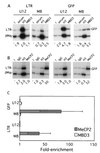
MeCP2 is recruited to the methylated provirus. Formaldehyde-cross-linked chromatin was prepared from MEL clones M8 and U12 without isopycnic centrifugation (as described in Materials and Methods). (A) Chromatin was immunoprecipitated with goat preimmune serum or antibodies specific for AcH3 (H3) or MBD3. Duplex PCR was conducted with the 5′ LTR or GFP gene primers described in Fig. 5, in combination with a control primer pair specific for the actively transcribed β-major promoter region (βMaj) of the endogenous β-globin locus. A representative acrylamide gel is shown, along with the levels of enrichment, as measured by PhosphorImager analysis, relative to the input (I) sample. The goat-derived MBD3 antibody shows no significant enrichment relative to the control goat serum. In contrast, concurrent immunoprecipitation with the AcH3 antibody shows a 7- to 10-fold difference in abundance between the unmethylated and the methylated proviruses, confirming the fidelity of the chromatin preparation. (B) Immunoprecipitation with control rabbit immunoglobulin G or antibodies specific for AcH3 (H3) or MeCP2 reveals that MeCP2 is significantly enriched in the 5′ LTR and the GFP gene of the methylated clone. (C) Quantification of duplex PCR products from two chromatin preparations (three independent MeCP2 or MBD3 immunoprecipitations) confirms these results. The mean and standard error of the mean for the enrichment are plotted (see the text). The x axis is set at 1, which reflects no enrichment.

Similar articles

Cited by

References

    1. Amir R E, Van den Veyver I B, Wan M, Tran C Q, Francke U, Zoghbi H Y. Rett syndrome is caused by mutations in X-linked MECP2, encoding methyl-CpG-binding protein 2. Nat Genet. 1999;23:185–188. - PubMed
    1. Anderson M T, Tjioe I M, Lorincz M C, Parks D R, Herzenberg L A, Nolan G P, Herzenberg L A. Simultaneous fluorescence-activated cell sorter analysis of two distinct transcriptional elements within a single cell using engineered green fluorescent proteins. Proc Natl Acad Sci USA. 1996;93:8508–8511. - PMC - PubMed
    1. Baer A, Schübeler D, Bode J. Transcriptional properties of genomic transgene integration sites marked by electroporation or retroviral infection. Biochemistry. 2000;39:7041–7049. - PubMed
    1. Bestor T H. The host defence function of genomic methylation patterns. Novartis Found Symp. 1998;214:187–195. - PubMed
    1. Bestor T H, Tycko B. Creation of genomic methylation patterns. Nat Genet. 1996;12:363–367. - PubMed

Publication types

MeSH terms

LinkOut - more resources