. 2001 Dec;21(23):7933-43.
doi: 10.1128/MCB.21.23.7933-7943.2001.

Individual somatic H1 subtypes are dispensable for mouse development even in mice lacking the H1(0) replacement subtype

Affiliations

Individual somatic H1 subtypes are dispensable for mouse development even in mice lacking the H1(0) replacement subtype

Y Fan et al. Mol Cell Biol. 2001 Dec.

Abstract

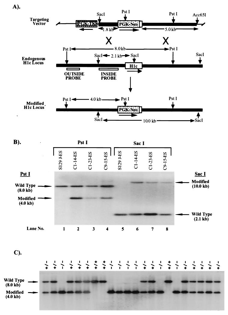
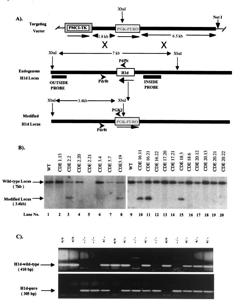
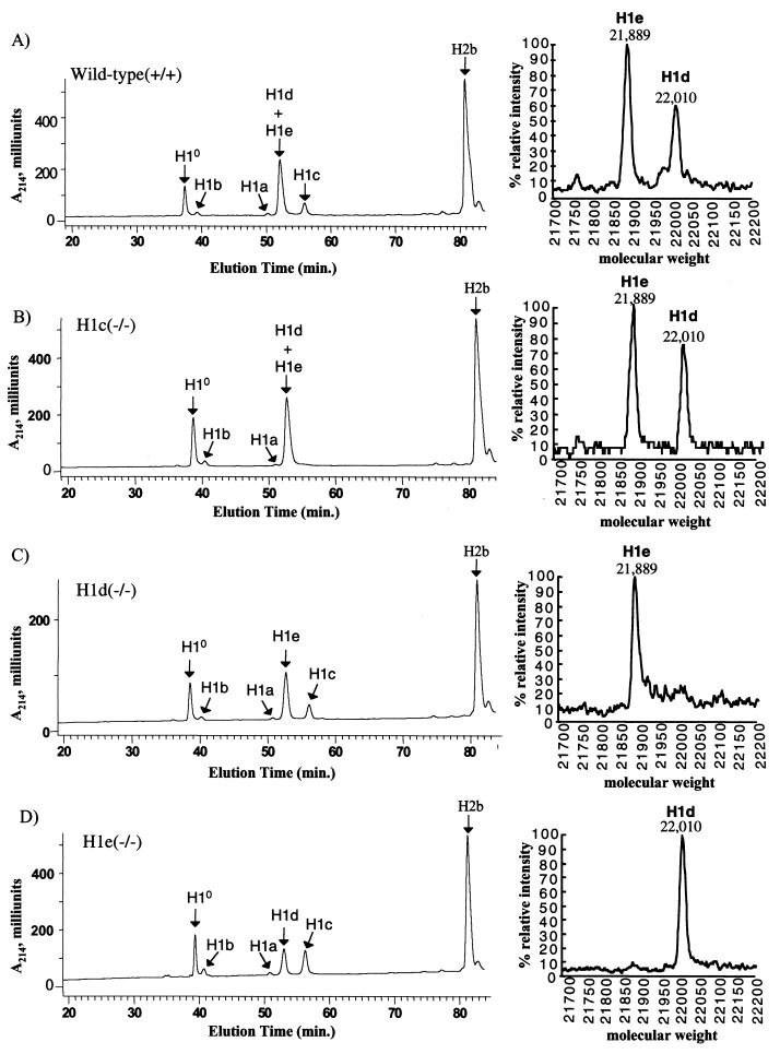
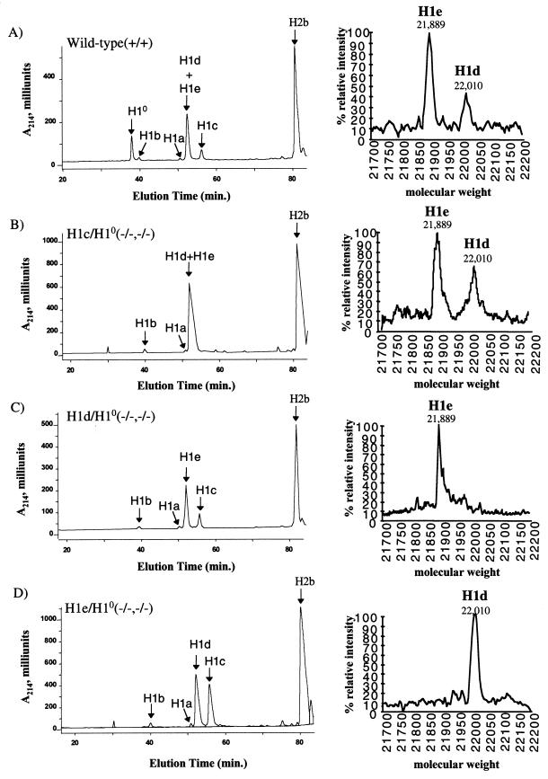
H1 linker histones are involved in facilitating the folding of chromatin into a 30-nm fiber. Mice contain eight H1 subtypes that differ in amino acid sequence and expression during development. Previous work showed that mice lacking H1(0), the most divergent subtype, develop normally. Examination of chromatin in H1(0-/-) mice showed that other H1s, especially H1c, H1d, and H1e, compensate for the loss of H1(0) to maintain a normal H1-to-nucleosome stoichiometry, even in tissues that normally contain abundant amounts of H1(0) (A. M. Sirotkin et al., Proc. Natl. Acad. Sci. USA 92:6434-6438, 1995). To further investigate the in vivo role of individual mammalian H1s in development, we generated mice lacking H1c, H1d, or H1e by homologous recombination in mouse embryonic stem cells. Mice lacking any one of these H1 subtypes grew and reproduced normally and did not exhibit any obvious phenotype. To determine whether one of these H1s, in particular, was responsible for the compensation present in H1(0-/-) mice, each of the three H1 knockout mouse lines was bred with H1(0) knockout mice to generate H1c/H1(0), H1d/H1(0), or H1e/H1(0) double-knockout mice. Each of these doubly H1-deficient mice also was fertile and exhibited no anatomic or histological abnormalities. Chromatin from the three double-knockout strains showed no significant change in the ratio of total H1 to nucleosomes. These results suggest that any individual H1 subtype is dispensable for mouse development and that loss of even two subtypes is tolerated if a normal H1-to-nucleosome stoichiometry is maintained. Multiple compound H1 knockouts will probably be needed to disrupt the compensation within this multigene family.


Figures

FIG. 1
FIG. 1
Targeted disruption of the H1c gene in mouse ES cells and mice. (A) Homologous-recombination strategy in ES cells. The H1c targeting vector (top) was constructed by removing a 566-bp ApaI/MscI segment from a 6.4-kb EcoRI genomic fragment cloned in pGEM-3Z (Promega), containing the H1c coding region (open box), and inserting by blunt-end ligation a 1.8-kb ApaI/HindIII fragment (stippled box) from PGK-NEO. The 7.2-kb H1c/PGK-NEO insert was subsequently released by SalI digestion and inserted into the XhoI site of pPGK-TK (19). To increase the length of homology within the short arm (5′ of PGK-NEO), the 0.8-kb ClaI-XhoI short-arm fragment was removed and replaced with a ClaI-XhoI 1.8-kb homologous H1c 5′ region fragment from H1c plasmid subclone HS7 (22). The transcriptional orientations of the genes are indicated by arrows. A homologous recombination event (X's) between the targeting vector and the endogenous H1c locus (middle) results in production of a modified H1c locus (bottom) in which a segment from 58 bp 5′ of the translation initiation codon to codon 170 was replaced with PGK-NEO. (B) Identification of ES cell clones containing the modified H1c allele. After an initial screening of pools of two ES cell clones, ES cell DNA (10 μg) from individual clones was digested with PstI (lanes 1 to 4) and Southern blot hybridized with the outside probe (A). Correct targeting was confirmed (lanes 5 to 8) by SacI digestion of ES cell DNA and blot hybridization with the inside probe (A). Results obtained with DNA from untransfected ES cells are shown in lanes 1 and 5. The expected positions of the hybridizing fragments from the unmodified (wild-type) and modified H1c loci and their respective sizes are indicated. (C) Genotype analysis of offspring from parents heterozygous for the modified H1c allele. Siblings that were heterozygous for the modified H1c allele were bred, and 15 μg of tail DNA from offspring was digested with PstI and blot hybridized with the inside probe (Fig. 1A). The deduced genotype of each animal is indicated above each lane. The expected positions of the hybridizing fragment from the wild-type and modified loci and their corresponding sizes are indicated.
FIG. 2
FIG. 2
Targeted disruption of the H1d gene in mouse ES cells and mice. (A) Homologous recombination strategy in ES cells. To generate the H1d targeting vector (top), the Scramble plasmid system (Lexicon) was used. The positive and negative selectable marker genes PGK-PURO (striped box) and PMCI-TK cassettes (shaded box) were inserted into vector 901 at the AscI and RsrII sites, respectively. The H1d targeting vector (top) was then constructed by inserting a 6.5-kb H1d 5′ ClaI/EcoRI fragment (in which the EcoRI site lies 616 bp 5′ of the H1d ATG codon) and a 1.8-kb PCR-amplified H1d 3′ fragment (beginning 120 bp 3′ of the stop codon) into the SmaI/NotI and KpnI sites, respectively, in modified vector 901. A homologous recombination event (X's) between the targeting vector and the endogenous H1d locus (middle) results in production of a modified H1d locus (bottom), in which a 1.4-kb fragment, including the entire H1d coding sequence (open box), 616 bp of the 5′ noncoding sequence, and 120 bp of the 3′ noncoding sequence, is removed. (B) Identification of ES cell clones containing the modified H1d allele. ES cell DNA (10 μg) was digested with XbaI and blot hybridized with the outside probe (A). The expected positions of the hybridizing fragments from the unmodified (wild-type) and modified H1d loci and their respective sizes are indicated. Lanes 1 and 9 contained DNA from wild-type ES cells. Clones analyzed in lanes 3, 8, 10, 11, and 15 underwent a homologous recombination event. (C) Genotype analysis of offspring from parents heterozygous for the modified H1d allele. Siblings that were heterozygous for the modified H1d allele were bred, and 1 μg of tail DNA from offspring was used for PCR analysis with the following primers shown in panel A: H1d wild-type allele, the H1d 5′ sequence-specific primers (Pdf5t [5′ AAGCCTAAAGCTTCTAAGCCG 3′] and Pdr8t [5′ CTAGAGAACCCCCCTAATGC 3′]; predicted band size, 410 bp); H1d null allele (H1d-Puro): the PGK-PURO gene-specific primer (PGK2 [5′ GCTGCTAAAGCGCATGCTCCA 3′]) and the H1d 5′ sequence-specific primer (Pdr8t [5′ CTAGAGAACCCCCCTAATGC 3′]; predicted band size, 305 bp). The deduced genotype of each animal is indicated above each lane. The migration of PCR products from the wild-type and modified loci and their corresponding sizes are indicated.
FIG. 3
FIG. 3
Targeted disruption of the H1e gene in mouse ES cells and mice. (A) Homologous recombination strategy in ES cells. The H1e targeting vector (top) was constructed by removing a 720-bp MscI fragment from a 6.5-kb EcoRI H1e genomic DNA fragment cloned in pGEM-3Z (Promega), containing the H1e coding region (open box), and inserting by blunt-end ligation a 1.8-kb ApaI/HindIII fragment (shaded box) from pPGK-NEO. A 1.8-kb SalI/XhoI fragment from pMCI-TK was inserted into the SalI site at the 5′ end of the gene. A homologous recombination event (X's) between the targeting vector and the endogenous H1e locus (middle) results in production of a modified H1e locus (bottom), in which a 720-bp fragment, including the entire H1e coding sequence, along with 49 bp of the 5′ noncoding sequence and 11 bp of the 3′ noncoding sequence, is removed. (B) Identification and confirmation of ES cell clones containing the modified H1e allele. ES cell DNA (10 μg) was digested with EcoRI, followed by Southern blot analysis using the outside probe shown in panel A. The expected positions of the hybridizing fragments from the unmodified (wild-type) and modified H1c loci and their respective sizes are indicated. Clones analyzed in lanes 1, 4, 5, 8, 9, 11, 13, 15, 17, 18, and 23 underwent a homologous recombination events. (C) Genotype analysis of offspring from parents heterozygous for the modified H1e allele. Siblings that were heterozygous for the modified H1e allele were bred, and 15 μg of tail DNA from offspring was digested with EcoRI, blotted, and hybridized with the inside probe shown in panel A. The deduced genotype of each animal is indicated above each lane. The wild-type and modified loci and their corresponding sizes are indicated.
FIG. 4
FIG. 4
Reverse-phase HPLC analysis of histones from wild-type and H1c, H1d, and H1e homozygous mutant mice. (Left side) Approximately 100 μg of total histone extract of chromatin from livers of 20-week-old mice was fractionated by reverse phase HPLC as previously described (23). Panels: A, wild-type; B, homozygous H1c mutant; C, homozygous H1d mutant; D, homozygous H1e mutant. The identity of the histone subtype(s) in each peak has been reported previously (3, 15, 25). (Right side) Fractions eluting between 52 and 54 min (corresponding to the peak marked H1d+H1e) were collected and subjected to TOF-MS analysis. The identity of the two H1 subtypes detected in this analysis was demonstrated in reference .
FIG. 5
FIG. 5
Reverse-phase HPLC analysis of histones from wild-type mice and three types of doubly homozygous mutant mice. Approximately 100 μg of total histone extract of chromatin from livers of 20-week-old mice were analyzed as described in the legend to Fig. 4. Panels: A, wild type; B, H1c/H10 homozygous double mutant; C, H1d/H10 homozygous double mutant; D, H1e/H10 homozygous double mutant.

Similar articles

Cited by

  • Histone h1 depletion impairs embryonic stem cell differentiation.
    Zhang Y, Cooke M, Panjwani S, Cao K, Krauth B, Ho PY, Medrzycki M, Berhe DT, Pan C, McDevitt TC, Fan Y. Zhang Y, et al. PLoS Genet. 2012;8(5):e1002691. doi: 10.1371/journal.pgen.1002691. Epub 2012 May 10. PLoS Genet. 2012. PMID: 22589736 Free PMC article.
  • Silencing the genome with linker histones.
    Hansen JC. Hansen JC. Proc Natl Acad Sci U S A. 2020 Jul 7;117(27):15388-15390. doi: 10.1073/pnas.2009513117. Epub 2020 Jun 19. Proc Natl Acad Sci U S A. 2020. PMID: 32561644 Free PMC article. No abstract available.
  • Mammalian linker-histone subtypes differentially affect gene expression in vivo.
    Alami R, Fan Y, Pack S, Sonbuchner TM, Besse A, Lin Q, Greally JM, Skoultchi AI, Bouhassira EE. Alami R, et al. Proc Natl Acad Sci U S A. 2003 May 13;100(10):5920-5. doi: 10.1073/pnas.0736105100. Epub 2003 Apr 28. Proc Natl Acad Sci U S A. 2003. PMID: 12719535 Free PMC article.
  • Aberrant Function of the C-Terminal Tail of HIST1H1E Accelerates Cellular Senescence and Causes Premature Aging.
    Flex E, Martinelli S, Van Dijck A, Ciolfi A, Cecchetti S, Coluzzi E, Pannone L, Andreoli C, Radio FC, Pizzi S, Carpentieri G, Bruselles A, Catanzaro G, Pedace L, Miele E, Carcarino E, Ge X, Chijiwa C, Lewis MES, Meuwissen M, Kenis S, Van der Aa N, Larson A, Brown K, Wasserstein MP, Skotko BG, Begtrup A, Person R, Karayiorgou M, Roos JL, Van Gassen KL, Koopmans M, Bijlsma EK, Santen GWE, Barge-Schaapveld DQCM, Ruivenkamp CAL, Hoffer MJV, Lalani SR, Streff H, Craigen WJ, Graham BH, van den Elzen APM, Kamphuis DJ, Õunap K, Reinson K, Pajusalu S, Wojcik MH, Viberti C, Di Gaetano C, Bertini E, Petrucci S, De Luca A, Rota R, Ferretti E, Matullo G, Dallapiccola B, Sgura A, Walkiewicz M, Kooy RF, Tartaglia M. Flex E, et al. Am J Hum Genet. 2019 Sep 5;105(3):493-508. doi: 10.1016/j.ajhg.2019.07.007. Epub 2019 Aug 22. Am J Hum Genet. 2019. PMID: 31447100 Free PMC article.
  • Spermatid-specific linker histone HILS1 is a poor condenser of DNA and chromatin and preferentially associates with LINE-1 elements.
    Mishra LN, Shalini V, Gupta N, Ghosh K, Suthar N, Bhaduri U, Rao MRS. Mishra LN, et al. Epigenetics Chromatin. 2018 Aug 1;11(1):43. doi: 10.1186/s13072-018-0214-0. Epigenetics Chromatin. 2018. PMID: 30068355 Free PMC article.

References

    1. Bates D L, Thomas J O. Histones H1 and H5: one or two molecules per nucleosome? Nucleic Acids Res. 1981;9:5883–5894. - PMC - PubMed
    1. Brannan C I, Gilbert D J, Ceci J D, Matsuda Y, Chapman V M, Mercer J A, Eisen H, Johnston L A, Copeland N G, Jenkins N A. An interspecific linkage map of mouse chromosome 15 positioned with respect to the centromere. Genomics. 1992;13:1075–1081. - PubMed
    1. Brown D T, Sittman D B. Identification through overexpression and tagging of the variant type of the mouse H1e and H1c genes. J Biol Chem. 1993;268:713–718. - PubMed
    1. Dong Y, Sirotkin A M, Yang Y S, Brown D T, Sittman D B, Skoultchi A I. Isolation and characterization of two replication-dependent mouse H1 histone genes. Nucleic Acids Res. 1994;22:1421–1428. - PMC - PubMed
    1. Drabent B, Saftig P, Bode C, Doenecke D. Spermatogenesis proceeds normally in mice without linker histone H1t. Histochem Cell Biol. 2000;113:433–442. - PubMed

Publication types

MeSH terms

LinkOut - more resources