. 2001 Dec;21(23):8007-21.
doi: 10.1128/MCB.21.23.8007-8021.2001.

Construction and analysis of mouse strains lacking the ubiquitin ligase UBR1 (E3alpha) of the N-end rule pathway

Affiliations

Construction and analysis of mouse strains lacking the ubiquitin ligase UBR1 (E3alpha) of the N-end rule pathway

Y T Kwon et al. Mol Cell Biol. 2001 Dec.

Abstract

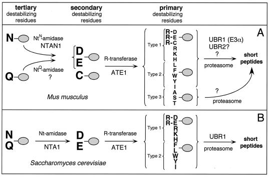
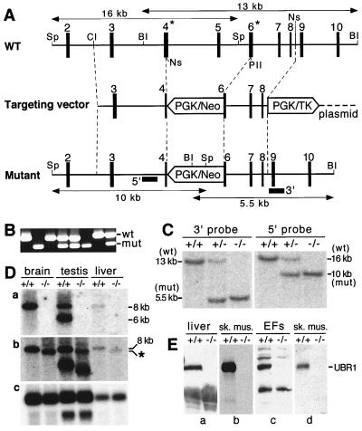
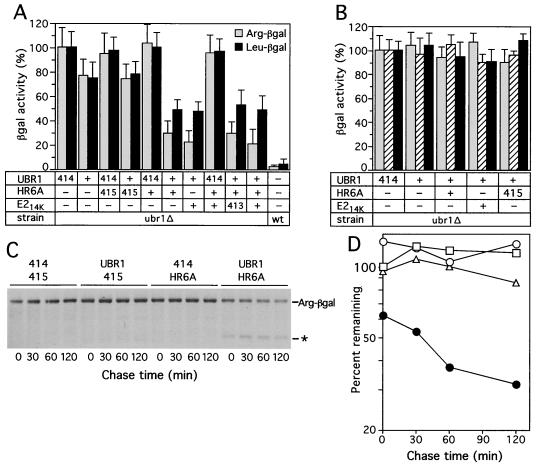
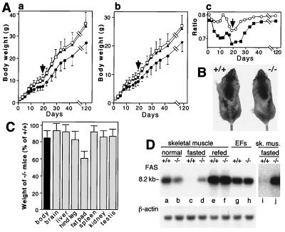
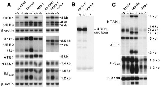
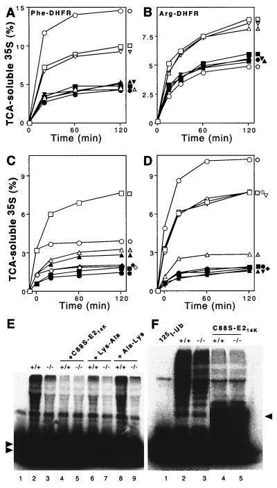
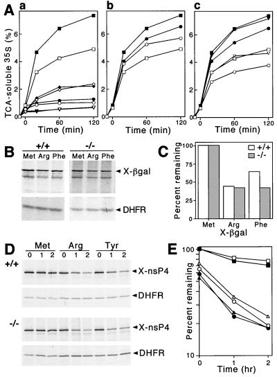
The N-end rule relates the in vivo half-life of a protein to the identity of its N-terminal residue. In the yeast Saccharomyces cerevisiae, the UBR1-encoded ubiquitin ligase (E3) of the N-end rule pathway mediates the targeting of substrate proteins in part through binding to their destabilizing N-terminal residues. The functions of the yeast N-end rule pathway include fidelity of chromosome segregation and the regulation of peptide import. Our previous work described the cloning of cDNA and a gene encoding the 200-kDa mouse UBR1 (E3alpha). Here we show that mouse UBR1, in the presence of a cognate mouse ubiquitin-conjugating (E2) enzyme, can rescue the N-end rule pathway in ubr1Delta S. cerevisiae. We also constructed UBR1(-/-) mouse strains that lacked the UBR1 protein. UBR1(-/-) mice were viable and fertile but weighed significantly less than congenic +/+ mice. The decreased mass of UBR1(-/-) mice stemmed at least in part from smaller amounts of the skeletal muscle and adipose tissues. The skeletal muscle of UBR1(-/-) mice apparently lacked the N-end rule pathway and exhibited abnormal regulation of fatty acid synthase upon starvation. By contrast, and despite the absence of the UBR1 protein, UBR1(-/-) fibroblasts contained the N-end rule pathway. Thus, UBR1(-/-) mice are mosaics in regard to the activity of this pathway, owing to differential expression of proteins that can substitute for the ubiquitin ligase UBR1 (E3alpha). We consider these UBR1-like proteins and discuss the functions of the mammalian N-end rule pathway.


Figures

FIG. 1
FIG. 1
(A) The N-end rule pathway in mammals (12, 32, 33). N-terminal residues are indicated by single-letter abbreviations for amino acids. The ovals denote the rest of a protein substrate. The Asn-specific N-terminal amidase (NtN-amidase) NTAN1 converts N-terminal Asn into Asp (19, 32). N-terminal Gln is deamidated by a distinct NtQ-amidase, NTAQ1, which remains to be characterized. In mammals, the secondary destabilizing N-terminal residues Asp, Glu, and Cys are arginylated by the Arg-tRNA-protein transferases (R-transferases) encoded by ATE1 ( and Y. T. Kwon, A. Kashina, and A. Varshavsky, unpublished data). The set of primary destabilizing N-terminal residues—Arg, Lys, His, Phe, Leu, Trp, Tyr, and Ile—is recognized in mammals by at least three distinct E3 enzymes of similar binding specificities, including the UBR1-encoded E3α and UBR2 (see Discussion). N-terminal Ala, Ser, and Thr are primary destabilizing residues in mammals but are stabilizing residues in S. cerevisiae (18, 22). An E3 that recognizes these N-terminal residues remains to be characterized. In mammals, either of the two highly similar Ub-conjugating (E2) enzymes, HR6A and HR6B (E214K), can be a component of E2-E3 complexes (Ub ligases) that mediate ubiquitylation of N-end rule substrates. The term “Ub ligase” is used to denote either an E2-E3 complex or its specific E3 component. A targeted, multi-Ub chain-bearing substrate is degraded by the 26S proteasome. (B) The N-end rule pathway in the yeast S. cerevisiae differs from its mammalian counterpart by the presence of a single Nt-amidase, NTA1, which mediates deamidation of N-terminal Asn or Gln (6) by Cys being a stabilizing residue; by the absence of E3 that recognizes N-terminal Ala, Ser, and Thr; and by the presence of a single E3, UBR1, that recognizes other primary destabilizing N-terminal residues (71).
FIG. 2
FIG. 2
Construction of UBR1−/− mice. (A) Top diagram, a restriction map of the ∼24-kb 5′-proximal region of the ∼120-kb mouse UBR1 gene. Middle diagram, the targeting vector. Bottom diagram, the deletion and/or disruption UBR1 allele. Exons are denoted by solid vertical rectangles. The directions of transcription of the neomycin (neo) and the thymidine kinase (tk) genes are indicated. Homologous recombination resulted in the replacement of the UBR1 exons 4 to 6 with the neo cassette. Exon 4, marked by an asterisk, contains Gly147 and Asp150, the residues that are essential, in S. cerevisiae UBR1, for binding type 1 destabilizing N-terminal residues (see Results). Exon 6, also marked by an asterisk, contains Asp233 and His236, the residues that are essential, in S. cerevisiae UBR1, for binding type 2 destabilizing N-terminal residues (see Results). Probes used for Southern hybridization are indicated by solid rectangles. Restriction sites: Sp, SphI; Cl, ClaI; BI, BamHI, Ns, NsiI, PII, PvuII. (B) PCR analysis of mouse tail DNA. The primers were 5′-GCCACTTGTGTAGCGCCAAGTGCCAG-3′ (for neo; forward), 5′-GAGATAGGAAACTGCATGCGCTGC-3′ (for UBR1; forward), and 5′-CAAGAGTGCAACAGTTACCACATG-3′ (for UBR1; reverse). DNA bands corresponding to the wild-type (wt) and mutant (mut) UBR1 alleles are indicated on the right. (C) Southern analysis of BamHI-cut (3′ probe) and SphI-cut (5′ probe) tail DNA from +/+, UBR1+/−, and UBR1−/− mice. The 3′ probe detected 13- and 5.5-kb UBR1 fragments in BamHI-cut DNA corresponding to the wild-type and mutant UBR1 alleles, respectively. The 5′ probe detected 16- and 10-kb fragments in the same SphI-cut alleles. The depicted organization of the deletion and/or disruption UBR1−/− allele was additionally verified using Southern analysis with other restriction endonucleases (data not shown). (D) Northern analysis. Total electrophoretically fractionated RNA from brain, testis, or liver of +/+ and UBR1−/− mice was probed either with the UBR1 cDNA fragment (nucleotides 555 to 888) which was deleted in the UBR1−/− allele (gel a) or with the ∼2-kb cDNA fragment (nucleotides 116 to 2124) that contained both the deleted region (nucleotides 532 to 885) and its flanking sequences (gel b) or with the human β-actin cDNA fragment (gel c). (E) Immunoblot analysis of total extracts from liver, skeletal muscle, and EFs with affinity-purified antibody (38) against the N-terminal ∼35-kDa fragment of mouse UBR1 (gels a to c) or with affinity-purified antibody specific for the 2-1 UBR1 peptide (see Materials and Methods) encoded by the deleted region of UBR1 in the UBR1 allele (gel d).
FIG. 3
FIG. 3
Mouse UBR1, in the presence of mouse E214K or HR6A, can rescue the N-end pathway in ubr1Δ S. cerevisiae. (A) Relative enzymatic activities of βgal in ubr1Δ S. cerevisiae expressing different combinations of the following components. (i) Type 1 (Arg-βgal) or type 2 (Leu-βgal) N-end rule substrates; (ii) mouse UBR1 or the p414-MET25 vector alone (designated 414); and (iii) mouse E214K, mouse HR6A (homolog of E214K), both of them together, or vector(s) alone (p415-MET25, designated 415, and p413-MET25, designated 413). The activities of X-βgal test proteins in wild-type (UBR1) S. cerevisiae are shown in the rightmost column. 100%, the activity of X-βgal in ubr1Δ cells transformed with the p414-MET25 vector alone. (B) Same as for panel A but with type 3 N-end rule substrates, Ala-βgal (shaded bars), Ser-βgal (hatched bars), or Thr–β-gal (black bars). (C) Pulse-chase analysis of Arg-βgal (produced from Ub-Arg-βgal) in ubr1Δ S. cerevisiae coexpressing either mouse UBR1 and the p415-MET25 vector (denoted 415), mouse HR6A E2 enzyme and the p414-MET25 vector (denoted 414), mouse UBR1 and HR6A, or vectors alone. Time zero refers to the end of 5-min pulse. The asterisk indicates the ∼90-kDa, long-lived βgal cleavage product specific for short-lived X-βgal test proteins (3) in the pulse-chase with cells coexpressing mouse UBR1 and mouse HR6A. (Much smaller amounts of the 90-kDa species could also be detected in the pulse-chase with cells expressing mouse UBR1 alone.) (D) Quantitation of Arg-βgal degradation depicted in panel C (the decay curves shown are averages from two independent experiments). 100%, the initial amount of Arg-βgal in cells transformed with vectors alone (p414-MET25 and p415-MET25); □, vectors alone; ▵, UBR1 and p415-MET25 (vector); ○, HR6A and p414-MET25 (vector); ●, UBR1 and HR6A.
FIG. 4
FIG. 4
Growth retardation and altered fat metabolism in UBR1−/− mice. (A) Growth retardation in UBR1−/− mice of the 129/C57 strain background. Body masses of the offspring (total of 141 mice) from UBR1+/− heterozygous matings were determined from 1 day after birth until adulthood. To keep track of newborn pups before they could be distinguished using ear punching (at 3 weeks of age), the pups were marked by applying spots of paint every day. At week 3 the genotypes of pups were determined by using PCR with tail-derived DNA. (A, graph a) Male masses as a function of age: 18 +/+ (□), 34 UBR1+/− (▴), and 20 UBR1−/− mice (●) were used. (A, graph b) Same as for panel A, graph a, but with females: 18 +/+ (□), 35 UBR1+/− (▴), and 16 UBR1−/− mice (●) were used. (A, graph c) The mass ratios of UBR1−/− males to +/+ males (▪) and of UBR1−/− females to +/+ females (○) as a function of age. The arrows in graphs a to c denote the time of weaning (day 20). (B) A pair of 6-week-old +/+ and UBR1−/− male littermates. The mass of the UBR1−/− mouse shown here was 15% lower than that of its +/+ littermate. (C) Relative masses of organs and tissues of UBR1−/− mice, expressed as percentages of the weights of age-matched +/+ counterparts. Standard deviations are indicated. For determining the masses of hind legs and hind leg fat pads, 94 pairs of 2- to 4-month-old mice (69 male pairs and 25 female pairs) were used (63 +/+, 31 UBR1+/−, and 94 UBR1−/− mice, produced through matings of UBR1+/− mice). For other measurements in this panel, 24 pairs of 2- to 4-month-old mice (17 +/+, 7 UBR1+/−, and 24 UBR1−/− mice) were used. (D) Altered regulation of the FAS mRNA in skeletal muscle of UBR1−/− mice. Lanes a to f, Northern analysis of FAS mRNA from skeletal muscle of +/+ and UBR1−/− mice that were either fed ad libitum, fasted for 48 h, or refed for 24 h after the fast. Lanes g and h, FAS mRNA from growing EF cells (+/+ and UBR1−/−) in culture. Lanes i and j, the same as lanes c and d but with a longer autoradiographic exposure. A 32P-labeled 1.1-kb FAS cDNA fragment (nucleotides 535 to 1642; accession no. AAG02285) was used as a probe.
FIG. 5
FIG. 5
Expression analysis of mouse UBR1 and functionally related genes in +/+ and UBR1−/− mice. (A) Northern analysis of skeletal muscle mRNAs encoding UBR1, other components of the N-end rule pathway, and the UBR1 homologs UBR2 and UBR3. RNA was isolated from skeletal muscle of control, 48-h fasted, or 24-h refed +/+ and UBR1−/− mice. See Materials and Methods and the legend to Fig. 4D for the fasting and refeeding protocols. Northern blots were hybridized with 32P-labeled cDNA probes specific for the following genes: UBR1 (nucleotides 555 to 888, accession no. AF061555; this probe was specific for the deleted region in the UBR1 allele); UBR2 (a 0.3-kb probe encompassing the region homologous to the one deleted in the UBR1−/− allele; Y. T. Kwon and A. Varshavsky, unpublished data); UBR3 (a 0.4-kb probe; Y. T. Kwon and A. Varshavsky, unpublished data); ATE1 (nucleotides 638 to 1734, accession no. AF079098); NTAN1 (nucleotides 34 to 900, accession no. U57692); mHR6B (E214K) (nucleotides 115 to 569, accession no. U57690); and β-actin. (B) The level of UBR1 protein in skeletal muscle is not significantly altered upon fasting. Total extracts (∼70 μg per lane) from skeletal muscles of normally fed or 48-h fasted +/+ and UBR1−/− mice were analyzed by immunoblotting, using affinity-purified antibody (38) against the N-terminal ∼35-kDa fragment of mouse UBR1. (C) Northern analysis of ATE1, NTAN1, and mHR6B (E214K) expression in the brain, liver, and testis of +/+ and UBR1−/− mice by using DNA probes described in the legend to panel A.
FIG. 6
FIG. 6
Extracts from skeletal muscle of UBR1−/− mice lack the N-end rule pathway. Degradation of 35S-labeled, purified Ub-X-DHFR test proteins in extracts from skeletal muscle of +/+ and UBR1−/− littermates (strain 129 background) was monitored by measuring TCA-soluble 35S (see Materials and Methods). Open and closed symbols denote, respectively, the data with +/+ and UBR1−/− extracts. (A) Degradation of Phe-DHFR, a type 2 N-end rule substrate. □ and ▪, no dipeptide inhibitor; ○ and ●, Lys-Ala; ▵ and ▴, Phe-Ala; ▿ and ▾, Ala-Lys (control dipeptide). (B) Degradation of Arg-DHFR, a type 1 substrate. □ and ▪, no dipeptide inhibitor; ○ and ●, Lys-Ala; ▵ and ▴, Phe-Ala; ▿ and ▾, Ala-Lys. (C and D) The N-end rule-specific degradation of Phe-DHFR is ATP-dependent. Reaction mixtures were incubated at 37°C for 10 min without ATP followed by assays either in the absence or the presence of added ATP. (Time zero corresponds to the end of 10-min preincubation in the absence of ATP to allow deubiquitylation of Ub-X-DHFRs.) (C) Degradation of Phe-DHFR in the presence (□ and ▪) or absence (○ and ●) of added ATP, and of Met-DHFR (not an N-end substrate) in the presence (▵ and ▴) or absence (⋄ and ♦) of ATP. (D) Effect of Lys-Ala, a type 1 dipeptide inhibitor, on the ATP-dependent degradation of Phe-DHFR, a type 2 N-end rule substrate. □ and ▪, no inhibitor; ○ and ●, Lys-Ala; ▵ and ▴, Phe-Ala; ⋄ and ♦, Ala-Lys; ▿ and ▾, Ala-Phe. (E) Conjugation of unlabeled Ub to 125I-α-lactalbumin (a type 1 N-end rule substrate) is decreased in the extract from UBR1−/− muscle. Some of the samples contained either the dominant-negative E214K(C88S) mutant protein (at 2 μM), 2 mM Lys-Ala, or 2 mM Ala-Lys. Double arrowheads on the left indicate the excess of unconjugated 125I-α-lactalbumin. Lane 1, 125I-α-lactalbumin alone. (F) Conjugation of 125I-Ub to endogenous proteins is marginally decreased in a UBR1−/− muscle extract compared to that in the wild-type extract. Fraction II (see Materials and Methods) of muscle extracts from +/+ and UBR1−/− mice was supplemented with AMP-PNP, and the formation of 125I-Ub-protein con- jugates was assayed by SDS-PAGE in the ab- sence (lanes 2 and 3) or presence (lanes 4 and 5) of 2 μM E214K(C88S). Arrowhead on the right indicates the position of 125I-Ub-E214K(C88S) conjugate. Lane 1, 125I-Ub alone. The results shown in panels A through D are typical of those obtained in at least five independent experiments, using three independently produced muscle extract preparations, and two independent EF cell extracts (see Fig. 7), in combination with two independent preparations of [35S]Ub-X-DHFR.
FIG. 7
FIG. 7
The N-end rule pathway is retained in UBR1−/− EF cells. (A) Degradation of 35S-labeled Ub-X-DHFR proteins in extracts from +/+ and UBR1−/− EF cell lines was monitored by measuring TCA-soluble 35S (see Materials and Methods). Open and closed symbols denote the data with +/+ and UBR1−/− extracts, respectively. Reaction mixtures were incubated at 37°C for 10 min without ATP, and then ATP-dependent degradation of X-DHFR was initiated by the addition of ATP (time zero). No ATP was added to control samples. (A, graph a) ATP-dependent degradation of Phe-DHFR, a type 2 N-end rule substrate, is faster in extracts from UBR1−/− EF cells. Squares and circles, Phe-DHFR with or without ATP, respectively; diamonds and triangles, Met-DHFR (not an N-end rule substrate) with or without ATP, respectively. (A, graph b) Lys-Ala, a type 1 dipeptide inhibitor, does not enhance degradation of the type 2 N-end rule substrate Phe-DHFR in UBR1−/− EF extracts. □ and ▪, no inhibitor; ○ and ●, Lys-Ala. (A, graph c) Effect of Phe-Ala, a type 2 dipeptide inhibitor, on the ATP-dependent degradation of Phe-DHFR. □ and ▪, no inhibitor; ○ and ●, Phe-Ala; ▿ and ▾, Ala-Phe. (B) Immortalized +/+ and UBR1−/− EF cells were transiently transfected with pRC/dhaUbXnsP4βgal (X = Met, Arg, or Phe) expressing X–β-gal test proteins as parts of UPR-based DHFRh-UbR48-X-nsP4βgal fusions (39, 66, 68, 69). Cells were labeled for 1 h with 35S-methionine–cysteine followed by immunoprecipitation and SDS-PAGE analysis of an X–β-gal test protein and the DHFR-based reference protein. (C) Quantitation of the patterns shown in panel B using PhosphorImager. Note a small but significant additional destabilization of Phe-DHFR in UBR1−/− EF cells. (D) Pulse-chase analysis of the N-end rule pathway in +/+ and UBR1−/− EF cells. Immortalized +/+ and UBR1−/− EF cells were transiently transfected with pcDNA3flagDHFRhaUbXnsP4flag (X = Met, Arg, or Tyr) expressing fDHFRh-UbR48-X-nsP4f, a UPR-based fusion yielding, cotranslationally, the fDHFRh-UbR48 reference protein (denoted DHFR on the right) and X-nsP4f (X-nsP4-flag) test protein, denoted X-nsP4 on the right. Cells were labeled for 10 min with 35S-methionine–cysteine followed by a chase for 0, 1, and 2 h in the presence of cycloheximide, preparation of extracts, immunoprecipitation, SDS-PAGE, autoradiography, and quantitation, essentially as described previously (39). (E) Quantitation of the patterns shown in panel D using a PhosphorImager. The amounts of 35S in an X-nsP4f relative to 35S in the fDHFRh-UbR48 reference protein at the same time points were plotted as percentages of this ratio for Met-nsP4f (bearing a stabilizing N-terminal residue) at time zero. □ and ▪, Met-nsP4; ○ and ●, Arg-nsP4; ▵ and ▴, Tyr-nsP4.

Similar articles

Cited by

References

    1. Alagramam K, Naider F, Becker J M. A. Recognition component of the ubiquitin system is required for peptide transport in Saccharomyces cerevisiae. Mol Microbiol. 1995;15:225–234. - PubMed
    1. Ausubel F M, Brent R, Kingston R E, Moore D D, Smith J A, Seidman J G, Struhl K, editors. Current protocols in molecular biology. New York, N.Y: Wiley-Interscience; 2000.
    1. Bachmair A, Finley D, Varshavsky A. In vivo half-life of a protein is a function of its amino-terminal residue. Science. 1986;234:179–186. - PubMed
    1. Bachmair A, Varshavsky A. The degradation signal in a short-lived protein. Cell. 1989;56:1019–1032. - PubMed
    1. Baker R T, Varshavsky A. Inhibition of the N-end rule pathway in living cells. Proc Natl Acad Sci USA. 1991;87:2374–2378. - PMC - PubMed

Publication types

MeSH terms