. 2001 Dec;21(23):8143-56.
doi: 10.1128/MCB.21.23.8143-8156.2001.

Cell density and phosphorylation control the subcellular localization of adenomatous polyposis coli protein

Affiliations

Cell density and phosphorylation control the subcellular localization of adenomatous polyposis coli protein

F Zhang et al. Mol Cell Biol. 2001 Dec.

Abstract

Loss of functional adenomatous polyposis coli protein (APC) leads to uncontrolled proliferation of colonic epithelial cells, as evidenced by polyp formation, a prelude to carcinogenesis. As a tumor suppressor, APC targets the oncogene beta-catenin for proteasome-mediated cytoplasmic degradation. Recently, it was demonstrated that APC also interacts with nuclear beta-catenin, thereby reducing beta-catenin's activity as a transcription cofactor and enhancing its nuclear export. The first objective of this study was to analyze how cellular context affected APC distribution. We determined that cell density but not cell cycle influenced APC's subcellular distribution, with predominantly nuclear APC found in subconfluent MDCK and intestinal epithelial cells but both cytoplasmic and nuclear APC in superconfluent cells. Redistribution of APC protein did not depend on continual nuclear export. Focusing on the two defined nuclear localization signals in the C-terminal third of APC (NLS1(APC) and NLS2(APC)), we found that phosphorylation at the CK2 site increased and phosphorylation at the PKA site decreased NLS2(APC)-mediated nuclear translocation. Cell density-mediated redistribution of beta-galactosidase was achieved by fusion to NLS2(APC) but not to NLS1(APC). Both the CK2 and PKA sites were important for this density-mediated redistribution, and pharmacological agents that target CK2 and PKA instigated relocalization of endogenous APC. Our data provide evidence that physiological signals such as cell density regulate APC's nuclear distribution, with phosphorylation sites near NLS2(APC) being critical for this regulation.


Figures
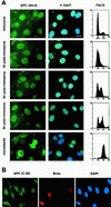
FIG. 1
FIG. 1
APC is predominantly nuclear throughout the MDCK cell cycle. (A) FACS analysis revealed that most MDCK cells were arrested in G0/G1 after an 18-h incubation with mimosine. Many cells harvested 3 h after mimosine removal had entered S phase. By 6 h following mimosine removal, the majority of cells were in S phase. After 9 h, most cells had a 4c DNA content, suggesting that they were in G2/M. The majority of cells showed a 4c DNA content after nocodazole treatment, suggesting arrest in M. Using immunofluorescence microscopy and a monoclonal antibody against APC (Ab-4), APC was located in the cytoplasm and predominantly in the nucleus throughout the cell cycle (left panels, green). Nuclei were visualized by DAPI staining (blue). Bar, 20 μm. (B) Asynchronous MDCK cells were pulse-labeled with BrdU prior to fixation. Cells in S phase were identified using an anti-BrdU antibody (red). APC was identified using a polyclonal antibody raised against APC (C-20, green). Nuclei were visualized with DAPI (blue).
FIG. 2
FIG. 2
The subcellular distribution of APC in MDCK cells is influenced by cell density. The distribution of APC in subconfluent and superconfluent MDCK cells was determined using immunofluorescence confocal microscopy and four different antibodies raised against APC. Nuclei were visualized with TO-PRO-3. Bar, 20 μm. FACS analysis confirmed that subconfluent cells were distributed throughout the cell cycle whereas superconfluent cells were mostly in G0/G1.
FIG. 3
FIG. 3
Redistribution of APC does not depend on sustained nuclear export. Super- and subconfluent MDCK cells were treated with LMB prior to fixation and staining with αAPC, Ab-1 (left panel) and RanBP-1 (right panel). LMB treatment resulted in an increase in nuclear APC in subconfluent but not superconfluent cells. RanBP-1 relocated from the cytoplasm to the nucleus in both super- and subconfluent cells following LMB treatment. Bar, 20 μm.
FIG. 4
FIG. 4
The adjacent CDK2 site does not regulate the NLS1APC-mediated nuclear translocation of a β-Gal chimera. β-Gal fusion proteins were expressed in mouse L cells and detected using immunofluorescence microscopy. Nuclei were visualized with DAPI. Areas of overlap between the β-Gal fusion protein (green) and the nuclei (blue) appear in aqua. Bar, 10 μm. For each construct, 100 cells were scored for β-Gal fusion protein localization. The results of three independent experiments are presented as the incidence of nuclear β-Gal staining ± standard deviations as follows: β-Gal, 27% ± 6%; β-Gal-NLS1APC, 73% ± 11%; β-Gal-mNLS1APC, 30% ± 13%; β-Gal-NLS1APCmCDKS/A, 58% ± 10%; β-Gal-NLS1APCmCDKS/D, 83% ± 6%.
FIG. 5
FIG. 5
The CK2APC site upstream of NLS2APC modifies nuclear import of a β-Gal-NLS2APC chimera. (A) Alignment of NLS2APC with NLSSV40 T-ag reveals similarity of NLS sequence as well as the adjacent phosphorylation sites. β-Gal chimeras were expressed in L cells (B) or MDCK cells (C) and stained as described for Fig. 4. β-Gal-NLS2APC and β-Gal-NLS2APCmCK2S/A localized to both the cytoplasm and nucleus. β-Gal-NLS2APCmCK2S/D was predominantly nuclear. Bars, 10 μm (B) and 20 μm (C). A negative charge, which mimics phosphorylation of Ser2034 in the potential CK2APC site increases NLS2APC-mediated nuclear import in both L cells and MDCK cells. The results of three independent experiments are presented as the incidence of MDCK cells with predominantly cytoplasmic β-Gal staining as follows: β-Gal-NLS2APC, 8%; β-Gal-NLS2APCmCK2S/D, 2%; β-Gal-NLS2APCmCK2S/A, 22%.
FIG. 6
FIG. 6
Cell density influences NLS2APC- but not NLS1APC-mediated nuclear translocation of β-Gal in MDCK cells. β-Gal fusion proteins expressed in MDCK cells grown to different densities were localized as previously described. β-Gal was predominantly cytoplasmic in both superconfluent and subconfluent MDCK cells. β-Gal-NLSSV40 T-ag was predominantly nuclear under both conditions. β-Gal-NLS1APC was evenly distributed in the nucleus and cytoplasm of over half of the super-confluent and subconfluent MDCK cells, with slightly more nuclear localization in superconfluent cells. β-Gal-NLS2APC was predominantly nuclear in subconfluent cells, but was more evenly distributed between the nucleus and cytoplasm in superconfluent cells. MDCK cells from at least three independent experiments, scored for protein localization, were placed in the following categories: cytoplasmic > nuclear (white bar), cytoplasmic = nuclear (grey bar), and cytoplasmic < nuclear (black bar).
FIG. 7
FIG. 7
The potential CK2 and PKA sites control β-Gal-NLS2APC distribution in a manner influenced by cell density. (A) If either the CK2APC or the PKAAPC site is turned off, then nuclear translocation of β-Gal-NLS2APC is impaired. β-Gal fusion proteins were expressed in MDCK cells grown to different cell densities and were located as previously described. β-Gal-NLS2APCmCK2S/A and β-Gal-NLS2APCmPKAS/D were both predominantly cytoplasmic in superconfluent MDCK cells with a slight increase in nuclear accumulation when cells were subconfluent. (B) Mutation of either the CK2APC or the PKAAPC site to the on position did not abolish the cell density-influenced β-Gal-NLS2APC redistribution. β-Gal-NLS2APCmCK2S/D and β-Gal-NLS2APCmPKAS/A were both nuclear in subconfluent cells but had more cytoplasmic localizations in superconfluent cells (see quantification in graph). Bar, 20 μm.
FIG. 8
FIG. 8
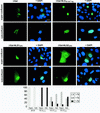
If both CK2APC and PKAAPC switches are constitutively turned on, then the cell density-influenced redistribution of β-Gal-NLS2APC is abolished. β-Gal-NLS2APCmCK2S/DmPKAS/A was expressed in MDCK cells grown to different cell densities and was located as described previously. β-Gal-NLS2APCmCK2S/DmPKAS/A was predominantly nuclear in both superconfluent and subconfluent cells (see results displayed in graphic form below images). Bar, 20 μm.
FIG. 9
FIG. 9
PKA and CK2 activities affect endogenous APC in MDCK and intestinal epithelial cells. MDCK (A) or IEC-6 (B) cells were treated for 30 min with various drug combinations prior to fixation and immunofluorescence microscopy with αAPC Ab-1 or C-20, respectively. Superconfluent cells (left panels) were treated with CK2 agonist (insulin and epidermal growth factor) and PKA inhibitors (Rp diastereomer of cAMP and 4-cyano-3-methylisoquinoline). Subconfluent cells were treated with CK2 inhibitor (DRB) and PKA agonists (8-bromo-cAMP and dibutylyl-cAMP). Both treatments altered localization of endogenous APC. Bar, 20 μm.
FIG. 10
FIG. 10
Model for the cell density-influenced regulation of APC's subcellular distribution. The CK2APC and PKAAPC sites function as switches promoting nuclear localization (on) or impeding nuclear localization (off). The CK2APC switch is on when the CK2APC site serine is phosphorylated. The PKAAPC switch is on when the PKAAPC site is dephosphorylated. (A) In subconfluent and/or proliferating cells, APC primarily exists in the form with both switches on and is therefore localized predominantly to the nucleus. (B) Superconfluent and/or quiescent cells contain APC with either or both switches turned off, resulting in an increased cytoplasmic pool of APC.

Similar articles

Cited by

References

    1. Aukema H M, Davidson L A, Pence B C, Jiang Y H, Lupton J R, Chapkin R S. Butyrate alters activity of specific cAMP-receptor proteins in a transgenic mouse colonic cell line. J Nutr. 1997;127:18–24. - PubMed
    1. Baeg G, Matsumine A, Kuroda T, Bhattacharjee R N, Miyashiro I, Toyoshima K, Akiyama T. The tumour suppressor gene product APC blocks cell cycle progression from G0/G1 to S phase. EMBO J. 1995;14:5618–5625. - PMC - PubMed
    1. Behrens J, Jerchow B A, Wurtele M, Grimm J, Asbrand C, Wirtz R, Kuhl M, Wedlich D, Birchmeier W. Functional interaction of an axin homolog, conductin, with β-catenin, APC, and GSK3β. Science. 1998;280:596–599. - PubMed
    1. Conti E, Uy M, Leighton L, Blobel G, Kuriyan J. Crystallographic analysis of the recognition of a nuclear localization signal by the nuclear import factor karyopherin-α. Cell. 1998;94:193–204. - PubMed
    1. Dingwall C, Laskey R A. Nuclear targeting sequences–a consensus? Trends Biochem Sci. 1991;16:478–481. - PubMed

Publication types

MeSH terms

Substances