. 2001 Dec;21(23):8168-83.
doi: 10.1128/MCB.21.23.8168-8183.2001.

Glycosylation defects and virulence phenotypes of Leishmania mexicana phosphomannomutase and dolicholphosphate-mannose synthase gene deletion mutants

Affiliations

Glycosylation defects and virulence phenotypes of Leishmania mexicana phosphomannomutase and dolicholphosphate-mannose synthase gene deletion mutants

A Garami et al. Mol Cell Biol. 2001 Dec.

Abstract

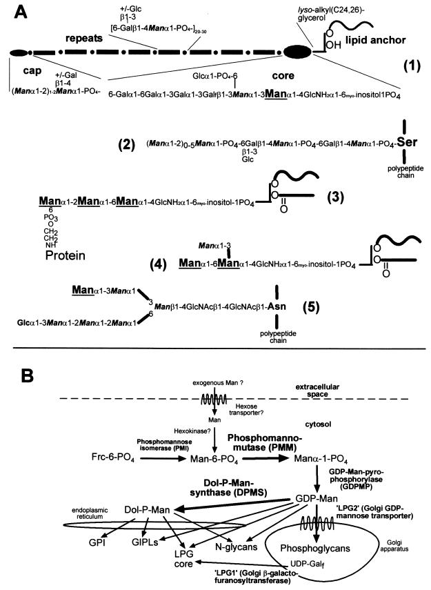
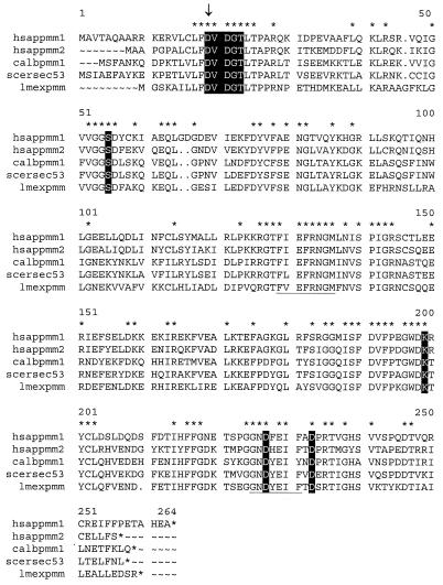
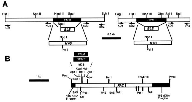
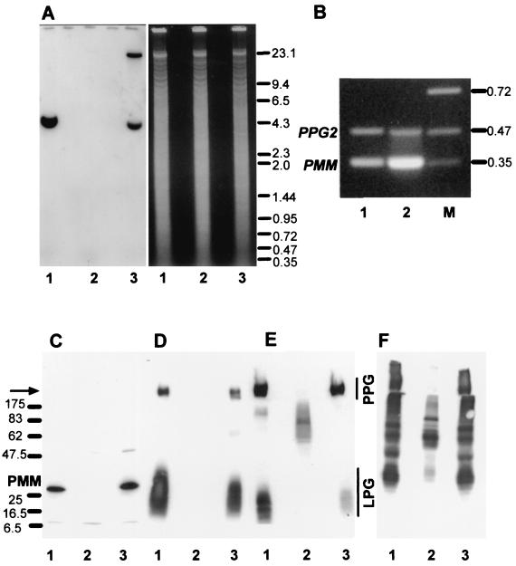
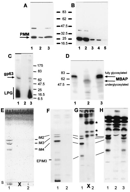
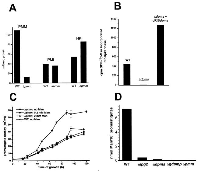
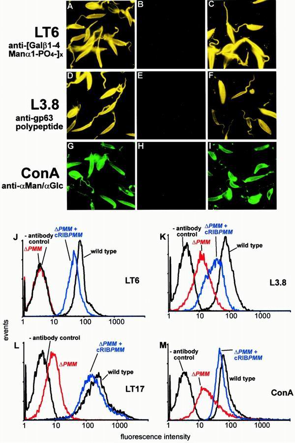
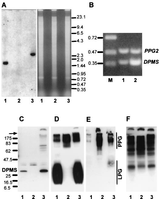
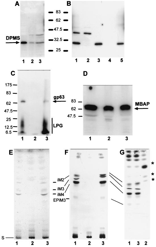
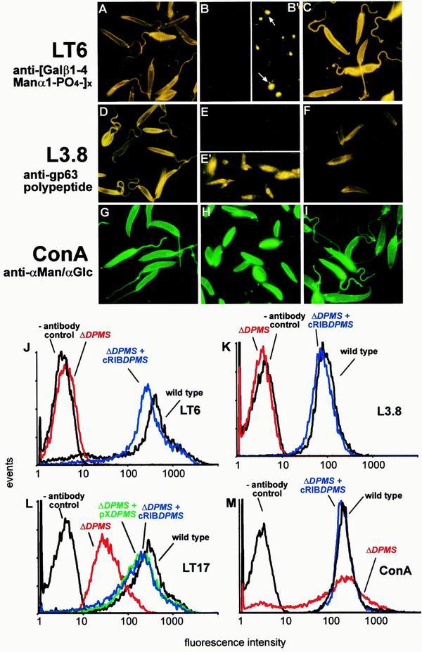
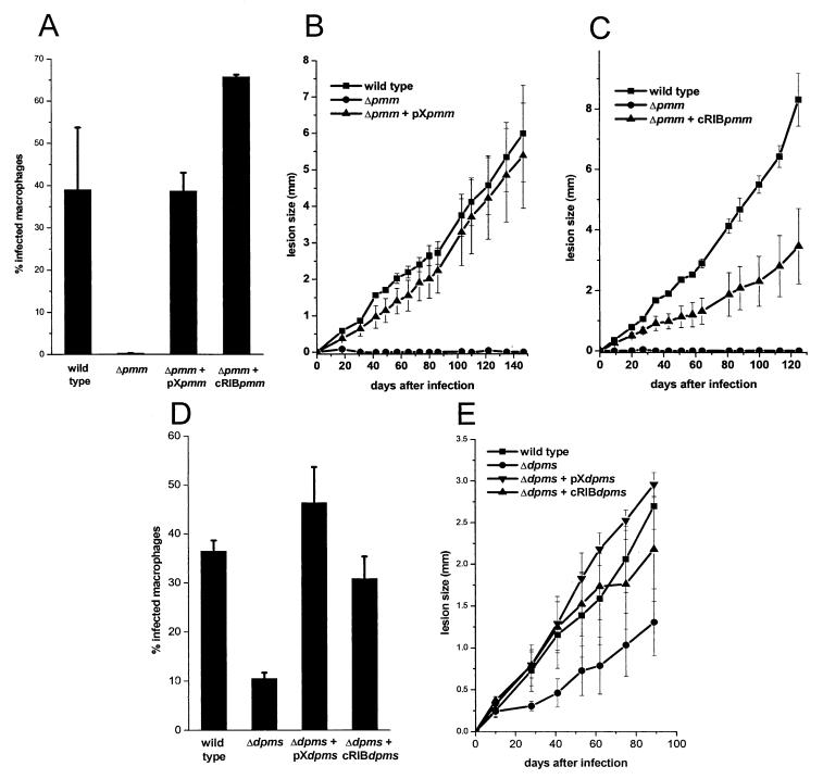
Leishmania parasites synthesize an abundance of mannose (Man)-containing glycoconjugates thought to be essential for virulence to the mammalian host and for viability. These glycoconjugates include lipophosphoglycan (LPG), proteophosphoglycans (PPGs), glycosylphosphatidylinositol (GPI)-anchored proteins, glycoinositolphospholipids (GIPLs), and N-glycans. A prerequisite for their biosynthesis is an ample supply of the Man donors GDP-Man and dolicholphosphate-Man. We have cloned from Leishmania mexicana the gene encoding the enzyme phosphomannomutase (PMM) and the previously described dolicholphosphate-Man synthase gene (DPMS) that are involved in Man activation. Surprisingly, gene deletion experiments resulted in viable parasite lines lacking the respective open reading frames (DeltaPMM and DeltaDPMS), a result against expectation and in contrast to the lethal phenotype observed in gene deletion experiments with fungi. L. mexicana DeltaDPMS exhibits a selective defect in LPG, protein GPI anchor, and GIPL biosynthesis, but despite the absence of these structures, which have been implicated in parasite virulence and viability, the mutant remains infectious to macrophages and mice. By contrast, L. mexicana DeltaPMM are largely devoid of all known Man-containing glycoconjugates and are unable to establish an infection in mouse macrophages or the living animal. Our results define Man activation leading to GDP-Man as a virulence pathway in Leishmania.


Figures

FIG. 1
FIG. 1
(A) Structure and biosynthesis of Man-containing L. mexicana glycoconjugates. (1), LPG; (2), PPG phosphoglycans; (3), protein GPI anchor; (4), GIPL iM3 (as an example); (5), protein N-glycan. Man residues added from Dol-P-Man are enlarged and underlined, while Man residues added from GDP-Man are in italics and bold. The indication of GDP-Man and Dol-P-Man as Man donors for the biosynthesis of different Leishmania glycoconjugates is based on earlier studies (references and and references therein). (B) Man biosynthesis and activation pathways and glycoconjugate synthesis in L. mexicana. Bold arrows mark the enzymes that are the topic of this study.
FIG. 2
FIG. 2
Alignment of L. mexicana PMM (lmexpmm) with amino acid sequences from various organisms: H. sapiens PMM1 and PMM2 (hsappmm1 and hsappmm2; 27, 28); C. albicans PMM (calbpmm1); S. cerevisiae Sec53p (scersec53; 23). Amino acids conserved in PMM of all four species are indicated by stars above the respective amino acids. Residues conserved in this new family of phosphotransferases that has been defined recently (4) are in white letters on black background, and the aspartic acid residue involved in catalysis (4) is marked by an arrow. Amino acid sequences used for the construction of degenerate oligonucleotide primers are underlined.
FIG. 3
FIG. 3
Targeted gene replacement and gene addback of the PMM and DPMS alleles. (A) Restriction maps of the PMM and DPMS loci. The resistance genes BLE and HYG and the primer binding sites (KO1 to KO4) used for the construction of gene deletion cassettes are indicated. (B) Restriction map of the chromosomal gene addback cassette for genetic rescue of the L. mexicana ΔPMM and ΔDPMS mutants.
FIG. 4
FIG. 4
Analysis of L. mexicana wild type, a ΔPMM mutant, and a PMM gene addback mutant by Southern blotting, RT-PCR, and immunoblotting. (A) Southern blot analysis of PstI restriction enzyme-digested chromosomal DNA (10 μg) from L. mexicana wild type (lanes 1), a ΔPMM mutant (lanes 2). and a ΔPMM + cRIBPMM gene addback mutant (lanes 3). The digested DNAs were separated on an ethidium bromide-containing 0.7% agarose gel (right), blotted onto a nylon membrane, and incubated with a DIG-labeled PMM ORF probe (left). The sizes of DNA standards are indicated in kilobases. (B) Amplification of PMM mRNA from L. mexicana log-phase promastigote (lane 1) and amastigote (lane 2) by RT-PCR from total RNA. The loading was normalized to the coamplified cDNA fragment derived from the PPG2 gene, whose mRNA is approximately equally abundant in L. mexicana promastigotes and amastigotes (13). The sizes of DNA standards (lane M) are indicated in kilobases. (C to F) SDS-PAGE and immunoblotting of L. mexicana wild type and ΔPMM mutant total-cell lysates. Lanes 1, wild type; lanes 2, ΔPMM; lanes 3, ΔPMM + cRIBPMM. Each lane was loaded with 106 promastigotes (∼4 μg of protein). (C) Blot was probed with affinity-purified rabbit anti-L. mexicana PMM antibodies. The same or identically loaded blots were then stripped and probed with MAb LT6 (directed against [6Galβ1-4Manα1-PO4]x) (D), LT17(directed against [6(Glcβ1-3)Galβ1-4Manα1-PO4]x [x = unknown]) (E), and MAb L7.25 (directed against [Manα1-2]0-2Manα1-PO4) (F). The molecular masses and relative positions of standard proteins and the positions of PMM, LPG, and PPG are indicated. The arrow marks the border between stacking and separating gels.
FIG. 5
FIG. 5
Analysis of L. mexicana wild type, a ΔPMM mutant, and a PMM gene addback mutant by SDS-PAGE and immunoblotting, SDS-PAGE and fluorography, and TLC analysis. (A) SDS-PAGE and immunoblotting of total-cell lysates of L. mexicana promastigotes (lane 1, 2.5 × 10⁶ parasites, corresponding to ∼10 μg of protein) and lesion-derived amastigotes (lane 2, 2.5 × 10⁶ parasites, corresponding to ∼3.5 μg of protein; lane 3, 7 × 10⁶ parasites, corresponding to ∼10 μg of protein). The blots were probed with affinity-purified rabbit anti-L. mexicana PMM antibodies. (B) SDS-PAGE and immunoblotting of total-cell lysates of L. mexicana promastigotes fractionated by ultracentrifugation: lane 1, total-cell lysate of 2.5 × 10⁶ parasites, corresponding to ∼10 μg of protein; lane 2, first ultracentrifugation supernatant; lane 3, first ultracentrifugation pellet; lane 4, second ultracentrifugation supernatant; lane 5, second ultracentrifugation pellet. Equivalent sample volumes were loaded, and the blots were probed with affinity-purified rabbit anti-L. mexicana PMM antibodies. (C) SDS-PAGE and fluorography of delipidated total promastigote lysates from [3H]myo-inositol-labeled L. mexicana wild type (lane 1), ΔPMM (lane 2), and ΔPMM + cRIBΔPMM (lane 3). Each lane was loaded with 2.5 × 10⁷ delipidated promastigotes labeled overnight with [3H]myo-inositol. The positions of 14C-labeled protein markers, LPG, and the major GPI-anchored surface metalloproteinase gp63 are indicated. (D) SDS-PAGE and immunoblotting of promastigote lysates (2 × 10⁷ lysates, corresponding to ∼80 μg of protein) of wild type (lane 1), ΔPMM (lane 2), and ΔPMM + cRIBPMM (lane 3). The blots were probed with affinity-purified rabbit anti-L. mexicana MBAP antibodies. The molecular masses and relative positions of standard proteins and the positions of PMM and MBAP are indicated (A to D). (E to H) Silica gel 60 HPTLC analysis of the predominant promastigote glycolipids of L. mexicana in wild type and ΔPMM mutant promastigotes. Lanes 1, wild type; lanes 2, ΔPMM; lane 3, ΔPMM + cRIBPMM. (E) Total lipids from 2 × 10⁸ promastigotes visualized by orcinol/H2SO4 spraying. (F) Fluorography of total lipids from 2.5 × 10⁷ [3H]Man-labeled promastigotes (approximately 100,000 cpm). (G) Fluorography of total lipids from 5 × 10⁶ [3H]GlcNH2-labeled promastigotes (approximately 100,000 cpm). (H) Fluorography of total lipids from 5 × 10⁶ [3H]myo-inositol-labeled promastigotes (approximately 100,000 cpm). Bars, positions of abundant L. mexicana GIPLs (30); S, start of TLCs; ∗, new [3H]GlcNH2-labeled compounds accumulating in the ΔPMM mutant; X, two lanes loaded with samples irrelevant to this study.
FIG. 6
FIG. 6
Enzyme activities, growth curve, and Man content of L. mexicana wild-type (WT) and mutant promastigotes. (A) Enzymatic activity of phosphomannomutase (PMM), phosphomannose isomerase (PMI), and hexokinase (HK) in freeze/thaw/sonication lysates of wild-type L. mexicana and a ΔPMM mutant. (B) Synthesis of lipid-bound [14C]Man by microsomal fractions of L. mexicana wild-type, ΔDPMS, and ΔDPMS + cRIBDPMS promastigotes. The bars represent the average of duplicate assays. (C) Growth curves of L. mexicana wild-type and ΔPMM mutant promastigotes with and without Man supplementation of the medium. (D) Man content of membranes from L. mexicana wild-type and several mutant promastigotes, as determined by gas chromatography-mass spectrometry.
FIG. 7
FIG. 7
Immuno-/lectin-fluorescence microscopy and FACS analysis of Leishmania wild-type, ΔPMM mutant, and PMM gene addback mutant promastigotes. (A, D, G) L. mexicana wild type; (B, E, H) L. mexicana ΔPMM; (C, F, I) L. mexicana ΔPMM + cRIBΔPMM. Exposure times within rows are identical. The cells were not permeabilized after fixation. The MAbs and lectins used are indicated by the labeling of rows. (J to M) FACS analysis of live L. mexicana promastigotes. The parasite lines and the MAbs and lectins used are indicated in each panel.
FIG. 8
FIG. 8
Analysis of L. mexicana wild type, a ΔDPMS mutant, and a DPMS gene addback mutant by Southern blotting, RT-PCR, and immunoblotting. (A) Southern blot analysis of SalI restriction enzyme-digested chromosomal DNA (10 μg) from L. mexicana wild type (lanes 1), a ΔDPMS mutant (lanes 2), and a ΔDPMS + cRIBDPMS gene addback mutant (lanes 3). The digested DNAs were separated on an ethidium bromide-containing 0.7% agarose gel (right), blotted onto a nylon membrane, and incubated with a DIG-labeled DPMS ORF probe (left). The sizes of DNA standards are indicated in kilobases. (B) Amplification of DPMS mRNA from L. mexicana log-phase promastigote (lane 1) and amastigote (lane 2) by RT-PCR from total RNA. The loading was normalized to the coamplified cDNA fragment derived from the PPG2 gene, whose mRNA is approximately equally abundant in L. mexicana promastigotes and amastigotes (13). The sizes of DNA standards are indicated in kilobases. (C to F) SDS-PAGE and immunoblotting of L. mexicana wild-type, ΔDPMS mutant, and DPMS gene addback total promastigote lysates. Lane 1, wild type; lane 2, ΔDPMS; lane 3 ΔDPMS + cRIBDPMS. Each lane was loaded with 106 promastigotes (∼4 μg of protein). (C) Blots probed with affinity-purified rabbit anti-L. mexicana DPMS antibodies. The same or identically loaded blots were then stripped and probed with MAb LT6 (directed against [6Galβ1-4Manα1-PO4]x) (D), MAb LT17(directed against [6(Glcβ1-3)Galβ1-4Manα1-PO4]x [x = unknown]) (E), and MAb L7.25 (directed against [Manα1-2]0-2Manα1-PO4) (F). The molecular masses and relative positions of standard proteins and the positions of DPMS, LPG, and PPG are indicated. The arrow marks the borders between stacking and separating gels.
FIG. 9
FIG. 9
Analysis of L. mexicana wild type, a ΔDPMS mutant, and a DPMS gene addback mutant by SDS-PAGE and immunoblotting, SDS-PAGE and fluorography, and TLC analysis. (A) SDS-PAGE and immunoblotting of total-cell lysates of L. mexicana promastigotes (lane 1, 2.5 × 10⁶ parasites, corresponding to ∼10 μg of protein) and lesion-derived amastigotes (lane 2, 2.5 × 10⁶ parasites, corresponding to ∼3.5 μg of protein; lane 3, 7 × 10⁶ parasites, corresponding to ∼10 μg of protein). The blots were probed with affinity-purified rabbit anti-L. mexicana DPMS antibodies. (B) SDS-PAGE and immunoblotting of total-cell lysates of L. mexicana promastigotes fractionated by ultracentrifugation: lane 1, total-cell lysate of 2.5 × 10⁶ parasites, corresponding to ∼10 μg of protein; lane 2, first ultracentrifugation supernatant; lane 3, first ultracentrifugation pellet; lane 4, second ultracentrifugation supernatant; lane 5, second ultracentrifugation pellet. Equivalent sample volumes were loaded, and the blots were probed with affinity-purified rabbit anti-L. mexicana DPMS antibodies. (C) SDS-PAGE and fluorography of delipidated total promastigote lysates from [3H]myo-inositol-labeled L. mexicana. Lane 1, wild type; lane 2, ΔDPMS; lane 3, ΔDPMS + cRIBDPMS. Each lane was loaded with 2.5 × 10⁷ delipidated promastigotes labeled overnight with [3H]myo-inositol. The positions of 14C-labeled protein markers, LPG ,and the major GPI-anchored surface metalloproteinase gp63 are indicated. (D) SDS-PAGE and immunoblotting of promastigote lysates (2 × 10⁷ lysates, corresponding to ∼80 μg of protein). Lane 1, wild type; lane 2, ΔDPMS; lane 3, ΔDPMS + cRIBDPMS. The blots were probed with affinity-purified rabbit anti-L. mexicana MBAP antibodies. The molecular masses and relative positions of standard proteins and the positions of DPMS and MBAP are indicated (A to D). (E to G) Silica gel 60 HPTLC analysis of the predominant promastigote glycolipids of L. mexicana in wild-type and ΔDPMS mutant promastigotes. Lanes 1, wild type; lanes 2, ΔDPMS; lanes 3, ΔDPMS + cRIBDPMS. (E) Total lipids from 2 × 10⁸ promastigotes were visualized by orcinol/H2SO4 spraying. (F) Fluorography of total lipids from 2.5 × 10⁷ [3H]Man-labeled promastigotes (approximately 100,000 cpm). (G) Fluorography of total lipids from 5 × 10⁶ [3H]GlcNH2-labeled promastigotes (approximately 100,000 cpm). Bars, positions of the abundant L. mexicana GIPLs (30); S, start of TLCs; ∗, new [3H]GlcNH2-labeled compounds accumulating in ΔDPMS mutant.
FIG. 10
FIG. 10
Immuno-/lectin-fluorescence microscopy and FACS analysis of Leishmania wild type, ΔDPMS mutant, and DPMS gene addback mutant promastigotes. (A, D, G) L. mexicana wild type; (B, B′, E, E′, H) L. mexicana ΔDPMS; (C, F, I) L. mexicana ΔDPMS + cRIBΔDPMS. Exposure times within rows are identical except for panels B′ and E′, which are approximately 20× overexposed compared to panels B and E, respectively. The cells were not permeabilized after fixation, except for panel E′, where the promastigotes were treated with 0.1% saponin throughout the labeling procedure. The MAbs and lectins used are indicated by the labeling of rows. The arrows in panel B′ indicate the positions of flagellar pockets. (J to M) FACS analysis of live L. mexicana promastigotes. The parasite lines and the MAbs and lectins used are indicated in each panel.
FIG. 11
FIG. 11
Analysis of macrophage and mouse infections by L. mexicana wild type and mutants. (A, D) Infection of peritoneal macrophages by L. mexicana wild type, ΔPMM, ΔPMM + pXPMM, ΔPMM + cRIBPMM (A) or by L. mexicana wild type, ΔDPMS, ΔDPMS + pXDPMS, ΔDPMS + cRIBDPMS (D). Peritoneal macrophages were infected at a ratio of two stationary phase promastigotes per cell. The percentage of infected macrophages (sample size, 300) was counted 6 days after the infection. The standard errors of duplicate experiments are indicated. (B, C, E) For in vivo infection experiments, BALB/c mice were challenged with 107 L. mexicana promastigotes in the right hind footpad. The swelling caused by L. mexicana wild type, ΔPMM, and ΔPMM + pXPMM (B), by L. mexicana wild type, ΔPMM, and ΔPMM + cRIBPMM (C), and by L. mexicana wild type, ΔDPMS, ΔDPMS + pXDPMS, or ΔDPMS + pRIBDPMS (E) was recorded. The infection experiments were performed in quadruplicates and the standard error is indicated.

Similar articles

Cited by

References

    1. Beverley S M, Turco S J. Lipophosphoglycan (LPG) and the identification of virulence genes in the protozoan parasite Leishmania. Trends Microbiol. 1998;6:35–40. - PubMed
    1. Boles E, Liebetrau W, Hofmann M, Zimmermann F K. A family of hexosephosphate mutases in Saccharomyces cerevisiae. Eur J Biochem. 1994;220:83–96. - PubMed
    1. Brittingham A, Morrison C J, McMaster W R, McGwire B S, Chang K P, Mosser D M. Role of the Leishmania surface protease gp63 in complement fixation, cell adhesion, and resistance to complement-mediated lysis. J Immunol. 1995;15:3102–3111. - PubMed
    1. Collet J, Strooban V, Pirard M, Delpierre G, Van Schaftingen E. A new class of phosphotransferases phosphorylated on an aspartate residue in an amino-terminal DXDX(T/V) motif. J Biol Chem. 1998;273:14107–14112. - PubMed
    1. Colussi P A, Taron C H, Mack J C, Orlean P. Human and Saccharomyces cerevisiae dolichol phosphate mannose synthases represent two classes of the enzyme, but both function in Schizosaccharomyces pombe. Proc Natl Acad Sci USA. 1997;94:7873–7878. - PMC - PubMed

MeSH terms

Substances

Associated data

LinkOut - more resources