. 2001 Dec;21(23):8184-8.
doi: 10.1128/MCB.21.23.8184-8188.2001.

The mouse snail gene encodes a key regulator of the epithelial-mesenchymal transition

Affiliations

The mouse snail gene encodes a key regulator of the epithelial-mesenchymal transition

E A Carver et al. Mol Cell Biol. 2001 Dec.

Abstract

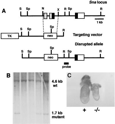
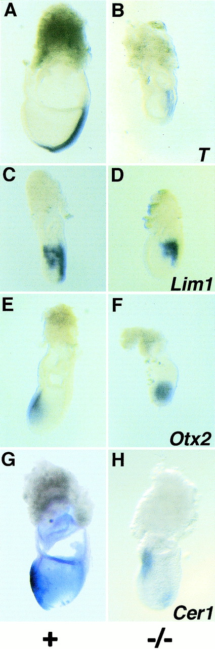
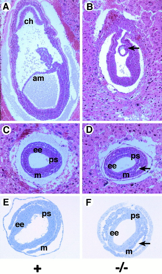
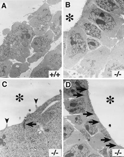
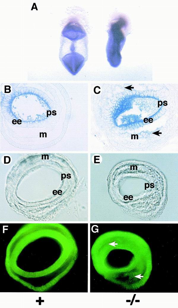
Snail family genes encode DNA binding zinc finger proteins that act as transcriptional repressors. Mouse embryos deficient for the Snail (Sna) gene exhibit defects in the formation of the mesoderm germ layer. In Sna(-/-) mutant embryos, a mesoderm layer forms and mesodermal marker genes are induced but the mutant mesoderm is morphologically abnormal. Lacunae form within the mesoderm layer of the mutant embryos, and cells lining these lacunae retain epithelial characteristics. These cells resemble a columnar epithelium and have apical-basal polarity, with microvilli along the apical surface and intercellular electron-dense adhesive junctions that resemble adherens junctions. E-cadherin expression is retained in the mesoderm of the Sna(-/-) embryos. These defects are strikingly similar to the gastrulation defects observed in snail-deficient Drosophila embryos, suggesting that the mechanism of repression of E-cadherin transcription by Snail family proteins may have been present in the metazoan ancestor of the arthropod and mammalian lineages.


Figures

FIG. 1
FIG. 1
Targeted disruption of the mouse Sna gene. (A) Targeting scheme. The upper line shows the genomic organization of the Sna gene (14). The three exons are indicated by boxes. The region encoding the amino terminus of the Sna protein is indicated by gray boxes, the region encoding the zinc fingers is indicated by black boxes, and the 3′ untranslated region is indicated by a white box. The middle line represents the structure of the targeting vector. The lower line represents the predicted structure of the Sna locus following homologous recombination of the targeting vector. The probe used for Southern blot analysis is indicated. N, NruI; R, EcoRI; S, SalI; Sp, SphI; X, XbaI; TK, thymidine kinase. (B) DNAs isolated from targeted ES cells were digested with SphI, blotted, and hybridized with the indicated probe. Wild-type (wt) and mutant hybridization bands are indicated. Three independently targeted ES cell clones are shown. (C) Whole-mount morphology of a Sna−/− embryo (right) and a control littermate embryo (left) at E7.5. In all figures, normal littermate embryos were either Sna+/− or Sna+/+ and are indicated with a plus sign.
FIG. 2
FIG. 2
Analysis of marker gene expression in Sna−/− mutant embryos at E7.5. (A and B) T expression. T is expressed in axial mesoderm cells, and expression extends rostrally from the node (A). In the Sna−/− embryo, T is expressed, but at lower levels than in the control embryos, and does not extend as far rostrally in the embryo (B). (C and D) Lim1 expression. Lim1 is expressed in the primitive streak and the mesodermal wings (C). Lim1 is expressed in these tissues in the Sna−/− embryo (D). (E and F) Otx2 expression. Otx2 is expressed in the visceral endoderm and epiblast, and expression is gradually restricted to the anterior third of the embryo as the primitive streak extends (E). In the Sna−/− mutant, Otx2 expression is not restricted to the anterior portion of the embryo (F). (G and H) Cer1 expression. Cer1 is expressed in the anterior visceral endoderm and the definitive endoderm (G). In the Sna−/− embryos, Cer1 expression is reduced (H). All embryos are oriented with the anterior side towards the left.
FIG. 3
FIG. 3
Morphological abnormalities in the Sna−/− mutant mesoderm. (A and B) Sagittal sections of embryos at E7.5. In the Sna−/− mutant embryo (B), a posterior amniotic fold forms (arrow) but no amnion or chorion is formed. (C to F) Transverse sections of embryos at E7.5. In Sna−/− mutant embryos, lacunae form within the mesoderm layers (arrows in D and F). Mesoderm cells lining these lacunae exhibit an epithelial morphology. (A to D) Hematoxylin-and eosin-stained paraffin sections. (E and F) Toluidine blue-stained plastic sections. Abbreviations: am, amnion; ch, chorion; ee, embryonic ectoderm; m, mesoderm; ps, primitive streak.
FIG. 4
FIG. 4
Apical-basal polarity and adhesive junctions in the Sna−/− mutant mesoderm. Transmission electron microscopic images for analysis of wild-type (A) and Sna−/− (B to D) embryos at E7.5. are shown. (A) Mesoderm cells in wild-type embryos exhibit a typical mesenchymal morphology. (B) In Sna−/− embryos, mesoderm cells lining the lacunae exhibit an ordered, columnar morphology. (B to D) The lumens of the lacunae are indicated with asterisks. (C) Mesoderm cells in the mutant embryos have microvilli (arrowheads) at the apical surface and exhibit electron-dense adhesive junctions (arrow) between the cells. (D) Adhesive junctions are numerous in the Sna−/− mutant mesoderm. The positions of intercellular adhesive junctions are indicated by arrows. Approximate magnifications: (A and B) ×4,000, (C) ×20,000, (D) ×3,000.
FIG. 5
FIG. 5
E-cadherin expression is retained in the mesoderm of Sna−/− mutant embryos. (A) Whole-mount in situ hybridization with E-cadherin antisense riboprobes of a control littermate (left) and a Sna−/− embryo (right). (B and C) Plastic sections of embryos treated as described for panel A. E-cadherin RNA expression is downregulated in the mesoderm of the control littermate embryo (B), but expression is retained (arrows) in the mesoderm of the Sna−/− embryo (C). (D to G) Immunofluorescence with anti-E-cadherin monoclonal antibody. (D and E) Nomarski optics. (F and G) Fluorescence optics. In the Sna−/− embryo (G), E-cadherin protein expression is retained in the mesoderm layer (arrows). Abbreviations: ee, embryonic ectoderm; m, mesoderm; ps, primitive streak.

Similar articles

Cited by

References

    1. Alberga A, Boulay J-L, Kempe E, Dennefeld C, Haenlin M. The snail gene required for mesoderm formation in Drosophila is expressed dynamically in derivatives of all three germ layers. Development. 1991;111:983–992. - PubMed
    1. Barnes J D, Crosby J L, Jones C M, Wright C V E, Hogan B L M. Embryonic expression of Lim-1, the mouse homolog of Xlim-1, suggests a role in lateral mesoderm differentiation and neurogenesis. Dev Biol. 1994;161:168–178. - PubMed
    1. Batlle E, Sancho E, Franci C, Dominguez D, Monfar M, Baulida J, Garcia de Herreros A. The transcription factor Snail is a repressor of E-cadherin gene expression in epithelial tumour cells. Nat Cell Biol. 2000;2:84–89. - PubMed
    1. Belo J A, Bouwmeester T, Leyns L, Kertesz N, Gallo M, Follettie M, De Robertis E M. Cerberus-like is a secreted factor with neutralizing activity expressed in the anterior primitive endoderm of the mouse gastrula. Mech Dev. 1997;68:45–57. - PubMed
    1. Boulay J L, Dennefeld C, Alberga A. The Drosophila developmental gene snail encodes a protein with nucleic acid binding fingers. Nature. 1987;330:395–398. - PubMed

Publication types

MeSH terms