. 2001 Dec;21(23):8225-35.
doi: 10.1128/MCB.21.23.8225-8235.2001.

Regulation of the Forkhead transcription factor AFX by Ral-dependent phosphorylation of threonines 447 and 451

Affiliations

Regulation of the Forkhead transcription factor AFX by Ral-dependent phosphorylation of threonines 447 and 451

N D De Ruiter et al. Mol Cell Biol. 2001 Dec.

Abstract

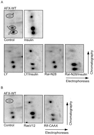
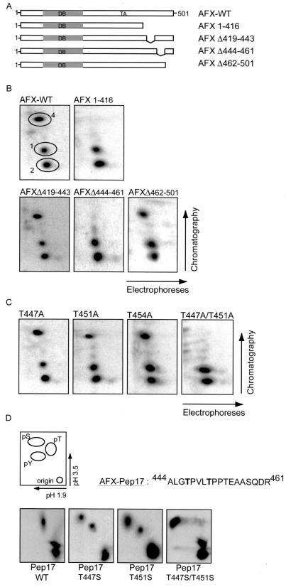
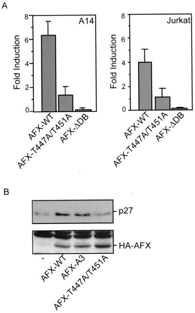
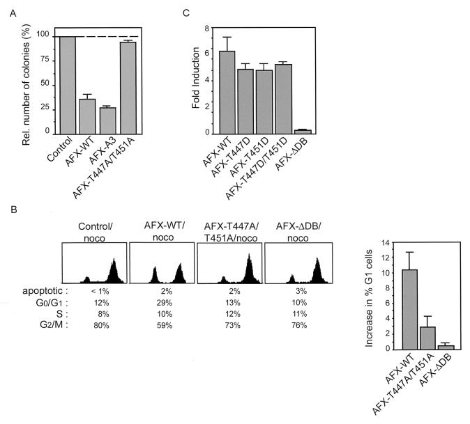
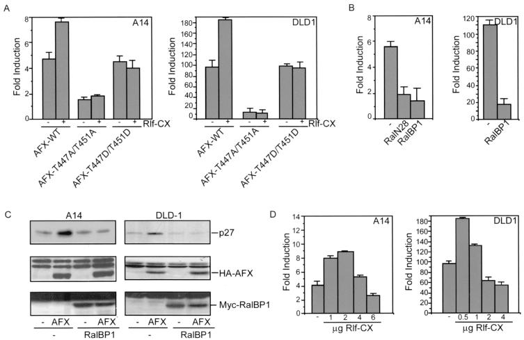
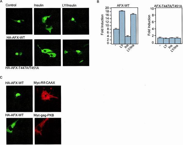
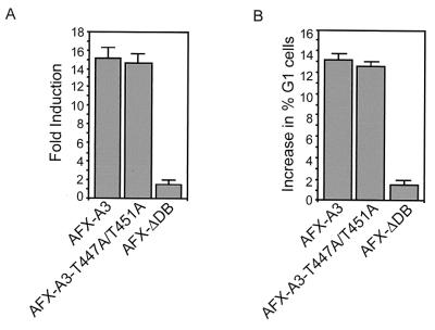
AFX is a Forkhead transcription factor that induces a G(1) cell cycle arrest via upregulation of the cell cycle inhibitor p27(Kip1). Previously we have shown that protein kinase B (PKB) phosphorylates AFX causing inhibition of AFX by nuclear exclusion. In addition, Ras, through the activation of the RalGEF-Ral pathway, induces phosphorylation of AFX. Here we show that the Ras-Ral pathway provokes phosphorylation of threonines 447 and 451 in the C terminus of AFX. A mutant protein in which both threonines are substituted for alanines (T447A/T451A) still responds to PKB-regulated nuclear-cytoplasmic shuttling, but transcriptional activity and consequent G(1) cell cycle arrest are greatly impaired. Furthermore, inhibition of the Ral signaling pathway abolishes both AFX-mediated transcription and regulation of p27(Kip1), while activation of Ral augments AFX activity. From these results we conclude that Ral-mediated phosphorylation of threonines 447 and 451 is required for proper activity of AFX-WT. Interestingly, the T447A/T451A mutation did not affect the induction of transcription and G(1) cell cycle arrest by the PKB-insensitive AFX-A3 mutant, suggesting that Ral-mediated phosphorylation plays a role in the regulation of AFX by PKB.


Figures

FIG. 1
FIG. 1
PKB-independent phosphorylation of AFX by the Ras-Ral pathway. (A) In vivo-phosphorylated HA-AFX was immunoprecipitated from 32P-labeled A14 cells which were left untreated, stimulated with insulin for 30 min, pretreated with LY294002 for 10 min prior to insulin stimulation, or cotransfected with 2 μg of pMT2-HA-Ral-N28. Following exposure to film, bands were cut out of the blot and processed for two-dimensional phosphopeptide mapping using trypsin as the digestive enzyme. Positions of peptides 1, 2, and 4 are indicated by number. (B) 32P-labeled HA-AFX was isolated from transfected A14 cells coexpressing either pSVE-Ras-V12 or pcDNA3-Myc-Rlf-CAAX and analyzed as described for panel A.
FIG. 2
FIG. 2
The Ras-Ral-induced phosphorylation sites in AFX. (A) Deletion mutants of AFX. Abbreviations: DB, DNA binding domain; TA, transactivating domain. (B) 32P-labeled pMT2-HA-AFX-WT, pMT2-HA-AFX-1–416, pMT2-HA-AFX-Δ419–443, pMT2-HA-AFX-Δ444–461, or pMT2-HA-AFX-Δ462–501 AFX was isolated from A14 cells which were cotransfected with p-SVE-RasV12 and analyzed as described in Fig. 1A. The same results were obtained when cells were cotransfected with Rlf-CAAX (data not shown). (C) 32P-labeled pMT2-HA-AFX-T447A, pMT2-HA-AFX-T451A, pMT2-HA-AFX-T454A, or pMT2-HA-AFX-T447A/T451A was isolated from A14 cells which were cotransfected with p-SVE-RasV12 and analyzed as described in Fig. 1A. The same results were obtained when cells were cotransfected with Rlf-CAAX (data not shown). (D) A14 cells were cotransfected with either 2 μg of pCMV-GFP-Pep17-WT, pCMV-GFP-Pep17-T447S, pCMV-GFP-Pep17-T451S, or pCMV-GFP-Pep17-T447S/T451S in combination with 2 μg of p-SVE-RasV12 (or pcDNA3-Myc-Rlf-CAAX [data not shown]). Following exposure to film, bands were cut out of the blot and processed for phosphoamino acid analysis. Positions of phosphoamino acids are as indicated.
FIG. 3
FIG. 3
AFX-T447A/T451A has strongly reduced activity regarding both transcription and upregulation of p27Kip1 protein levels. (A) A14 cells were transfected with 2 μg of either pMT2-HA-AFX, pMT2-HA-AFX-T447A/T451A, or pMT2-HA-AFX-ΔDB in combination with 0.1 μg of p1205LUC reporter construct. The next day, the cells were allowed to recover in the presence of 10% FCS for 6 h and put on serum-free medium for 16 h. Lysates were made 40 h after transfection, and luciferase activity was determined. In each experiment, duplicate dishes were analyzed. Transfection efficiency was monitored by cotransfection of a cytomegalovirus (CMV)-LacZ construct and measuring β-galactosidase activity. Jurkat T cells were transfected by electroporation. Cells were subjected to electroporation with 10 μg of either pMT2-HA-AFX-WT, pMT2-HA-AFX-T447A/T451A, or pMT2-HA-AFX-ΛDB, with 4 μg of pCMV-p27Kip1-Luc reporter construct and 4 μg of pCMV-LacZ in a total amount of DNA of 50 μg equalized with empty vector plasmid. Cells were analyzed for luciferase activity 48 h after transfection. The increases in luciferase activity are shown as fold induction over the activity in cells transfected with control plasmid alone. The results represent the averages of at least four independent experiments. The error bars represent the standard deviations of the values. Protein expression levels were regularly controlled by immunoblotting. (B) A14 cells were transfected with 2 μg of empty vector—pMT2-HA-AFX-WT, pMT2-HA-AFX-A3, or pMT2-HA-AFX-T447A/T451A—in combination with 2 μg of pCMV-CD20. Transfected CD20-positive cells were isolated by MACS. Samples were analyzed for p27Kip1 protein levels (top panel) and expression of the different AFX constructs (bottom panel).
FIG. 4
FIG. 4
AFX-T447A/T451A has greatly impaired growth suppressive effects. (A) A14 cells were transfected with 1 μg of empty vector, pMT2-HA-AFX-WT, pMT2-HA-AFX-A3 or pMT2-HA-AFX-T447A/T451A in combination with 0.1 μg of pBabe-puro. Cells were cultured for two weeks in DMEM 10% FCS supplemented with 5 μg of Puromycin per ml. Puromycin-resistant colonies were scored after 2 weeks of selection. (B) A14 cells were transfected with pEGFP in combination with 2 μg of empty vector, pMT2-HA-AFX-WT, pMT2-HA-AFX-T447A/T451A, or pMT2-HA-AFX-ΛDB. Cells were grown overnight with nocodazole (250 ng/ml). DNA profiles of GFP-positive cells were analyzed on a fluorescence-activated cell sorter. The absolute increase in percentage of A14 cells in the G0/G1 phase upon expression of the indicated proteins is presented in a graph. The error bars represent the standard deviations of the values. Protein expression levels were controlled by immunoblotting. (C) A14 cells were transfected with 2 μg of either pMT2-HA-AFX, pMT2-HA-AFX-T447D, pMT2-HA-AFX-T451D, pMT2-HA-AFX-T447D/T451D, or pMT2-HA-AFX-ΔDB in combination with 0.1 μg of p1205LUC reporter construct and analyzed as described in the legend of Fig. 3A. The increases in luciferase activity are shown as fold inductions over the activity in cells transfected with control plasmid alone. The results represent the averages of at least four independent experiments. The error bars represent the standard deviations of the values. Protein expression levels were regularly controlled by immunoblotting.
FIG. 5
FIG. 5
Inhibition of the Ral pathway inhibits AFX activity. (A) A14 cells were transfected with 4 μg of pMT2-HA-AFX, pMT2-HA-AFX-T447A/T451A, or pMT2-HA-AFX-T447D/T451D in combination with 1 μg of pMT2-HA-Rlf-CAAX and 0.1 μg of p1205LUC reporter construct. DLD1 cells were transfected with 0.5 μg of either pMT2-HA-AFX, pMT2-HA-AFX-T447A/T451A, or pMT2-HA-AFX-T447D/T451D in combination with 0.5 μg of pMT2-HA-Rlf-CAAX and 0.1 μg of p1205LUC reporter construct. Samples were analyzed as described in the legend to Fig. 3A. The increases in luciferase activity are shown as fold induction over the activity in cells transfected with control plasmid alone. In each experiment, duplicate dishes were analyzed. The results represent the averages of at least three independent experiments. The error bars represent the standard deviations of the values. Protein expression levels were regularly controlled by immunoblotting. (B) A14 cells were transfected with 4 μg of pMT2-HA-AFX in combination with 2 μg of either pMT2-HA-RalN28 or pRK5-Myc-RalBP1ΔGAP and 0.1 μg of p1205LUC reporter construct. DLD1 cells were transfected with 0.5 μg of pMT2-HA-AFX in combination with 1 μg of pRK5-Myc-RalBP1ΔGAP and 0.1 μg of p1205LUC reporter construct. Samples were analyzed as described in the legend to Fig. 3A. The increases in luciferase activity are shown as fold induction over the activity in cells transfected with control plasmid alone. In each experiment, duplicate dishes were analyzed. The results represent the averages of at least three independent experiments. The error bars represent the standard deviations of the values. Protein expression levels were regularly controlled by immunoblotting. (C) A14 cells were transfected with 2 μg of empty vector or pMT2-HA-AFX in combination with 2 μg of pRK5-Myc-RalBP1ΔGAP and 2 μg of pCMV-CD20. DLD1 cells were transfected with 0.5 μg of empty vector (−) or pMT2-HA-AFX in combination with 1 μg of pRK5-Myc-RalBP1ΔGAP and 1 μg of pCMV-CD20. Transfected CD20-positive cells were isolated by MACS. Samples were analyzed for p27Kip1 protein levels (top panel), expression of AFX (middle panel), and expression of Myc-RalBP1ΔGAP (bottom panel). (D) A14 cells were transfected with 4 μg of pMT2-HA-AFX in combination with 1,2, 4, or 6 μg of pMT2-HA-Rlf-CAAX and 0.1 μg of p1205LUC reporter construct. DLD1 cells were transfected with 0.5 μg of pMT2-HA-AFX in combination with 0.5, 1, 2, or 4 μg of pMT2-HA-Rlf-CAAX and 0.1 μg of p1205LUC reporter construct. Samples were analyzed as described in the legend to Fig. 3A. The increases in luciferase activity are shown as fold induction over the activity in cells transfected with control plasmid alone. In each experiment, duplicate dishes were analyzed. The result depicted in this figure is a representative example of at least three independent experiments. The error bars represent the standard deviations of the duplicate values in this particular experiment. Protein expression levels were regularly controlled by immunoblotting.
FIG. 6
FIG. 6
The Ras-Ral signaling pathway does not induce a change in the steady-state distribution of AFX from the nucleus to the cytoplasm. (A) A14 cells were transfected with 0.2 μg of either pMT2-HA-AFX-WT or pMT2-HA-AFX-T447A/T451A. The next day, the cells were allowed to recover in the presence of 10% FCS for 6 h and put on serum-free medium for 16 h. Cells were stimulated with insulin (30 min) and LY294002 (10 min prior to insulin treatment) as indicated. Cells were fixed and stained for AFX using the 12CA5 monoclonal antibody. (B) A14 cells were transfected with 2 μg of either pMT2-HA-AFX or pMT2-HA-AFX-T447A/T451A in combination with 0.1 μg of p1205LUC reporter construct. The next day, the cells were allowed to recover in the presence of 10% FCS for 6 h and put on serum-free medium for 16 h. Cells were stimulated with insulin (30 min) and LY294002 (10 min prior to insulin treatment) as indicated. Lysates were made 40 h after transfection, and luciferase activity was determined. In each experiment, duplicate dishes were analyzed. Transfection efficiency was monitored by cotransfection of a CMV-LacZ construct and measuring β-galactosidase activity. (C) A14 cells were transfected with 0.2 μg of pMT2-HA-AFX-WT in combination with 0.5 μg of pcDNA3-Myc-Rlf-CAAX or pSG5-Myc-gag-PKB. The next day, the cells were allowed to recover in the presence of 10% FCS for 6 h and put on serum-free medium for 16 h. Cells were fixed and stained for AFX using the 12CA5 monoclonal antibody and 9E10 antibody to stain Rlf-CAAX- and gag-PKB-expressing cells.
FIG. 7
FIG. 7
Mutation of threonines 447 and 451 does not affect the activity of AFX-A3. (A) Jurkat T cells were transfected by electroporation. Cells were subjected to electroporation with 10 μg of either pMT2-HA-AFX-A3, pMT2-HA-AFX-A3-T447A/T451A, or pMT2-HA-AFX-ΛDB; 4 μg of pCMV-p27Kip1-Luc reporter construct; and 4 μg of pCMV-LacZ in a total of 50 μg of DNA equalized with empty vector plasmid. Cells were analyzed for luciferase activity 48 h after transfection. The increases in luciferase activity are shown as fold induction over the activity in cells transfected with control plasmid alone. The results represent the averages of at least four independent experiments. The error bars represent the standard deviations of the values. Protein expression levels were regularly controlled by immunoblotting. (B) A14 cells were transfected with pEGFP in combination with 2 μg of either empty vector, pMT2-HA-AFX-A3, pMT2-HA-AFX-A3-T447A/T451A, or pMT2-HA-AFX-ΛDB. Cells were grown overnight with nocodazole (250 ng/ml). DNA profiles of GFP-positive cells were analyzed on a fluorescence-activated cell sorter. The absolute increase in percentage of A14 cells in the G0/G1 phase upon expression of the indicated proteins is presented. The error bars represent the standard deviations of the values. Protein expression levels were controlled by immunoblotting.

Similar articles

Cited by

References

    1. Borkhardt A, Repp R, Haas O A, Leis T, Harbott J, Kreuder J, Hammermann J, Henn T, Lampert F. Cloning and characterization of AFX, the gene that fuses to MLL in acute leukemias with a t(X;11)(q13;q23) Oncogene. 1997;14:195–202. - PubMed
    1. Bos J L. All in the family? New insights and questions regarding interconnectivity of Ras, Rap1 and Ral. EMBO J. 1998;17:6776–6782. - PMC - PubMed
    1. Boyle W J, van der Geer P, Hunter T. Phosphopeptide mapping and phosphoamino acid analysis by two-dimensional separation on thin-layer cellulose plates. Methods Enzymol. 1991;201:110–149. - PubMed
    1. Brownawell A M, Kops G J, Macara I G, Burgering B M. Inhibition of nuclear import by protein kinase B (AKT) regulates the subcellular distribution and activity of the Forkhead transcription factor AFX. Mol Cell Biol. 2001;21:3534–3546. - PMC - PubMed
    1. Burgering B M, Coffer P J. Protein kinase B (c-Akt) in phosphatidylinositol-3-OH kinase signal transduction. Nature. 1995;376:599–602. - PubMed

Publication types

MeSH terms