. 2001 Nov 1;15(21):2852-64.
doi: 10.1101/gad.912401.

Hierarchical phosphorylation of the translation inhibitor 4E-BP1

Affiliations

Hierarchical phosphorylation of the translation inhibitor 4E-BP1

A C Gingras et al. Genes Dev. .

Abstract

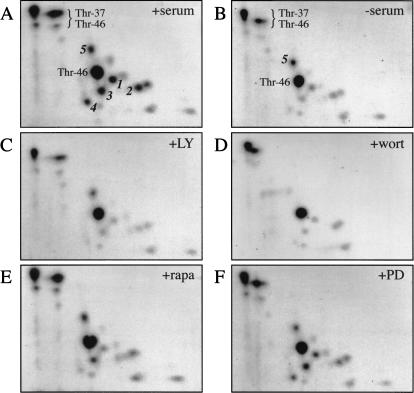
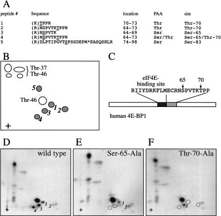
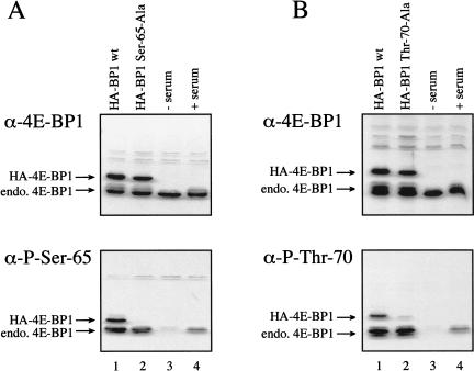
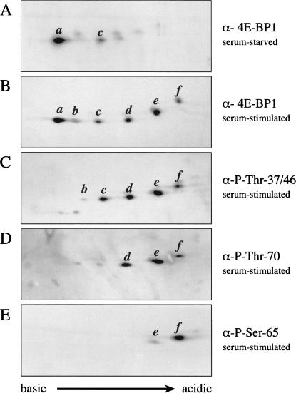
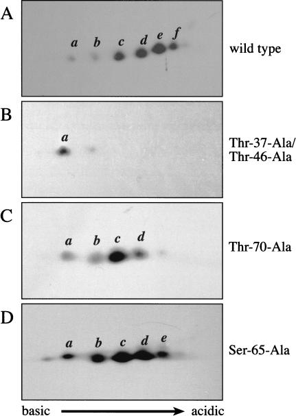
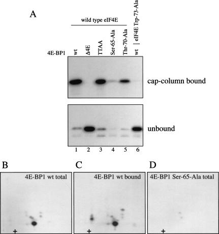
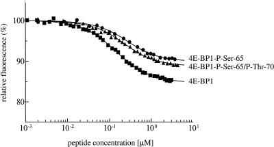
In most instances, translation is regulated at the initiation phase, when a ribosome is recruited to the 5' end of an mRNA. The eIF4E-binding proteins (4E-BPs) interdict translation initiation by binding to the translation factor eIF4E, and preventing recruitment of the translation machinery to mRNA. The 4E-BPs inhibit translation in a reversible manner. Hypophosphorylated 4E-BPs interact avidly with eIF4E, whereas 4E-BP hyperphosphorylation, elicited by stimulation of cells with hormones, cytokines, or growth factors, results in an abrogation of eIF4E-binding activity. We reported previously that phosphorylation of 4E-BP1 on Thr 37 and Thr 46 is relatively insensitive to serum deprivation and rapamycin treatment, and that phosphorylation of these residues is required for the subsequent phosphorylation of a set of unidentified serum-responsive sites. Here, using mass spectrometry, we identify the serum-responsive, rapamycin-sensitive sites as Ser 65 and Thr 70. Utilizing a novel combination of two-dimensional isoelectric focusing/SDS-PAGE and Western blotting with phosphospecific antibodies, we also establish the order of 4E-BP1 phosphorylation in vivo; phosphorylation of Thr 37/Thr 46 is followed by Thr 70 phosphorylation, and Ser 65 is phosphorylated last. Finally, we show that phosphorylation of Ser 65 and Thr 70 alone is insufficient to block binding to eIF4E, indicating that a combination of phosphorylation events is necessary to dissociate 4E-BP1 from eIF4E.


Figures

Figure 1
Figure 1
Sensitivity of 4E-BP1 phosphopeptides to serum deprivation and kinase inhibitor treatment. HEK 293 cells were starved of serum for 30 h. 32P-labeling was performed for 3.5 h. Kinase inhibitors were then added (for 30 min), followed by stimulation for 30 min with 10% dialyzed FBS. Phosphopeptide maps were prepared (see Materials and Methods). Previously identified phosphopeptides containing Thr 37 and/or Thr 46 are indicated. Phosphopeptides exhibiting sensitivity to serum-deprivation or kinase inhibitors are labeled 1–5. The various treatments are indicated in the top, right corner as follows: serum-stimulation (A, +serum), no stimulation (B, −serum), pre-treatment with LY294002 (C, +LY), wortmannin (D, +wort), rapamycin (E, +rapa), or PD098059 (F, +PD). A and B, which served here as controls, were published previously (Gingras et al. 1999a).
Figure 2
Figure 2
Identification of Ser 65 and Thr 70 as the serum-responsive, kinase inhibitor-sensitive phosphorylation sites. 4E-BP1 was immunoprecipitated from 2 × 108 serum-stimulated cells (1/20 labeled in vivo with [32P]orthophosphate), and subjected to phosphopeptide mapping. Peptides 1–5 (see Fig. 1A) were isolated from the plates and identified by LC-MS/MS. (A) Determination of the sequence of phosphopeptides 1–5, and of the identity of the phosphorylated residues by mass spectrometry. The phosphorylated residue is underlined; (*) oxidized methionine. Trypsin cleavage sites are indicated in parentheses. Phosphoaminoacid analysis (PAA) was performed in parallel. (Identification of phosphopeptides 2 and 4 was reported previously in Gygi et al. 1999a). (B) Schema of the phosphopeptides identified by two-dimensional phosphopeptide mapping. (C) Positions of Ser 65 and Thr 70 in 4E-BP1, relative to the eIF4E-binding site. (D–F) Confirmation of the identity of the phosphopeptides containing Ser 65 and Thr 70. HEK 293T cells expressing HA-tagged 4E-BP1 proteins were subjected to metabolic 32P labeling, and two-dimensional phosphopeptide mapping of the HA–4E-BP1 proteins was performed. Dotted circles indicate the absence of phosphopeptides, as compared with the wild-type protein
Figure 3
Figure 3
Phosphospecific antibodies directed against Ser 65 and Thr 70 confirm the serum sensitivity of the phosphorylation at these sites. (A) Western blotting using an antibody to total 4E-BP1 (top), or a phosphospecific antibody directed against Ser 65 (bottom) was performed on extracts from HEK 293T cells transfected with HA–4E-BP1 wt (lane 1) or HA–4E-BP1 Ser 65-Ala (lane 2), or extracts from HEK 293 cells that were serum starved (lane 3), or serum starved, and then serum stimulated for 30 min (lane 4). (B) Western blotting was performed as in A, but using an HA–4E-BP1 Thr 70-Ala protein in lane 2, and a phosphospecific antibody directed against Thr 70 (bottom). The positions of the endogenous 4E-BP1, and the transfected proteins are indicated.
Figure 4
Figure 4
Phosphorylation of endogenous 4E-BP1 in vivo is an ordered process, culminating in the phosphorylation of Ser 65. Two-dimensional IEF/SDS-PAGE (see Materials and Methods) was performed on lysates from serum-starved (A), or serum-stimulated (B–E) HEK 293 cells. Western blotting was then performed using antisera directed against the entire 4E-BP1 protein (A,B), or a series of phosphospecific antibodies raised against 4E-BP1 Thr 37/Thr 46 (C), Thr 70 (D), or Ser 65 (E). Western blots C–E were subsequently reprobed with the antisera raised against the entire 4E-BP1 protein to confirm the identity of the isoforms.
Figure 5
Figure 5
Effects of mutating individual phosphorylation sites on 4E-BP1 phosphorylation. HA-tagged 4E-BP1 proteins were expressed in HEK 293T cells. At 48 h after transfection, lysates were prepared and analysed by two-dimensional IEF/SDS-PAGE, as above. Western blots were prepared using an antibody directed against the HA tag.
Figure 6
Figure 6
Phosphorylation of Ser 65 alone is insufficient to effect release from eIF4E. Bacterially expressed wild-type and mutant GST–4E-BP1 proteins were phosphorylated for 15 min with recombinant ERK2 in the presence of [γ-32P]ATP. Labeled 4E-BP1 proteins were incubated for 1 h with equimolar concentrations of recombinant GST–eIF4E proteins. (A) Complexes were recovered on an m7GDP-sepharose column (cap-column bound), and analyzed by SDS-PAGE and autoradiography. Fractions not retained on the cap column (unbound) were analyzed similarly. Δ4E is a 4E-BP1 mutant deleted in the binding site for eIF4E, and TTAA is the double point mutant Thr 37-Ala/Thr 46-Ala. Trp 73-Ala is an eIF4E mutant unable to bind to 4E-BP1. (B–D) Phosphopeptide mapping of ERK2-phosphorylated 4E-BP1. (B) Phosphopeptide map of 4E-BP1 phosphorylated in vitro by ERK2. (C) Phosphopeptide map of 4E-BP1 phosphorylated in vitro by ERK2, and recovered on a cap column. (D) Phosphopeptide map of the Ser 65-Ala mutant phosphorylated in vitro.
Figure 7
Figure 7
4E-BP1 peptides phosphorylated on Ser 65 and Thr 70 interact with eIF4E. Increasing concentrations of an unphosphorylated 4E-BP1 peptide (residues 51–67; ▪), a monophosphorylated peptide (Ser 65; ●), or a diphosphorylated peptide (Ser 65 and Thr 70; ▴) were incubated with eIF4E, and the binding affinity measured using fluorescence quenching of eIF4E. The eIF4E concentration in the experiment shown was 0.1 μM. The peptide concentration range at which the saturation stage is achieved determines the binding constant. It is similar for each of the three peptides, indicating that the binding affinities are nearly equal, with Kd values of ∼100–200 nM. The twofold difference is not significant at the nanomolar level of the binding constant.

References

    1. Beretta L, Gingras A-C, Svitkin YV, Hall MN, Sonenberg N. Rapamycin blocks the phosphorylation of 4E-BP1 and inhibits cap-dependent initiation of translation. EMBO J. 1996;15:658–664. - PMC - PubMed
    1. Blachut-Okrasinska E, Bojarska E, Niedzwiecka A, Chlebicka L, Darzynkiewicz E, Stolarski R, Stepinski J, Antosiewicz JM. Stopped-flow and Brownian dynamics studies of electrostatic effects in the kinetics of binding of 7-methyl-GpppG to the protein eIF4E. Eur Biophys J. 2000;29:487–498. - PubMed
    1. Brunn GJ, Fadden P, Haystead TAJ, Lawrence JC., Jr The mammalian target of rapamycin phosphorylates sites having a (Ser/Thr)-Pro motif and is activated by antibodies to a region near its COOH terminus. J Biol Chem. 1997a;272:32547–32550. - PubMed
    1. Brunn GJ, Hudson CC, Sekulic A, Williams JM, Hosoi H, Houghton PJ, Lawrence JC, Jr, Abraham RT. Phosphorylation of the translational repressor PHAS-I by the mammalian target of rapamycin. Science. 1997b;277:99–101. - PubMed
    1. Burnett PE, Barrow RK, Cohen NA, Snyder SH, Sabatini DM. RAFT1 phosphorylation of the translational regulators p70 S6 kinase and 4E-BP1. Proc Natl Acad Sci. 1998;95:1432–1437. - PMC - PubMed

Publication types

MeSH terms