. 2001 Nov;11(11):1826-32.
doi: 10.1101/gr.203001.

A short pseudoautosomal region in laboratory mice

Affiliations

A short pseudoautosomal region in laboratory mice

J Perry et al. Genome Res. 2001 Nov.

Abstract

The pseudoautosomal region (PAR) of mammalian sex chromosomes is a small region of sequence identity that is the site of an obligatory pairing and recombination event between the X and Y chromosomes during male meiosis. During female meiosis, X chromosomes can pair and recombine along their entire length; recombination in the PAR is therefore approximately 10x greater in male meiosis compared with female meiosis. A consequence of the presence of the PAR in two copies in males and females is that genes in the region escape the process of X-inactivation. Although the structure and gene content of the human PAR at Xq/Yq is well understood, the mouse PAR, which appears to be of independent evolutionary origin, is poorly characterized. Here we describe a yeast artificial chromosome (YAC) contig covering the distal part of the mouse X chromosome, which we have used to define the pseudoautosomal boundary, that is, the point of divergence of X-specific and X-Y-identical sequences. In addition, we have investigated the size of the mouse PAR by integrating a unique restriction endonuclease recognition site just proximal to the pseudoautosomal boundary by homologous recombination. Restriction digestion of this modified DNA and pulsed field gel electrophoresis reveal that the PAR in these cells is approximately 700 kb. Thus, the mouse PAR, although small in size, has retained essential sex chromosome pairing functions despite its rapid rate of evolution.


Figures

Figure 1
Figure 1
Isolation of the mouse pseudoautosomal boundary (PAB). (A) A genomic contig covering the distal part of the mouse X chromosome consisting of overlapping YAC and bacterial artificial chromosomes (BAC) clones covering the region proximal of, and including, the PAB. Boxed regions of YACs indicate the presence of X chromosomal DNA, whereas lines indicate the presence of non–X chromosomal DNA. The STSs derived from these YACs and their relative distances are indicated below the line. The suffix L or R on the STS name indicates the left or right arm of the YAC from which the STS is derived. The position of additional genetic markers localized to the contig are also indicated. TEL and CEN indicate telomeric and centromeric directions, respectively. The figure is not to scale; however, the total walk covers ∼2.5 Mb. (B) Schematic representation of a 10-kb genomic fragment spanning the mouse PAB, showing markers isolated from this region. Black boxes indicate markers derived from Fxy exon sequence.
Figure 2
Figure 2
Structure of the mouse pseudoautosomal boundary (PAB). Sequence of 400 bp spanning the mouse PAB on the X chromosome, showing the point the X-Y identical pseudoautosomal region (PAR) diverges into X-unique sequence (black triangle). Identical nucleotides are indicated by asterisks.
Figure 3
Figure 3
A Mus musculus pseudoautosomal marker is X unique in Mus spretus. The pseudoautosomal marker, DXYCbl1, was amplified by polymerase chain reaction (PCR) from genomic DNA derived from B6 (B6), SPE (SPE), (B6 × SPE)F1 male mice, and an admixture of B6 + SPE DNA. PCR fragments were sequenced directly, and chromatograms are displayed.
Figure 4
Figure 4
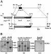
Introduction of an I-SceI restriction site proximal to the pseudoautosomal boundary (PAB). (A) Targeting strategy for the insertion of an I-SceI restriction site proximal to the PAB: wild-type Fxy allele (i), Fxy targeting vector (ii), and targeted Fxy allele (iii). The region of the gene surrounding exon two (black box) of the Fxy gene (top panel) was replaced by a LacZ reporter gene (diagonal lines), which is in frame with the initiation codon (ATG), and a neo selection cassette (dark gray box) flanked by two LoxP sites (black triangles). An negative selection gene, Diphtheria toxin (dotted box), and an ampicillin resistance gene (light grey box), are located upstream of the 5′ homology region. The bottom panel shows the correctly targeted allele and the position of two probes used for Southern hybridization (probes A and B). Restriction enzyme sites indicated are A, AscI; B, BamHI; Bx, BstXI; En, EcoNI; I, I-SceI; N, NotI; and P, PacI. (B) Analysis of targeted clones by Southern hybridization. Genomic DNA from wild-type (WT) and targeted embryonic stem cells (Perry and Ashworth 1999) digested with restriction enzymes and hybridized to probe A (i), probe B (ii), or a probe derived from the neo gene (iii). Sizes estimated from molecular weight markers are indicated on the right.
Figure 5
Figure 5
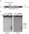
Direct sizing of the pseudoautosomal region (PAR). (A) Schematic representation of the genomic structure of the Fxy gene, indicating the position of the pseudoautosomal boundary (PAB) and the integrated I-SceI site. Exons are indicated by roman numerals, and the initiation and termination codons (ATG and TGA, respectively) are shown. (B) Southern analysis of the size of the PAR in 129 mice. Wild-type embryonic stem cell (ES) and targeted embryonic stem cell (Targeted) genomic DNA was incubated with (+) or without (−) the restriction enzyme I-SceI and were hybridized to the pseudoautosomal probes Mov15 or Fxy3. Size, estimated from molecular weight markers (Hansenula wingei chromosomes), is indicated on the right.
Figure 6
Figure 6
The mouse pseudoautosomal region (PAR). (A) A physical map of the PAR of laboratory mice (129Sv) incorporating a previously reported study (Kipling et al. 1996a) as well as results presented here. The location of the Fxy gene is indicated by a gray line. PAB indicates the position of the pseudoautosomal boundary; TEL,of the telomere. The presence of two repeat units containing the 3′ of Fxy, DXYMov15, PAR-4, and the interstitial telomeric repeat (TTAGGG)n are also indicated. The positions of a PacI site and the Steroid sulphatase (Sts) gene have been taken from a study by Kipling et al. (1996a). (B) Schematic of the genomic organization of the Fxy gene in laboratory mice. Exons are indicated by gray boxes. The first three exons are X unique, whereas the remaining exons are pseudoautosomal. Intron sizes, where known, are indicated (kb). PAB indicates the position of the pseudoautosomal boundary.

References

    1. Birren B, Lai E. Pulsed field gel electrophoresis: A practical guide. San Diego: Academic Press; 1993.
    1. Burgoyne PS. Genetic homology and crossing over in the X and Y chromosomes of mammals. Hum Genet. 1982;61:85–90. - PubMed
    1. Burgoyne PS, Mahadevaiah SK, Sutcliffe MJ, Palmer SJ. Fertility in mice requires X-Y pairing and a Y-chromosomal “spermiogenesis” gene mapping to the long arm. Cell. 1992;71:391–398. - PubMed
    1. Church GM, Gilbert W. Genomic sequencing. Proc Natl Acad Sci. 1984;81:1991–1995. - PMC - PubMed
    1. Ciccodicola A, D'Esposito M, Esposito T, Gianfrancesco F, Migliaccio C, Miano MG, Matarazzo MR, Vacca M, Franze A, Cuccurese M, et al. Differentially regulated and evolved genes in the fully sequenced Xq/Yq pseudoautosomal region. Hum Mol Genet. 2000;9:395–401. - PubMed

Publication types

LinkOut - more resources