

SNP genotyping by multiplexed solid-phase amplification and fluorescent minisequencing
- PMID: 11691857
- PMCID: PMC311152
- DOI: 10.1101/gr.205001
SNP genotyping by multiplexed solid-phase amplification and fluorescent minisequencing
Abstract
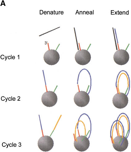
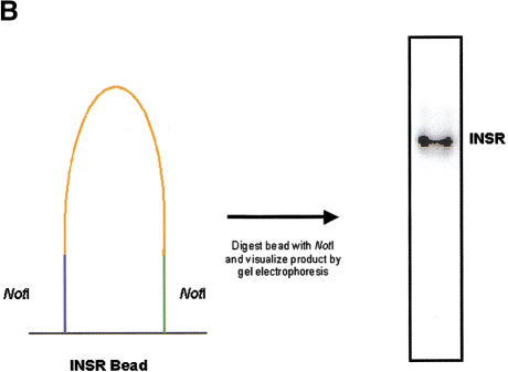
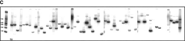
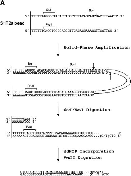
The emerging role of single-nucleotide polymorphisms (SNPs) in clinical association and pharmacogenetic studies has created a need for high-throughput genotyping technologies. We describe a novel method for multiplexed genotyping of SNPs that employs PCR amplification on microspheres. Oligonucleotide PCR primers were designed for each polymorphic locus such that one of the primers contained a recognition site for BbvI (a type IIS restriction enzyme), followed by 11 nucleotides of locus-specific sequence, which reside immediately upstream of the polymorphic site. Following amplification, this configuration allows for any SNP to be exposed by BbvI digestion and interrogated via primer extension, four-color minisequencing. Primers containing 5' acrylamide groups were attached covalently to the solid support through copolymerization into acrylamide beads. Highly multiplexed solid-phase amplification using human genomic DNA was demonstrated with 57 beads in a single reaction. Multiplexed amplification and minisequencing reactions using bead sets representing eight polymorphic loci were carried out with genomic DNA from eight individuals. Sixty-three of 64 genotypes were accurately determined by this method when compared to genotypes determined by restriction-enzyme digestion of PCR products. This method provides an accurate, robust approach toward multiplexed genotyping that may facilitate the use of SNPs in such diverse applications as pharmacogenetics and genome-wide association studies for complex genetic diseases.
Figures
Similar articles
-
A multiplexing single nucleotide polymorphism typing method based on restriction-enzyme-mediated single-base extension and capillary electrophoresis.Anal Biochem. 2004 Jun 15;329(2):220-9. doi: 10.1016/j.ab.2004.01.038. Anal Biochem. 2004. PMID: 15158480
-
Fluorescent microsphere-based readout technology for multiplexed human single nucleotide polymorphism analysis and bacterial identification.Hum Mutat. 2001 Apr;17(4):305-16. doi: 10.1002/humu.28. Hum Mutat. 2001. PMID: 11295829
-
Multiplexed single nucleotide polymorphism genotyping by oligonucleotide ligation and flow cytometry.Cytometry. 2000 Feb 1;39(2):131-40. Cytometry. 2000. PMID: 10679731
-
Multiplex genotyping for thrombophilia-associated SNPs by universal bead arrays.Methods Mol Biol. 2009;496:59-72. doi: 10.1007/978-1-59745-553-4_6. Methods Mol Biol. 2009. PMID: 18839105 Review.
-
From gels to chips: "minisequencing" primer extension for analysis of point mutations and single nucleotide polymorphisms.Hum Mutat. 1999;13(1):1-10. doi: 10.1002/(SICI)1098-1004(1999)13:1<1::AID-HUMU1>3.0.CO;2-I. Hum Mutat. 1999. PMID: 9888384 Review.
Cited by
-
Co-phylogeographic structure in a disease-causing parasite and its oyster host.Parasitology. 2024 Jun;151(7):671-678. doi: 10.1017/S0031182024000611. Epub 2024 May 21. Parasitology. 2024. PMID: 38769826 Free PMC article.
-
Multiplex isothermal solid-phase recombinase polymerase amplification for the specific and fast DNA-based detection of three bacterial pathogens.Mikrochim Acta. 2014;181(13-14):1715-1723. doi: 10.1007/s00604-014-1198-5. Epub 2014 Feb 18. Mikrochim Acta. 2014. PMID: 25253912 Free PMC article.
-
g you The direct determination of haplotypes from extended regions of genomic DNA.BMC Genomics. 2010 Apr 6;11:223. doi: 10.1186/1471-2164-11-223. BMC Genomics. 2010. PMID: 20370899 Free PMC article.
-
Rapid Visual LAMP Method for Detection of Genetically Modified Organisms.ACS Omega. 2023 Aug 1;8(32):29608-29614. doi: 10.1021/acsomega.3c03567. eCollection 2023 Aug 15. ACS Omega. 2023. PMID: 37599972 Free PMC article.
-
The role of DNA diffusion in solid phase polymerase chain reaction with gel-immobilized primers in planar and capillary microarray format.Biomicrofluidics. 2009 Dec 1;3(4):44112. doi: 10.1063/1.3271461. Biomicrofluidics. 2009. PMID: 20216974 Free PMC article.
References
-
- Adams, C. and Kron, S. 1997. Method for performing amplification of nucleic acid with two primers bound to a single solid support. US Patent No. 5641658.
-
- Brenner S, Johnson M, Bridgham J, Golda G, Lloyd DH, Johnson D, Luo S, McCurdy S, Foy M, Ewan M, et al. Gene expression analysis by massively parallel signature sequencing (MPSS) on microbead arrays. Nat Biotechnol. 2000;18:630–634. - PubMed
MeSH terms
Substances
LinkOut - more resources
Full Text Sources
Other Literature Sources