. 2001 Nov 1;536(Pt 3):809-23.
doi: 10.1111/j.1469-7793.2001.00809.x.

Interaction between synaptic excitation and slow afterhyperpolarization current in rat hippocampal pyramidal cells

Affiliations

Interaction between synaptic excitation and slow afterhyperpolarization current in rat hippocampal pyramidal cells

B Lancaster et al. J Physiol. .

Abstract

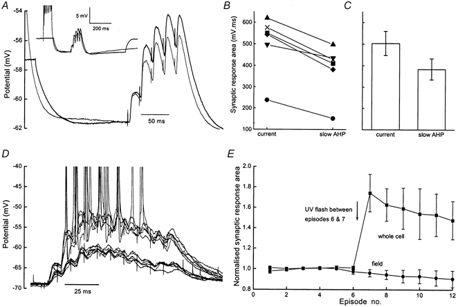
1. Whole cell recordings from CA1 pyramidal cells were performed to investigate the interaction between excitatory postsynaptic potentials (EPSPs) or currents (EPSCs), and the slow Ca(2+)-dependent K(+) current, I(sAHP). Blockers of the slow afterhyperpolarization (sAHP) such as isoprenaline (ISO) or noradrenaline (NA) reduced the hyperpolarization that followed a short train of EPSPs, and slowed the decay of summated EPSPs or EPSCs. 2. ISO/NA action on synaptic responses was observed in the absence of action potentials, but was curtailed by Ca(2+) chelation (10 mM EGTA in the electrode) and was not observed with a caesium-based recording solution. This suggests the involvement of an ISO/NA-sensitive Ca(2+)-dependent K(+) current without a requirement for regenerative spiking. 3. An ISO/NA-sensitive sAHP was observed following both NMDA and non-NMDA receptor-mediated EPSP trains in nominally zero Mg(2+) medium. Isoprenaline sensitivity was blocked by hyperpolarization during EPSPs or by isradipine, suggesting a requirement for voltage-dependent Ca(2+) influx during EPSPs. The data indicate that bursts of EPSPs can activate voltage-gated Ca(2+) channels, which trigger I(sAHP) during synaptic responses. 4. A decrease in EPSP temporal summation occurred during both spike-evoked sAHPs and persistent activation of sAHP conductance following internal dialysis with diazo-2 (2 mM). At constant membrane potential, diazo-2 caused a decrease in membrane time constant and input resistance and accelerated the rate of EPSP decay. Photolysis of diazo-2 or application of NA reduced the resting sAHP conductance, causing an increased membrane time constant and input resistance in association with an increase in EPSP half-width. 5. These results indicate that short bursts of EPSPs can activate a Ca(2+)-dependent K(+) current resembling I(sAHP), and that activation of this current reduces the postsynaptic response to high-frequency synaptic input. The findings imply that modulation of I(sAHP) can regulate synaptic efficacy and may influence the threshold for tetanus-induced synaptic plasticity.


Figures

Figure 1
Figure 1. Bursts of excitatory synaptic input generate noradrenaline-sensitive current
A, averages of 8 consecutive traces before and after noradrenaline (10 μm) application which reduced the hyperpolarization following 5 EPSPs (50 Hz). EPSPs were preceded by brief current pulses to monitor input resistance. Inset, the inhibitory action of noradrenaline (10 μm, applied for 3 min) on the depolarization-evoked IsAHP following an 80 ms voltage command to −10 mV from Vh −60 mV. B, summary data of the action of noradrenaline on the hyperpolarization following a short train of EPSPs as in A (n = 6). C, in voltage clamp, sequential application of 10 μm bicuculline methiodide (bath-applied for 6 min) and noradrenaline (bath-applied for 3 min) caused a slower decay of the inward current following the final EPSC (Vh−55 mV). Da, NMDA receptor-mediated synaptic responses (in 4 μm NBQX, 10 μm bicuculline methiodide) to paired stimuli. The decay of the response is prolonged by 4 μm isoprenaline (Vh−54 mV). Db, a digital subtraction of the two traces in Da, represents the isoprenaline-sensitive current.
Figure 4
Figure 4. An isoprenaline-sensitive slow afterhyperpolarization (sAHP) can be evoked by NMDA receptor-mediated EPSPs
The slices were maintained at room temperature in a nominally Mg2+-free medium designed to isolate NMDA receptor-mediated responses (see text). A, a train of five evoked EPSPs (at 50 Hz) was followed by a sAHP (Control). Bath application of 10 μm isoprenaline (ISO) inhibited the sAHP. To the right, the traces before and after application of ISO are shown on an expanded scale. The vertical dotted lines indicate the integration period over which the sAHP area was measured (columns in C). In each case, the cell was held at −60 mV prior to stimulation. The insets in A and B show the EPSP trains at an expanded time scale. Scale bars: 10 mV, 100 ms. B, when the identical train of five EPSPs was superimposed on a hyperpolarizing current pulse, there was no sAHP. Same cell as in A. The insets show the EPSP trains at an expanded time scale. Scale bars: 10 mV, 100 ms. C, summary data from 6 experiments of the kind shown in A and B. The sAHP area was measured by integration between the vertical dotted lines in right-hand panels of A and B. □, control medium. ▪, after isoprenaline application. D, summary data (n = 5) of EPSC train decay during voltage clamp experiments similar to Fig. 2B. Columns show the 90–10 % decay time of EPSCs under control conditions (□), following application of isradipine (10 μm, formula image
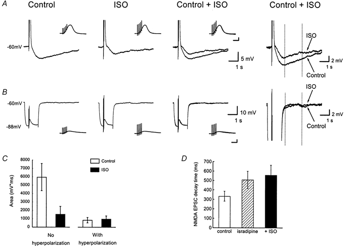
) and sequential application of isoprenaline (ISO, 10 μ
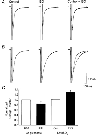
m, ▪).
Figure 2
Figure 2. Internal Cs+ blocks the noradrenergic modulation of EPSCs
A, examples of NMDA receptor-mediated EPSCs (5 at 50 Hz) recorded with a caesium gluconate-based intracellular solution to suppress K+ currents, before (Control) and during (ISO) application of 10 μm isoprenaline. There was no measurable difference in the synaptic current between the two conditions (superimposed records: Control and ISO). B, NMDA receptor-mediated EPSCs recorded with a KMeSO4-based intracellular solution before (Control) and during (ISO) application of 10 μm isoprenaline. Under these conditions, an isoprenaline-sensitive outward current was observed. C, summary data showing that the Cs+-based intracellular solution (n = 5) blocked the isoprenaline-sensitive increase in charge transfer observed with KMeSO4 electrodes (n = 9).
Figure 3
Figure 3. Internal EGTA inhibits the noradrenergic modulation of EPSCs
A, each sample trace contains (left to right) a 5 mV hyperpolarizing command (to monitor access and input resistance), the response to 5 stimuli (50 Hz) and IsAHP evoked by an 80 ms depolarization to −10 mV. The IsAHP was absent when electrodes contained 10 mm EGTA (right trace). B, expansions of the synaptic responses of the cells in A. The action of noradrenaline (10 μm) of prolonging the inward current (left trace) was absent in the presence of 10 mm EGTA internally (right trace). Dashed lines indicate the measurement windows used for integration (see text). C, noradrenaline-sensitive charge transfer (100–400 ms integral of subtracted traces) was significantly reduced by the presence of 10 mm EGTA (n = 7, P = 0.03, unpaired t test). D, the synaptic charge (0–100 ms integral) is not significantly altered by the EGTA (n = 7, P = 0.1, unpaired t test).
Figure 5
Figure 5. An isoprenaline-sensitive slow afterhyperpolarization (sAHP) can be evoked by non-NMDA receptor-mediated EPSPs
The slices were bathed in medium designed to isolate non-NMDA receptor-mediated synaptic responses (see text). A, a train of five EPSPs (50 Hz) was followed by a sAHP (Control). The sAHP was blocked by bath application of 10 μm isoprenaline (ISO). Superimposed and amplified traces are shown to the right. In each case, the cell was held at −60 mV prior to stimulation. The insets in A and B show the EPSP trains at an expanded time scale. Scale bars: 10 mV, 100 ms. B, when the identical train of five EPSPs was superimposed on a hyperpolarizing current pulse injected into the cell, there was no sAHP, although the amplitude of the EPSPs were enhanced by the hyperpolarization. Same cell as in A (EPSP trains with and without hyperpolarizing pulse were alternated before and after application of ISO). C, summary data from 3 experiments of the kind shown in A and B. The sAHP area was measured by integration between the vertical dotted lines in right-hand panels of A and B). □, control medium. ▪, after isoprenaline application.
Figure 6
Figure 6. Slow AHP conductance modulates excitatory synaptic responses
A, superimposed whole cell recordings (averages, n = 6) showing input resistance and EPSPs for the minute prior and subsequent to a UV flash, which reduces the steady state AHP conductance. After the UV flash, the input resistance increased and EPSPs showed a slower decay. Inset shows corresponding field recordings. B, experimental time course of input resistance measured at the end of a 30 ms (0.1 nA) hyperpolarization. Left panel in C, normalized input resistance in the minute before and after photolysis (pre-UV and post-UV respectively). Right panel in C, comparison of normalized membrane time constants before and after photolysis. D, whole cell EPSP half-width (normalized to the minute before a flash) decreases during onset of the K+ conductance and is increased following a UV flash. E–G, field EPSP half-width, whole cell EPSP slope and EPSP amplitude are unaffected by flash photolysis.
Figure 7
Figure 7. EPSPs during noradrenergic block of steady state K+ conductance
All graphs show summary data (mean ±s.e.m.) from the same 6 cells. A, long traces show superimposed whole cell recordings (average of 6 sweeps) before and after isoprenaline (2 μm) application, which increases input resistance and prolongs EPSP decay. The shorter traces are two corresponding field recordings which are unaltered by isoprenaline. B, summary data (n = 6) showing experimental time course of whole cell EPSP half-width, which increased from 15 to 21 ms following isoprenaline administration. C, experimental time course of field EPSP half-width was unaltered by isoprenaline. D, experimental time course of whole cell EPSP slope was unaltered by isoprenaline. E, input resistance before (Con, 61 MΩ) and after (ISO, 86 MΩ) isoprenaline action.
Figure 8
Figure 8. AHP conductance suppresses temporal integration
A, the traces are averages (n = 20) from a single recording in which the burst of EPSPs was alternately superimposed on the sAHP evoked by action potentials (thin trace) or a matching, passive hyperpolarization caused by current injection (thick trace). Inset shows a fuller time course with the synaptic response at the peak of the sAHP. B, area under the synaptic response in six cells when EPSP bursts were superimposed on hyperpolarization caused by current injection (current) and action potential evoked sAHP (slow AHP). C, mean values (±s.e.m.) of area under the EPSPs during the slow AHP or matching hyperpolarization. D, 12 superimposed traces showing the whole cell response to 10 stimuli (100 Hz) in the presence of 50 μmd-AP5 and 100 μm picrotoxin. In the presence of diazo-2, a UV flash (after trial 6) caused a substantial enhancement of the subsequent 6 trials. Membrane potential after the flash maintained by current injection. E, summary data (mean ±s.e.m., normalized to the first 6 responses) from 6 slices in which the area under the simultaneously recorded whole cell and field responses was calculated. A UV flash between episodes 6 and 7 (arrow) enhanced the whole cell responses from 101 ± 2 to 174 ± 18 %.

Similar articles

Cited by

References

    1. Alford S, Frenguelli BG, Schofield JG, Collingridge GL. Characterization of Ca2+ signals induced in hippocampal CA1 neurones by the synaptic activation of NMDA receptors. Journal of Physiology. 1993;469:693–716. - PMC - PubMed
    1. Andreasen M, Lambert JDC. Noradrenaline receptors participate in the regulation of GABAergic inhibition in area CA1 of the rat hippocampus. Journal of Physiology. 1991;439:649–669. - PMC - PubMed
    1. Andreasen M, Lambert JDC. The excitability of CA1 pyramidal cell dendrites is modulated by a local Ca2+-dependent K+-conductance. Brain Research. 1995;698:193–203. - PubMed
    1. Andreasen M, Lambert JDC. Somatic amplification of distally generated subthreshold EPSPs in rat hippocampal neurones. Journal of Physiology. 1999;519:85–100. - PMC - PubMed
    1. Bekkers JM. Distribution of slow AHP channels on hippocampal CA1 pyramidal neurons. Journal of Neurophysiology. 2000;83:1756–1759. - PubMed

Publication types

MeSH terms

LinkOut - more resources