. 2001 Nov;12(11):3340-52.
doi: 10.1091/mbc.12.11.3340.

GCP5 and GCP6: two new members of the human gamma-tubulin complex

Affiliations
Free PMC article

GCP5 and GCP6: two new members of the human gamma-tubulin complex

S M Murphy et al. Mol Biol Cell. 2001 Nov.
Free PMC article

Abstract

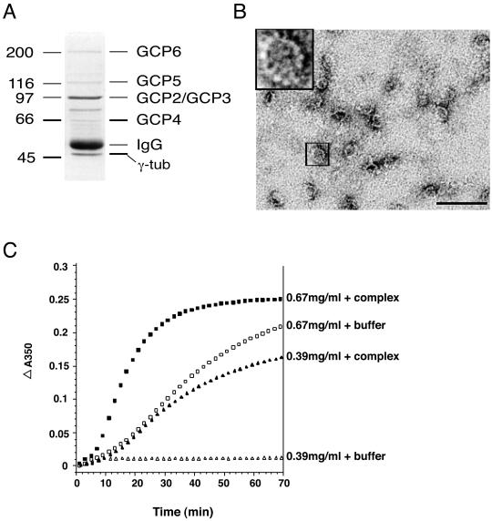
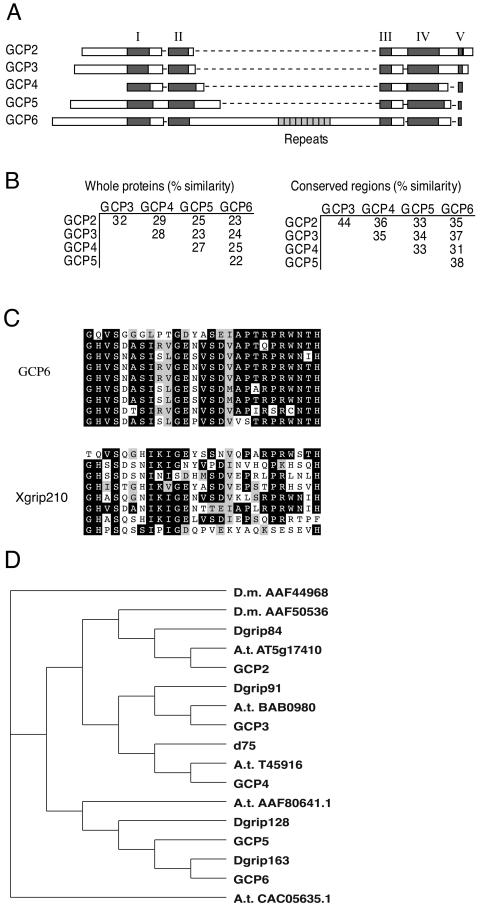
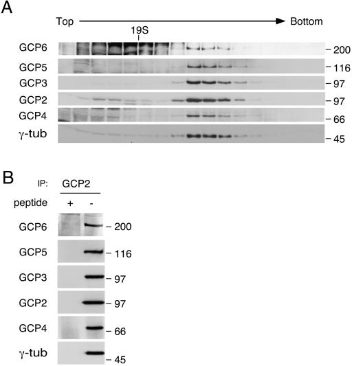
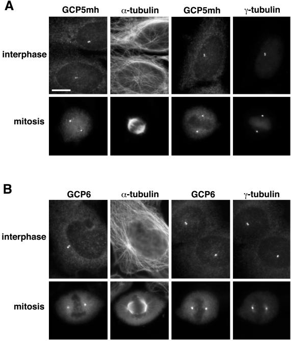
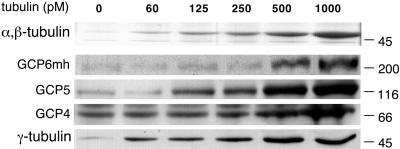
The gamma-tubulin complex is a large multiprotein complex that is required for microtubule nucleation at the centrosome. Here we report the purification and characterization of the human gamma-tubulin complex and the identification of its subunits. The human gamma-tubulin complex is a ring of ~25 nm, has a subunit structure similar to that reported for gamma-tubulin complexes from other species, and is able to nucleate microtubule polymerization in vitro. Mass spectrometry analysis of the human gamma-tubulin complex components confirmed the presence of four previously identified components (gamma-tubulin and gamma-tubulin complex proteins [GCPs] 2, 3, and 4) and led to the identification of two new components, GCP5 and GCP6. Sequence analysis revealed that the GCPs share five regions of sequence similarity and define a novel protein superfamily that is conserved in metazoans. GCP5 and GCP6, like other components of the gamma-tubulin complex, localize to the centrosome and associate with microtubules, suggesting that the entire gamma-tubulin complex takes part in both of these interactions. Stoichiometry experiments revealed that there is a single copy of GCP5 and multiple copies of gamma-tubulin, GCP2, GCP3, and GCP4 within the gamma-tubulin complex. Thus, the gamma-tubulin complex is conserved in structure and function, suggesting that the mechanism of microtubule nucleation is conserved.


Figures

Figure 1
Figure 1
Characterization of the purified human γ-tubulin complex. (A) A Coomassie Blue-stained gel showing the proteins from the purified γ-tubulin (γ-tub) complex. The identities of the bands are indicated to the right. Size in kilodaltons is indicated to the left. (B) Negative stain electron microscopy of the purified complex. The human γ-tubulin complex is shaped like an open ring or spiral and has a diameter of 25 nm. Bar, 100 nm. (C) Microtubule nucleation by the complex was assayed by turbidity. Addition of γ-tubulin decreased microtubule polymerization lag time when compared with buffer control. Open symbols represent microtubule polymerization in the absence of the complex. Filled symbols represent microtubule polymerization in the presence of the complex. Two different concentrations of tubulin are shown: squares represent 0.67 mg/ml and triangles represent 0.39 mg/ml.
Figure 2
Figure 2
The GCPs are related to each other in five regions of conserved sequence. (A) Schematic diagram showing sequence conservation between the GCPs. Conserved regions are shown in dark gray. Unique regions are shown in white. The repeat region of GCP6 is shown in light gray. Gaps are indicated by dashed lines. (B) Percentage of sequence similarity between the GCPs. The amino acid sequences of the GCPs range from 22–32% similarity, whereas the conserved regions range from 31–44% similarity. (C) Alignment of human GCP6 and Xenopus Xgrip210 repeat regions. Black boxes indicate identical amino acids. Gray boxes indicate similar amino acids. (D) A phylogenetic tree of GCP homologues from human, D. melanogaster (D.m.), and A. thaliana (A.t.). The human GCP2 (accession number AF042379) and GCP3 (AF042378), GCP4 (AJ249677) and the Drosophila Dgrip84 (AF118379), Dgrip91 (AF118380), Dgrip128 (AJ291604), Dgrip163 (AJ291605), and d75p (AJ249678) sequences were previously published. GCP5 (AF272884) and GCP6 (AF272887) are described here, and the other proteins were identified by BLAST searches against the respective genome databases. The previously uncharacterized sequences are indicated as either D.m. or A.t., followed by the accession number. The repeat region of GCP6 was excluded in the comparison. The sequences were aligned with the use of ClustalW with a gap penalty of 50.
Figure 3
Figure 3
GCP5 and GCP6 cosediment and coimmunoprecipitate with γ-tubulin (γ-tub). (A) GCP5 and GCP6 from U20S cell lysate were examined by sucrose gradient sedimentation. Fractions from a 10–40% gradient were immunoblotted for γ-tubulin, GCP2, GCP3, GCP4, GCP5, and GCP6. The GCPs cosedimented with γ -tubulin. The anti-GCP6 antibody also recognizes nonspecific proteins in the lower molecular weight part of the gradient. The position of thyroglobulin (19S) is indicated. (B) U2OS cell lysate immunoprecipitated (IP) with anti-GCP2 antibody in the presence (+) and absence (−) of the antigenic peptide was subjected to immunoblot analysis with the use of antibodies against γ-tubulin, GCP2, GCP3, GCP4, GCP5, and GCP6. Size in kilodaltons is indicated to the right.
Figure 4
Figure 4
(A) Cell lines stably expressing γ-tubulinmh, GCP2mh, GCP3mh, GCP4mh, GCP5mh, and GCP6mh were subjected to sucrose gradient sedimentation. Fractions were immunoblotted with anti-myc (myc) and anti-γ-tubulin (γ-tub) antibodies. Epitope-tagged proteins cosedimented with γ-tubulin. The position of thyroglobulin (19S) is indicated. (B) The γ-tubulin complex proteins were examined in pairwise combinations. Epitope-tagged proteins were immunoprecipitated with the anti-myc antibody in the presence (+) and absence (−) of the antigenic peptide. The immunoprecipitations were immunoblotted and probed with antibodies against other γ-tubulin complex proteins. Size in kilodaltons is indicated to the right.
Figure 5
Figure 5
GCP2 and GCP4 are present in multiple copies in the human γ-tubulin complex, whereas GCP5 is present in a single copy. Wild-type cells (wt) and cell lines stably expressing epitope-tagged GCPs were immunoprecipitated with anti-myc and anti-GCP2 antibodies. The immunoprecipitates (IP) were probed with antibodies against the endogenous protein corresponding to the protein tagged in that cell line. Immunoblots were also probed with antibodies against γ-tubulin or GCP2 as loading controls. There is less endogenous GCP4 on the blot because the GCP4mh cell line expresses higher levels of GCP4mh than the endogenous GCP4. Size in kilodaltons is indicated to the right.
Figure 6
Figure 6
Localization of GCP5 and GCP6 by immunofluorescence. (A) Images of GCP5mh cell line stained with antibodies against the myc epitope, α-tubulin, and γ-tubulin. (B) Images of U2OS cells stained with antibodies against GCP6, α-tubulin, and γ-tubulin. Both GCP5mh and GCP6 colocalize with γ-tubulin at the centrosomes of both interphase and mitotic cells. Bar, 10 μm.
Figure 7
Figure 7
GCP4, GCP5, and GCP6 copellet with taxol-stabilized microtubules. Equal amounts of GCP6mh cell lysates were incubated with increasing amounts of taxol-stabilized microtubules. Samples were centrifuged through a 10% glycerol cushion to pellet microtubules. Pellets were analyzed by SDS-PAGE and subsequent Coomassie Blue staining to examine α/β-tubulin and immunoblotting with antibodies against γ-tubulin, GCP4, GCP5, and myc. Size in kilodaltons is indicated to the right. Amount of microtubules in picomolar is indicated at the top.

References

    1. Bergen LG, Kuriyama R, Borisy GG. Polarity of microtubules nucleated by centrosomes and chromosomes of Chinese hamster ovary cells in vitro. J Cell Biol. 1980;84:151–159. - PMC - PubMed
    1. Burge C, Karlin S. Prediction of complete gene structures in human genomic DNA. J Mol Biol. 1997;268:78–94. - PubMed
    1. Detraves C, Mazarguil H, Lajoie MI, Julian M, Raynaud MB, Wright M. Protein complexes containing gamma-tubulin are present in mammalian brain microtubule protein preparations. Cell Motil Cytoskeleton. 1997;36:179–189. - PubMed
    1. Erickson HP, Stoffler D. Protofilaments and rings, two conformations of the tubulin family conserved from bacterial FtsZ to alpha/beta and gamma tubulin. J Cell Biol. 1996;135:5–8. - PMC - PubMed
    1. Evan GI, Lewis GK, Ramsay G, Bishop JM. Isolation of monoclonal antibodies specific for human c-myc proto-oncogene product. Mol Cell Biol. 1985;5:3610–3616. - PMC - PubMed

Publication types

Associated data