. 2001 Nov;12(11):3451-64.
doi: 10.1091/mbc.12.11.3451.

Pmel17 initiates premelanosome morphogenesis within multivesicular bodies

Affiliations
Free PMC article

Pmel17 initiates premelanosome morphogenesis within multivesicular bodies

J F Berson et al. Mol Biol Cell. 2001 Nov.
Free PMC article

Abstract

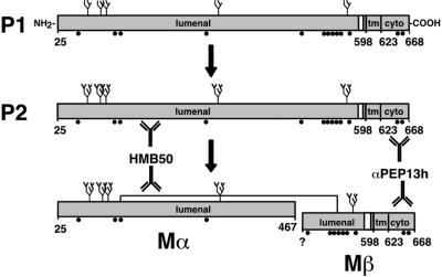
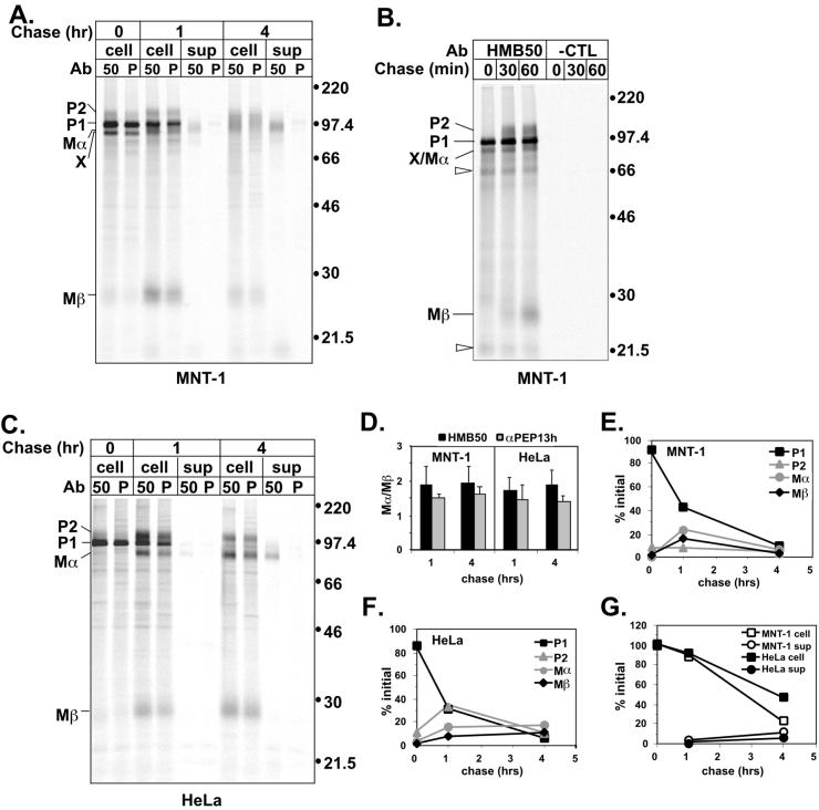
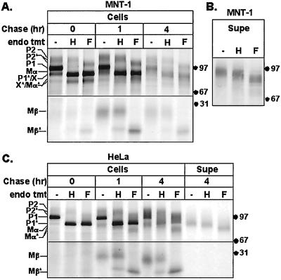
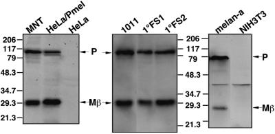
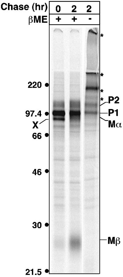
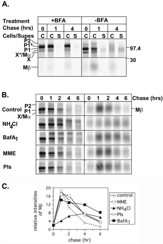
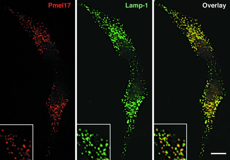
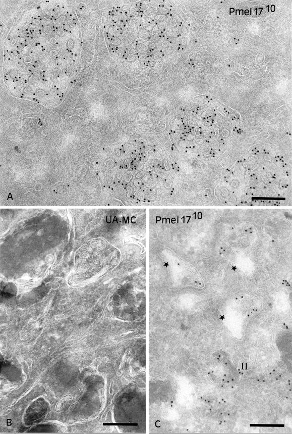
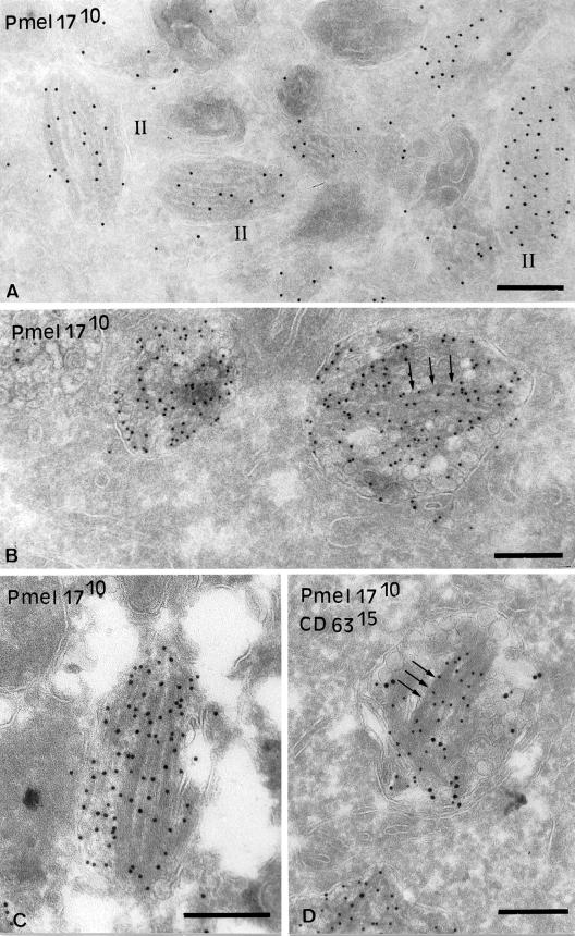
Melanosomes are tissue-specific organelles within which melanin is synthesized and stored. The melanocyte-specific glycoprotein Pmel17 is enriched in the lumen of premelanosomes, where it associates with characteristic striations of unknown composition upon which melanin is deposited. However, Pmel17 is synthesized as an integral membrane protein. To clarify its physical linkage to premelanosomes, we analyzed the posttranslational processing of human Pmel17 in pigmented and transfected nonpigmented cells. We show that Pmel17 is cleaved in a post-Golgi compartment into two disulfide-linked subunits: a large lumenal subunit, M alpha, and an integral membrane subunit, M beta. The two subunits remain associated intracellularly, indicating that detectable M alpha remains membrane bound. We have previously shown that Pmel17 accumulates on intralumenal membrane vesicles and striations of premelanosomes in pigmented cells. In transfected nonpigmented cells Pmel17 associates with the intralumenal membrane vesicles of multivesicular bodies; cells overexpressing Pmel17 also display structures resembling premelanosomal striations within these compartments. These results suggest that Pmel17 is sufficient to drive the formation of striations from within multivesicular bodies and is thus directly involved in the biogenesis of premelanosomes.


Figures

Figure 1
Figure 1
Schematic diagram of Pmel17 and proposed processed forms. The nomenclature for the bands P1, P2, Mα, and Mβ reflects their precursor/product relationships; P1 is the initial precursor, P2 is the processed precursor, and Mα and Mβ are the mature forms (see text). Antibody recognition sites are shown for HMB50 and αPEP13h; the precise recognition site for HMB50 is unknown. N-linked glycans (predicted from consensus sequences) are indicated as branched structures, and potential processing is indicated by increased branching. Disulfide-linkage of Mα and Mβ is indicated by a line; cysteine residues capable of disulfide bond formation are indicated by black circles. Amino acid 25 is the first residue of the proprotein after signal sequence removal. “598” and “623” denote the first and last residues of the transmembrane domain (tm). Assignment of amino acid 467 as the C terminus of Mα is tentative and based on Maresh et al. (1994b). ? indicates the exact N-terminus of Mβ is unknown. Cyto, cytoplasmic domain.
Figure 2
Figure 2
Metabolic pulse-chase analysis of Pmel17 in melanocyte and nonmelanocyte-derived cells. (A). MNT-1 cells were metabolically pulse-labeled with [35S]methionine/cysteine for 30 min and then chased for 0, 1, or 4 h. Cell lysates (cell) or culture medium (sup) were immunoprecipitated in parallel with HMB50 (50) and αPEP13h (P), and precipitated proteins were separated by SDS-PAGE and detected by PhosphorImager analysis. Migration of MW standards (kDa) is indicated to the right, and that of relevant bands, including P1, P2, Mα, Mβ, and band X, are indicated to the left. (B) MNT-1 cells were metabolically labeled as in A except for only 10 min and then chased for 0, 30, or 60 min. Cell lysates were immunoprecipitated with HMB50 or an isotype matched negative control antibody (7G7.B6; -CTL) and immunoprecipitates were analyzed as in A. Unfilled arrowheads indicate bands that were not consistently observed in all experiments. (C) HeLa cells were transiently transfected with Pmel17. Two days posttransfection, the cells were metabolically labeled and immunoprecipitates were analyzed as in A. (D) The ratio of HMB50- or αPEP13h-immunoprecipitable Mα to Mβ at the indicated time points is shown for MNT-1 and HeLa cells. Mα/Mβ is expected to be ∼2:1 based on methionine/cysteine content. The results represent the mean and SD of three experiments. (E and F) Maturation and degradation of the intracellular forms of Pmel17 from MNT-1 (E) and HeLa (F) cells was determined by quantitating the P1, P2, Mα, and Mβ bands immunoprecipitated by HMB50 in the experiments shown in part A and C, and expressing them as a percentage of the initial total Pmel17 from the pulse sample (0 h). This is representative of at least three experiments. (G) Degradation of total Pmel17 from MNT-1 or HeLa cells was determined by summing the intensities of the P1, P2, Mα, and Mβ bands immunoprecipitated by HMB50 from either the lysates or culture medium and expressing these values as a percentage of the total cell associated Pmel17 from the pulse sample. The data are from the experiments shown in A and C, and are representative of at least three experiments.
Figure 3
Figure 3
Endoglycosidase sensitivity of Pmel17 in melanocyte- and nonmelanocyte-derived cells. (A) MNT-1 cells were metabolically labeled as in Figure 2A, cell lysates were immunoprecipitated with HMB50, and precipitated proteins were mock (−), endoH (H), or endoF (F) treated before loading. Asterisk denotes bands resulting from cleavage of the indicated polypeptides by endoH or endoF. Only the relevant regions of the gel are shown. (B) Metabolically pulse labeled MNT-1 cells were chased for 1, 2, 4, and 6 h, and cell supernatants from each time point were pooled and immunoprecipitated with HMB50. Precipitated proteins were mock, endoH, or endoF treated as above. The major band comigrates with Mα, as in Figure 2A. (C) HeLa cells were transiently transfected with Pmel17 and metabolically labeled as in Figure 2C, and cell lysates (cells) or supernatants (Supe) were mock, endoH, or endoF treated as above.
Figure 4
Figure 4
Western blot analysis of Pmel17 processing in melanocytic and nonmelanocytic cells. Cell lysates from human melanoma lines (MNT-1 and 1011-mel), a mouse melanocyte line (melan-a), human primary foreskin melanocytes (1°FS1 and 1°FS2), and nonmelanocyte derived human (HeLa) and mouse (NIH-3T3) lines, as well as HeLa cells transiently transfected with Pmel17 (HeLa/Pmel), were fractionated by SDS-PAGE. Proteins were transferred to PVDF, probed with αPEP13h, and detected by ECF. Migration of MW standards (kDa) is indicated.
Figure 5
Figure 5
Nonreducing gel analysis of Pmel17. MNT-1 cells were metabolically labeled as in Figure 2A and chased for 0 or 2 h. N-ethylmaleimide was added to the final PBS wash of the labeling procedure. Cell lysates were prepared in the presence of iodoacetamide and immunoprecipitated with αPEP13h. Immunoprecipitated proteins were fractionated by SDS-PAGE in the presence (+) or absence (−) of β-mercaptoethanol (β-ME) and detected by PhosphorImager analysis. Asterisk indicates high MW forms of Pmel17. Migration of MW standards (kDa) is indicated to the left.
Figure 6
Figure 6
Effect of inhibitors on Pmel17 processing and degradation. (A) MNT-1 cells were metabolically labeled and chased as in Figure 2A, except that BFA was added to the starvation, pulse and chase media in half of the samples. Cell lysates (C) or culture medium (S) was immunoprecipitated with HMB50, and precipitated proteins were separated by SDS-PAGE and detected by PhosphorImager analysis. Shown separately are the upper and lower parts of the gel encompassing the critical regions. Migration of MW standards (kDa) is indicated to the right. (B) MNT-1 cells were metabolically labeled as in Figure 1A and chased for 0, 1, 2, 4, or 6 h in the presence of DMSO, NH4Cl, BafA1, MME, or a mix of E64, pepstatin A, and leupeptin (PIs). Cell lysates were immunoprecipitated with αPEP13h and analyzed as in A. The images on the left show the upper part of the gel including P1, P2, and Mα, and the images on the right show the lower part of the gel encompassing Mβ. The images on the right were exposed 3.5-fold longer than those on the left in order to better appreciate the differences among samples. (C) The relative intensities of Mβ in part B were quantitated and expressed in arbitrary units. A representative experiment is shown.
Figure 7
Figure 7
Immunofluorescence localization of Pmel17 in HeLa cells. HeLa cells transiently transfected with Pmel17 were fixed, permeabilized, and stained with HMB50 (left) and anti-lamp-1 (middle), followed by LRSC- and FITC-conjugated secondary antibodies, and cells were analyzed by IFM. (right) A merged image.
Figure 8
Figure 8
Localization of Pmel17 to intralumenal vesicles of MVBs. (A) Ultrathin cryosections of HeLa cells transiently transfected with Pmel17 were immunogold labeled with HMB50 (PAG10). The cytoplasm is filled with numerous MVBs. Labeling for Pmel17 is mainly restricted to the ILVs. (B) Ultrathin cryosections of MNT-1 cells retrieved with a mixture of methylcellulose (MC) and uranyl acetate (UA). Note the presence of ILVs beneath polymerized melanin in late stage melanosomes. (C) Ultrathin cryosections of MNT-1 cells were immunogold labeled with HMB50 (PAG10). Early premelanosomal structures (stage I, stars) with no visible striations and displaying ILVs (arrows) are shown. Bars, 200 nm.
Figure 9
Figure 9
Immunogold localization of Pmel17 to striations. (A) Ultrathin cryosections of MNT-1 cells were immunogold labeled with HMB50 (PAG10). Stage II premelanosomes with visible striations are shown. (B–D) Ultrathin cryosections of HeLa cells transiently overexpressing Pmel17 (cells were transfected with 2 μg DNA/6-well) were immunogold labeled with HMB50 (PAG10) (B and C) or HMB50 (PAG10) and anti-CD63 (PAG15) (D). (B) Note the presence of striated-like structures highly labeled for Pmel17 in the lumen of the MVBs (arrows). (C) Some of the striated-like structures are very well developed. (D) The ILVs and associated tubular structures contain both Pmel17 and CD63, whereas the fibrillar structures (arrows) are labeled only for Pmel17. Bars, 200 nm.

Similar articles

Cited by

References

    1. Adema GJ, de Boer AJ, Vogel AM, Loenen WAM, Figdor CG. Molecular characterization of the melanocyte lineage-specific antigen gp100. J Biol Chem. 1994;269:20126–20133. - PubMed
    1. Berson JF, Frank DW, Calvo PA, Bieler BM, Marks MS. A common temperature-sensitive allelic form of human tyrosinase is retained in the endoplasmic reticulum at the nonpermissive temperature. J Biol Chem. 2000;275:12281–12289. - PubMed
    1. Chakraborty AK, Platt JT, Kim KK, Kwon BS, Bennett DC, Pawelek JM. Polymerization of 5,6-dihydroxyindole-2-carboxylic acid to melanin by the pmel 17/silver locus protein. Eur J Biochem. 1996;236:180–188. - PubMed
    1. Dell'Angelica EC, Mullins C, Caplan S, Bonifacino JS. Lysosome-related organelles. FASEB J. 2000;14:1265–1278. - PubMed
    1. Donatien PD, Orlow SJ. Interaction of melanosomal proteins with melanin. Eur J Biochem. 1995;232:159–164. - PubMed

Publication types

MeSH terms