. 2001 Nov;12(11):3618-30.
doi: 10.1091/mbc.12.11.3618.

Tumor necrosis factor-alpha induces stress fiber formation through ceramide production: role of sphingosine kinase

Affiliations
Free PMC article

Tumor necrosis factor-alpha induces stress fiber formation through ceramide production: role of sphingosine kinase

A N Hanna et al. Mol Biol Cell. 2001 Nov.
Free PMC article

Abstract

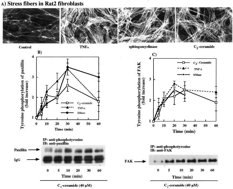
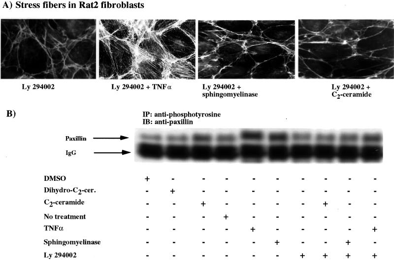
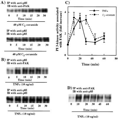
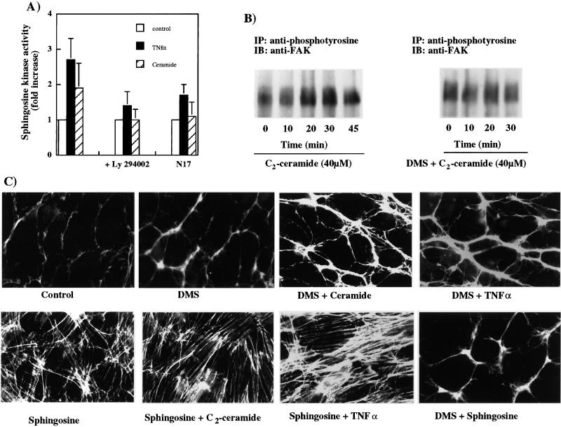
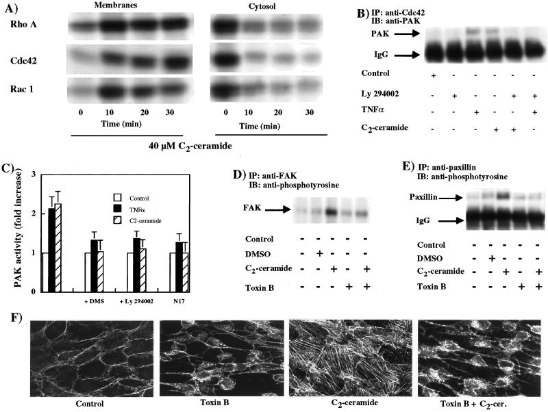
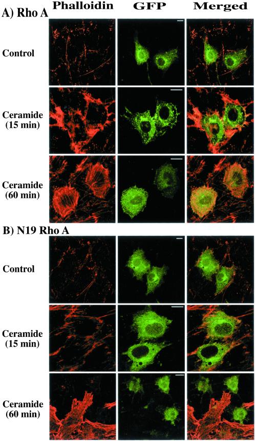
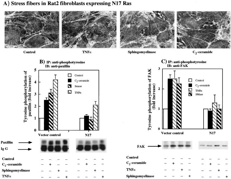
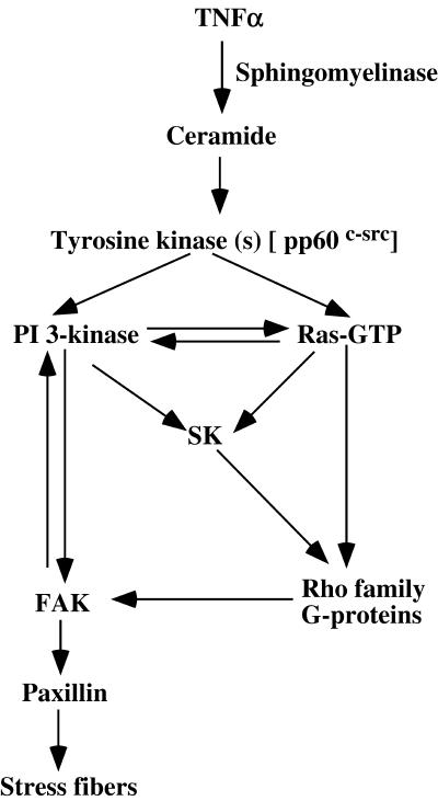
Tumor necrosis factor-alpha (TNF-alpha) is a proinflammatory cytokine that activates several signaling cascades. We determined the extent to which ceramide is a second messenger for TNF-alpha-induced signaling leading to cytoskeletal rearrangement in Rat2 fibroblasts. TNF-alpha, sphingomyelinase, or C(2)-ceramide induced tyrosine phosphorylation of focal adhesion kinase (FAK) and paxillin, and stress fiber formation. Ly 294002, a phosphatidylinositol 3-kinase (PI 3-K) inhibitor, or expression of dominant/negative Ras (N17) completely blocked C(2)-ceramide- and sphingomyelinase-induced tyrosine phosphorylation of FAK and paxillin and severely decreased stress fiber formation. The TNF-alpha effects were only partially inhibited. Dimethylsphingosine, a sphingosine kinase (SK) inhibitor, blocked stress fiber formation by TNF-alpha and C(2)-ceramide. TNF-alpha, sphingomyelinase, and C(2)-ceramide translocated Cdc42, Rac, and RhoA to membranes, and stimulated p21-activated protein kinase downstream of Ras-GTP, PI 3-K, and SK. Transfection with inactive RhoA inhibited the TNF-alpha- and C(2)-ceramide-induced stress fiber formation. Our results demonstrate that stimulation by TNF-alpha, which increases sphingomyelinase activity and ceramide formation, activates sphingosine kinase, Rho family GTPases, focal adhesion kinase, and paxillin. This novel pathway of ceramide signaling can account for approximately 70% of TNF-alpha-induced stress fiber formation and cytoskeletal reorganization.


Figures

Figure 1
Figure 1
Stress fiber formation by TNF-α, sphingomyelinase, and C2-ceramide. (A) Fibroblasts treated in the absence or presence of 10 ng/ml TNF-α, 0.1 unit/ml sphingomyelinase, or 40 μM C2-ceramide for 1 h. Cells were then fixed, permeabilized, and stress fibers were visualized with the use of Texas Red-conjugated phalloidin. The results were reproduced in two further independent experiments. (B and C) Tyrosine phosphorylation of paxillin and FAK, respectively, is shown for Rat2 fibroblasts treated with 40 μM C2-ceramide, 10 ng/ml TNF-α, or 0.1 unit/ml sphingomyelinase for various times. Results are means ± SEM of three independent experiments. The lower parts of B and C show representative Western blots for C2-ceramide-induced tyrosine phosphorylation of paxillin and FAK.
Figure 2
Figure 2
Involvement of PI 3-K in TNF-α-, sphingomyelinase-, and C2-ceramide-induced stress fiber formation. (A) Rat2 fibroblasts were pretreated for 60 min with 20 μM Ly 294002. Cells were then treated with or without 10 ng/ml TNF-α, 0.1 unit/ml sphingomyelinase, or 40 μM C2-ceramide for 60 min. Stress fibers were visualized with the use of Texas Red-conjugated phalloidin. These results can be compared directly to those shown in Figure 1A. (B) Tyrosine phosphorylation of paxillin after treatment of Rat2 fibroblasts with or without 20 μM Ly 294002 followed by adding 10 ng/ml TNF-α, 0.1 unit/ml sphingomyelinase, 40 μM C2-ceramide, or 40 μM dihydro-C2-ceramide for 30 min. Tyrosine phosphorylation of paxillin was assessed by with the use of anti-phosphotyrosine for immunoprecipitation and anti-paxillin for Western blotting. The results were reproduced in two further independent experiments.
Figure 3
Figure 3
TNF-α and C2-ceramide induce physical association between PI 3-kinase and FAK. (A and B) Physical association of FAK (A) with PI 3-K (B) after treatment of Rat2 fibroblasts with 40 μM C2-ceramide or 10 ng/ml TNF-α for various times. The lower blots show that similar amounts of PI 3-K were used to measure the association of FAK to PI 3-K. The results were reproduced in two further independent experiments. (C) PI 3-K activity associated with the anti-FAK precipitate after treatment of Rat2 fibroblasts with 10 ng/ml TNF-α or 40 μM C2-ceramide for various times. In a control experiment, no PI 3-K activity was detected in beads without anti-FAK antibody. Results are means ± SEM for three independent experiments. The blot in D is a representative Western blot to show the effect of TNF-α treatment on the detection of PI 3-K in anti-FAK immunoprecipitates.
Figure 4
Figure 4
TNF-α and C2-ceramide treatment increases sphingosine kinase activity that is upstream of the tyrosine phosphorylation of FAK and stress fiber formation. (A) Activation of sphingosine kinase by 40 μM C2-ceramide or 10 ng/ml TNF-α is attenuated by inhibiting PI 3-kinase with Ly 294002, or in fibroblasts expressing N17 Ras. Results are means ± SEM for three independent experiments. (B) Effects of treating Rat2 fibroblasts with DMS in inhibiting the effects of TNF-α and C2-ceramide in stimulating the tyrosine phosphorylation of FAK. (C) Distribution of stress fibers in Rat2 fibroblasts that were pretreated with 10 μM DMS and then treated with 10 ng/ml TNF-α, 40 μM C2-ceramide, or 20 μM sphingosine.
Figure 5
Figure 5
TNF-α and C2-ceramide treatment increases membrane association of Rho family GTPases, association of Cdc42 with PAK, and phosphorylation of FAK and paxillin. (A) Time courses for the translocations of Cdc42, Rac-1, and RhoA to membranes after treatment with 40 μM C2-ceramide. The results were reproduced in three independent experiments. (B) Physical association of Cdc42 with PAK after treatment of Rat2 fibroblasts with 10 ng/ml TNF-α or 40 μM C2-ceramide for various times in absence or presence of PI 3-kinase inhibitor, 20 μM Ly 294002. (C) PAK activation after treatment of fibroblasts with TNF-α and C2-ceramide for 30 min in the presence or absence of DMS, Ly 294002, or N17 Ras. Results are means ± ranges from two independent experiments. (D and E) Rat2 fibroblasts were treated with or without 0.5 ng/ml toxin B for 2 h in the presence or absence of 40 μM C2-ceramide. The tyrosine phosphorylations of FAK and paxillin were measured with the use of either anti-FAK or anti-paxillin for precipitation and anti-phosphotyrosine for immunoblotting. The blots in this figure were reproduced in a further experiment. (F) Effect of toxin B on ceramide-induced stress fiber formation in Rat2 fibroblasts.
Figure 6
Figure 6
Role of RhoA in C2-ceramide-induced stress fiber formation. This figure shows cytoskeletal rearrangement in fibroblasts transiently transfected with wild-type RhoA or inactive N19 RhoA, both attached to GFP, in the presence of 40 μM C2-ceramide for 15 min or for 1 h. Transfected cells were identified by the green fluorescence of GFP. Actin stress fibers were labeled with the use of Texas Red-conjugated phalloidin and colocalization with GFP is shown in yellow in the merged images. The size bar in the central panels represents 10 nm. The controls are from untreated cells at 15 min and the images were not significantly different for untreated cells incubated for 60 min (our unpublished results). The results were reproduced in three independent experiments and similar results were obtained with 10 ng/ml TNF-α (our unpublished results).
Figure 7
Figure 7
Role of Ras-GTP in TNF-α-, sphingomyelinase- and C2-ceramide-induced stress fiber formation and tyrosine phosphorylation of paxillin and FAK. Rat2 fibroblasts stably expressing N17 Ras were treated with or without 10 ng/ml TNF-α, 0.1 unit/ml sphingomyelinase or 40 μM C2-ceramide for 1 h and stress fibers were visualized. The results were reproduced in two further independent experiments. These images can be compared directly with the results for Rat2 fibroblasts shown in Figure 1A, or to those for fibroblasts transduced with the empty vector (our unpublished results). (B and C) Expression of N17 Ras inhibits the TNF-α-, sphingomyelinase-, and C2-ceramide-induced tyrosine phosphorylation of paxillin and FAK compared with fibroblasts transduced with the empty vector. Results are means ± SEM for three independent experiments.
Figure 8
Figure 8
Proposed scheme for the signaling pathway of TNF-α leading to stress fiber formation. The figure illustrates how ceramide formation can contribute to the signaling pathways whereby TNF-α induces the formation of stress fibers. The ceramide-mediated pathway depends upon the formation of Ras-GTP and the activations of PI 3-K and SK. In addition, TNF-α also induces the tyrosine phosphorylations of FAK and paxillin and stress fiber formation independently of the activation of Ras and PI 3-K.

References

    1. Abousalham A, Liossis C, O'Brien L, Brindley DN. Cell-permeable ceramides prevent the activation of phospholipase D by ADP-ribosylation factor and RhoA. J Biol Chem. 1997;272:1069–1075. - PubMed
    1. Albouz S, Boutry JM, Dubois G, Bourdon R, Hauw JJ, Baumann N. Lipid and lysosomal enzymes in human fibroblasts cultured with perhexiline maleate. Naunyn-Schmiedeberg's Arch Pharmacol. 1981;317:173–177. - PubMed
    1. Bradford MM. A rapid and sensitive method for the quantitation of microgram quantities of protein utilizing the principle of protein-dye binding. Anal Biochem. 1976;72:248–254. - PubMed
    1. Chant J, Stowers L. GTPase cascades choreographing cellular behavior: movement, morphogenesis, and more. Cell. 1995;81:1–4. - PubMed
    1. Chen HC, Appeddu PA, Isodo H, Guan JL. Phosphorylation of tyrosine 397 in focal adhesion kinase is required for binding phosphatidylinositol 3-kinase. J Biol Chem. 1996;271:26329–26334. - PubMed

Publication types

MeSH terms

Substances