. 2001 Nov;12(11):3668-79.
doi: 10.1091/mbc.12.11.3668.

In vivo role for actin-regulating kinases in endocytosis and yeast epsin phosphorylation

Affiliations
Free PMC article

In vivo role for actin-regulating kinases in endocytosis and yeast epsin phosphorylation

H A Watson et al. Mol Biol Cell. 2001 Nov.
Free PMC article

Abstract

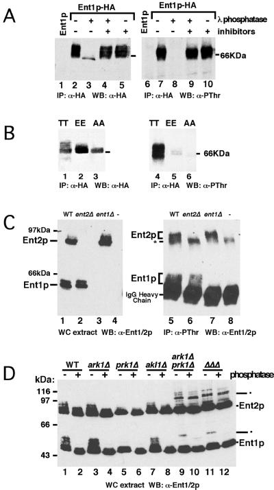
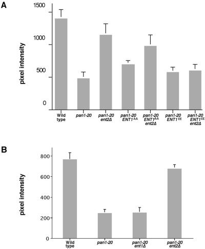
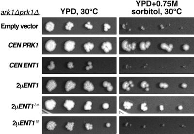
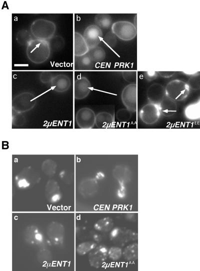
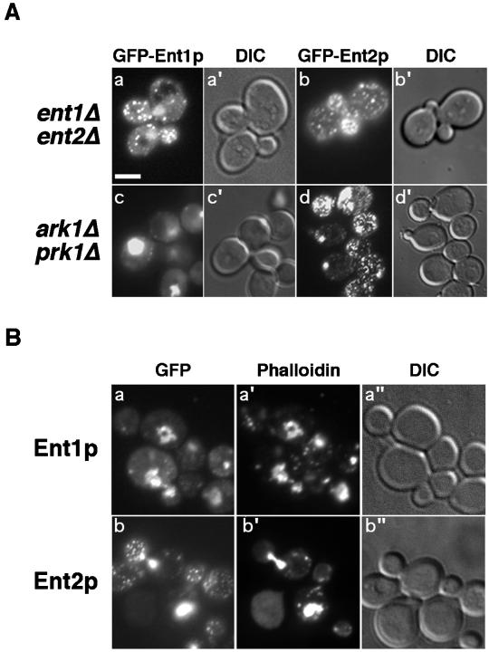
The yeast actin-regulating kinases Ark1p and Prk1p are signaling proteins localized to cortical actin patches, which may be sites of endocytosis. Interactions between the endocytic proteins Pan1p and End3p may be regulated by Prk1p-dependent threonine phosphorylation of Pan1p within the consensus sequence [L/I]xxQxTG. We identified two Prk1p phosphorylation sites within the Pan1p-binding protein Ent1p, a yeast epsin homologue, and demonstrate Prk1p-dependent phosphorylation of both threonines. Converting both threonines to either glutamate or alanine mimics constitutively phosphorylated or dephosphorylated Ent1p, respectively. Synthetic growth defects were observed in a pan1-20 ENT1(EE) double mutant, suggesting that Ent1p phosphorylation negatively regulates the formation/activity of a Pan1p-Ent1p complex. Interestingly, pan1-20 ent2 Delta but not pan1-20 ent1 Delta double mutants had improved growth and endocytosis over the pan1-20 mutant. We found that actin-regulating Ser/Thr kinase (ARK) mutants exhibit endocytic defects and that overexpressing either wild-type or alanine-substituted Ent1p partially suppressed phenotypes associated with loss of ARK kinases, including growth, endocytosis, and actin localization defects. Consistent with synthetic growth defects of pan1-20 ENT1(EE) cells, overexpressing glutamate-substituted Ent1p was deleterious to ARK mutants. Surprisingly, overexpressing the related Ent2p protein could not suppress ARK kinase mutant phenotypes. These results suggest that Ent1p and Ent2p are not completely redundant and may perform opposing functions in endocytosis. These data support the model that, as for clathrin-dependent recycling of synaptic vesicles, yeast endocytic protein phosphorylation inhibits endocytic functions.


Figures

Figure 1
Figure 1
Ent1p and Ent2p threonine phosphorylation requires Prk1p. (A) Epitope-tagged Ent1p (Ent1p-HA) was immunoprecipitated, with the use of mouse α-HA, from wild-type cell extracts treated with lambda protein phosphatase and/or phosphatase inhibitors and analyzed on immunoblots. Left panel, rabbit α-HA; right panel, rabbit α-phosphothreonine. Lanes 1 and 6 correspond to immunoprecipitates from wild-type cells expressing untagged Ent1p; lanes 2–5 and 7–10 correspond to immunoprecipitates from wild-type cells expressing Ent1p-HA. (B) HA-tagged ent1p point mutants in which the two putative target Thr residues were converted to Ala (T394A, T416A; AA) or Glu (T394E, T416E; EE) were expressed in wild-type cells. Wild-type (TT) or mutant forms of Ent1p-HA were immunoprecipitated with the use of mouse α-HA and analyzed by immunoblotting. Left panel, rabbit α-HA; right panel, rabbit α-phosphothreonine. Lanes 1–3 and 4–6 each correspond to wild-type Ent1p-HA, Ent1pEE-HA, and Ent1pAA-HA immunoprecipitates, respectively. (C) Rabbit polyclonal antisera raised against a region of protein conserved between Ent1p and Ent2p (rabbit α-Ent1/2p) recognizes endogenous Ent1p and Ent2p in both whole-cell extracts (left panel) and rabbit α-phosphothreonine immunoprecipitates. Left panel, immunoblot of whole-cell ex tracts from wild-type cells (lane 1), ent2Δ cells (lane 2), ent1Δ cells (lane 3), and negative control ent2Δ cells expressing an Ent1p construct lacking the region used for generating rabbit α-Ent1/2p (lane 4). Right panel, endogenous Ent1p and Ent2p were immunoprecipitated from cells with the use of α-phosphothreonine and analyzed by immunoblotting with rabbit α-Ent1/2p. Nonspecific background bands are indicated by asterisk. (D) Whole-cell extracts from five different ARK family deletion strains and wild-type cells were either mock treated (−) or treated (+) with protein phosphatase and analyzed on immunoblots with the use of rabbit α-Ent1/2p. Phosphatase-resistant and/or slower mobility bands are indicated (•).
Figure 2
Figure 2
Effects of ENT1AA, ENT1EE, and ent2Δ on pan1–20 growth and endocytic defects. Cells were grown to midlog phase in rich medium at 30°C, preshifted to 37°C for 30–45 min, and labeled with the lipophilic dye FM 4–64 at either 30 or 37°C for 15–20 min. After a 30–45 min chase period, cells were washed and observed with the use of fluorescence microscopy. (A) Pixel intensity values (35–50 cells per sample) refer to quantitation from a single focal plane of vacuolar membrane fluorescence after dye uptake and are as follows: wild-type, 1400 ± 130; pan1–20, 483 ± 90; pan1–20 ent2Δ, 1150 ± 170; pan1–20 ENT1AA, 700 ± 60; pan1–20 ent2Δ ENT1AA, 984 ± 160; pan1–20 ENT1EE, 565 ± 90; pan1–20 ent2Δ ENT1EE, 595 ± 100. (B) The FM 4–64 uptake assay at 37°C was performed as described above on wild-type, pan1–20, pan1–20 ent1Δ, and pan1–20 ent2Δ cells. Pixel intensities in this experiment were lower because of differences in labeling times and are as follows: wild-type, 765 ± 26; pan1–20, 244 ± 12; pan1–20 ent1Δ, 250 ± 17; and pan1–20 ent2Δ, 677 ± 12.
Figure 3
Figure 3
High copy ENT1 and ENT1AA can suppress ark1Δ prk1Δ sorbitol sensitivity. Cells transformed with empty vector, single copy (abbreviated, CEN)PRK1 plasmid, and single or high copy (abbreviated, 2 μ) ENT1, ENT1AA, and ENT1EE were grown overnight in selective medium. Serial dilutions of each strain were spotted onto plates containing either rich medium (YPD, left panels) or sorbitol (YPD + 0.75 M sorbitol, right panels) and grown at 30°C for 3 d.
Figure 4
Figure 4
Overexpressing ENT1 and ENT1AA but not ENT1EE partially suppresses the endocytosis and actin defects in ark1Δ prk1Δ mutant cells. (A) The fluorescent fluid-phase marker LY was used to monitor endocytosis from the medium to the vacuole interior, in cells transformed with (a) empty vector, (b) single copy PRK1, (c) 2 μ ENT1, (d) 2μ ENT1AA, and (e) 2μ ENT1EE. Vacuoles, long arrows; peripheral patches, short arrows. Bar, 2 μm. (B) Cells were grown overnight in selective media at 30°C and fixed, and filamentous actin was labeled with the use of Texas Red-conjugated phalloidin. Panel a corresponds to ark1Δ prk1Δ cells with empty vector, panels b–d correspond to ark1Δ prk1Δ with single copy PRK1, high copy ENT1, and ENT1AA, respectively.
Figure 5
Figure 5
Ent1p-GFP and Ent2p-GFP colocalize with actin clumps in ark1Δprk1Δ mutant cells. (A) GFP localization of Ent1p and Ent2p in ent1Δ ent2Δ and ark1Δ prk1Δ cells. ent1Δ ent2Δ and ark1Δ prk1Δ cells transformed with high copy plasmids encoding GFP-tagged Ent1p (GFP-Ent1p) or GFP-tagged Ent2p (GFP-Ent2p) were grown overnight in selective media and observed by fluorescence microscopy. Bar, 4 μm. (B) Ent1p-GFP and Ent2p-GFP colocalization with phalloidin-labeled ark1Δ prk1Δ cells. ark1Δ prk1Δ cells transformed with GFP-Ent1p or GFP-Ent2p were grown to midlog phase, harvested, and resuspended in permeabilization buffer containing Texas Red-conjugated phalloidin without fixation. Cells were incubated at room temperature for 10 min, followed by 20 min on ice, then observed by fluorescence microscopy.

References

    1. Ayscough KR. Endocytosis and the development of cell polarity in yeast require a dynamic F-actin cytoskeleton. Curr Biol. 2000;10:1587–1590. - PubMed
    1. Benmerah A, Poupon V, Cerf-Bensussan N, Dautry-Varsat A. Mapping of Eps15 domains involved in its targeting to clathrin-coated pits. J Biol Chem. 2000;275:3288–3295. - PubMed
    1. Carbone R, Fre S, Iannolo G, Belleudi F, Mancini P, Pelicci PG, Torrisi MR, Di Fiore PP. eps15 and eps15R are essential components of the endocytic pathway. Cancer Res. 1997;57:5498–5504. - PubMed
    1. Chen H, Fre S, Slepnev VI, Capua MR, Takei K, Butler MH, Di Fiore PP, De Camilli P. Epsin is an EH-domain-binding protein implicated in clathrin-mediated endocytosis. Nature. 1998;394:793–797. - PubMed
    1. Chen H, Slepnev VI, Di Fiore PP, De Camilli P. The interaction of epsin and Eps15 with the clathrin adaptor AP-2 is inhibited by mitotic phosphorylation and enhanced by stimulation-dependent dephosphorylation in nerve terminals. J Biol Chem. 1999;274:3257–3260. - PubMed

Publication types

MeSH terms

Substances