. 2001:2:10.
doi: 10.1186/1471-2091-2-10. Epub 2001 Oct 15.

Beta1 integrins show specific association with CD98 protein in low density membranes

Affiliations

Beta1 integrins show specific association with CD98 protein in low density membranes

T V Kolesnikova et al. BMC Biochem. 2001.

Abstract

Background: The CD98 (4F2, FRP-1) is a widely expressed cell surface protein heterodimer composed of a glycosylated heavy chain and a non-glycosylated light chain. Originally described as a T cell activation antigen, it was later shown to function in amino acid transport, cell fusion and homotypic cell aggregation. Several lines of evidence suggest its functional interaction with integrins but the biochemical basis for this interaction has been unclear.

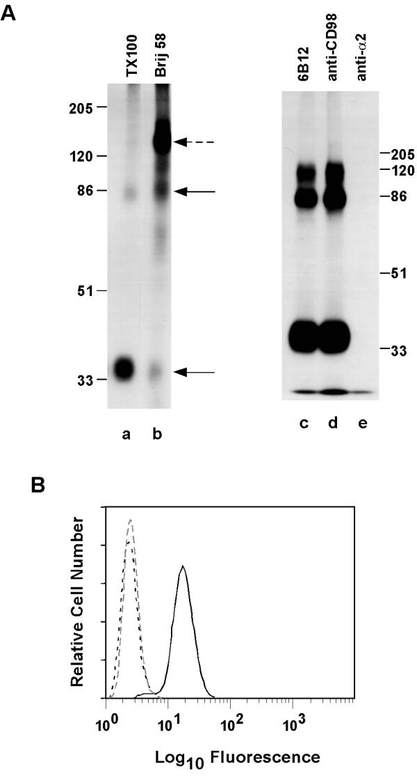
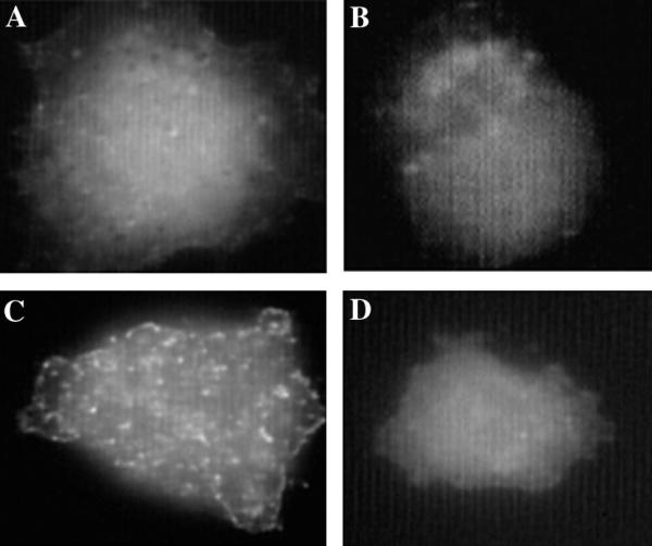
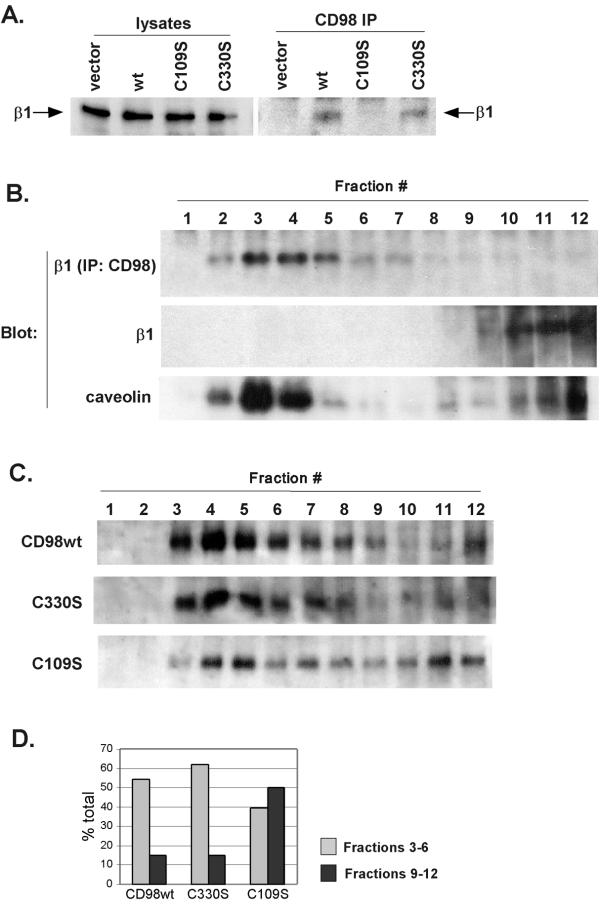
Results: We demonstrate that CD98 constitutively and specifically associates with beta1 integrins (alpha2beta1,alpha3beta1, alpha5beta1 and alpha6beta1), but minimally with alpha4beta1. Integrin-CD98 association was established by reciprocal immunoprecipitation experiments, and confirmed by CD98-induced clustering of alpha3beta1 but not alpha4beta1 on the surface of rhabdomyosarcoma cells. Integrin-CD98 association is independent of the alpha subunit cytoplasmic tail, is maintained in alpha3beta1 ligand-interaction deficient mutants, and is not inhibited by EDTA. Within the CD98 heavy chain, a C109S mutation (but not a C330S mutation) caused a loss of beta1 integrin association. The same C109S mutation also caused a loss of CD98 light chain association. Importantly, CD98 associated selectively with beta1 integrins present in low density "light membrane" fractions on a sucrose gradient. CD98 was not present in dense fractions that contained the majority of beta1 integrins. Notably, the C109S mutant of CD98, that did not associate with beta1 integrins, showed also a reduced localization into light membrane fractions.

Conclusions: We demonstrate that CD98 association with beta1 integrins is specific, occurs in the context of low density membranes, and may require the CD98 light chain.


Figures

Figure 1
Figure 1
Characterization of mAb 6B12. A. HT1080 cells were surface labeled with 125I, lysed in the indicated detergents (each at 1%), and then proteins were immunoprecipitated using mAb 6B12 (lanes a,b). Also, 125I-labeled Molt4 cells were lysed in 1% Triton XI 00, and labeled proteins were precipitated using mAb 6B12 (lane c), 4F2 (lane d), or anti-mAb A2-2E10 (lane e). Proteins were resolved on 10% SDS-PAGE under reduced conditions. B. Chinese hamster ovary cells transfected with CD98 cDNA (solid line), with β1 integrin cDNA (close dotted line), or mock transfected (dotted line) were each stained with mAb 6B12. Following FITC-labeled goat anti-mouse second antibody staining, flow cytometry was carried out using a FACScan Instrument (Becton Dickinson, Mountainview, CA) as previously described [30].
Figure 2
Figure 2
Selective co-precipitation of integrins with CD98. A. HT1080 cells were surface labeled with 125I, lysed in 1% Brij 58, then immunoprecipitated with anti-CD98 mAb 6B12. The CD98 immunoprecipitate was then dissociated using 1% Triton X-100 and reprecipitated with mAb to the indicated proteins. Numbers in parentheses above each lane indicate the mean fluorescence intensity of each cell surface antigen as determined by flow cytometry analysis. B. HT1080 cells were labeled overnight with 35S methionine, lysed in 1% Brij 58 then immunoprecipitated with mAb A3-IIF5. Immune complexes were dissociated using 1% Triton X100 and re-precipitated using anti-CD98 mAb 6B12 or anti-CD81 mAb M38. C. Lysates prepared as in Part A were analyzed by immunoprecipitation using mAbs A2-2E10, A3-IIF5, A5-PUJ2, and A6-BB respectively (left 4 lanes). Immunoprecipitation with mAb 6B12 was followed by elution with 1% Triton X100 and reprecipitation with A2-2E10, A3-IIF5, A5-PUJ2, and A6-BB mAb respectively (right 4 lanes). D. Cells transfected with wild type α3 or α3 integrin lacking a cytoplasmic tail (X3C0), were surface-labeled with 125I and lysed in 1% Brij 58. Lysates were immunoprecipitated with anti-α3 mAb A3-IIF5 (left 2 lanes) or first immunoprecipitated with mAb 6B12 then subjected to reprecipitation with mAb A3-IIF5 (right 2 lanes). E. Cells transfected with wild type α3 (lane a) or mutant α3 (W220A or Y218A) were cell surface biotinylated and lysed in 1% Brij 58. Lysates were immunoprecipitated with anti-α3 mAb A3-IIF5 (left 3 lanes) or first immunoprecipitated with mAb 4F2 and then subjected to reprecipitation with mAb A3-IIF5 (right 3 lanes).
Figure 3
Figure 3
Integrin clustering induced by CD98 cross-linking. RD A3 cells were grown on coverslips coated with fibronectin (20 μg/ml) for 48 hours. Cells were blocked with 20% goat serum and in some cases incubated with anti-CD98 mAb 4F2 (10 μg/ml) (C,D). Next, cells were washed, blocked with 20% goat serum and incubated with rabbit anti-mouse Ab (5 μg/ml). Finally, cells were fixed for 15 minutes with 2% paraformaldehyde, blocked with 20% goat serum and incubated with anti-α3 mAb A3X8 PUJ1 (10 μg/ml) (A, C) or anti-α4 mAb A4-PUJ1 (10 μg/ml) (B, D), each directly coupled with Alexa-488 (Molecular Probes). Coverslips were mounted with ProLong AntiFade Kit (Molecular Probes) and immunofluorescence examined using a Zeiss Axioscop equipped with optics for epifluorescence.
Figure 4
Figure 4
Correlation between CD98-integrin association and CD98 heavy chain-light chain dimerization. A. NIH 3T3 cells (107 cells/experiment) expressing wild type human CD98 heavy chain, CD98 heavy chain mutants (C109S or C330S) or control NIH 3T3 cells were lysed in 1% Brij 58 and then proteins were immunoprecipitated using 4F2 mAb. Cell lysates (left 4 lanes) and immunoprecipitates (right 4 lanes) were resolved in 8% SDS-PAGE, transferred to nitrocellulose membrane, probed with biotinylated KMI6 mAb (anti-mouse β1 integrin) followed by incubation with ExtrAvidin-HRP (Sigma) and developed using Renaissance Chemiluminescence Reagent (NEN Life Science Products, Boston, MA). B. HT1080 cells (107 cells/experiment) were lysed in sodium carbonate buffer, pH 11 and fractionated in isopycnic discontinuous sucrose gradient as described in Materials and Methods. Fractions were analyzed by SDS-PAGE electrophoresis followed by Western blot analysis using biotinylated TS2/16 mAb or polyclonal anti-caveolin antibody, or adjusted to pH 7.5 and immunoprecipitated with 4F2 mAb and then blotted with biotinylated TS2/16 mAb. C. NIH 3T3 cells (107 cells/experiment) expressing wild type human CD98 heavy chain, or mutant CD98 heavy chains (C109S or C330S) were cell surface biotinylated, lysed in sodium carbonate buffer, pH 11 and fractionated in sucrose gradients as described in Materials and Methods. Fractions were adjusted to pH 7.5 and immunoprecipitated using 4F2 mAb. Immunoprecipitated proteins were separated in 8% SDS-PAGE under non-reducing conditions and blotted onto nitrocellulose membrane. Blots were developed using ExtrAvidin-HRP (Sigma) and Renaissance Chemiluminescence Reagent (NEN Life Science Products, Boston, MA). For NIH3T3 cells, flow cytometry was used to demonstrate comparable levels of wild type CD98 (MFI = 27), C109S (MFI = 30), and C330S (MFI = 53) expression. Cells were 100% positive, with homogeneous peaks, well above background staining (MFI = 1). MFI=Mean Fluorescence Intensity. D. Relative distribution of CD98 wild type and C109S and C330S mutants in light (fractions 3–6) and dense (fractions 9–12) fractions from the Western blots shown in Fig. 4C. Densitometry was carried out using GeneGenius Bio Imaging System and analysis software, (Syngene, Frederick, MD). A typical experiment out of three performed is shown.

Similar articles

Cited by

References

    1. Haynes BF, Hemler ME, Mann DL, Eisenbarth GS, Shelhamer J, Mostowski HS, et al. Characterization of a monoclonal antibody (4F2) that binds to human monocytes and to a subset of activated lymphocytes. J Immunol. 1981;126:1409–1414. - PubMed
    1. Freidman AW, Diaz LAJ, Moore S, Schaller J, Fox DA. The human 4F2 antigen: evidence for cryptic and noncryptic epitopes and for a role of 4F2 in human T lymphocyte activation. Cell Immunol. 1994;154:253–263. doi: 10.1006/cimm.1994.1075. - DOI - PubMed
    1. Stonehouse TJ, Woodhead VE, Herridge PS, Ashrafian H, George M, Chain BM, et al. Molecular characterization of U937-dependent T-cell co-stimulation. Immunology. 1999;96:35–47. doi: 10.1046/j.1365-2567.1999.00670.x. - DOI - PMC - PubMed
    1. Diaz LAJ, Friedman AW, He X, Kuick RD, Hanash SM, Fox DA. Monocyte-dependent regulation of T lymphocyte activation through CD98. Int Immunol. 1997;9:1221–1231. doi: 10.1093/intimm/9.9.1221. - DOI - PubMed
    1. Tabata N, Ito M, Shimokata K, Suga S, Ohgimoto S, Tsurudome M, et al. Expression of fusion regulatory proteins (FRPs) on human peripheral blood monocytes. J Immunol. 1994;153:3256. - PubMed

Publication types

LinkOut - more resources