. 2001 Nov;159(5):1661-70.
doi: 10.1016/S0002-9440(10)63013-1.

In vivo imaging of physiological angiogenesis from immature to preovulatory ovarian follicles

Affiliations

In vivo imaging of physiological angiogenesis from immature to preovulatory ovarian follicles

B Vollmar et al. Am J Pathol. 2001 Nov.

Abstract

To develop a model for the study of physiological angiogenesis, we transplanted ovarian follicles onto striated muscle tissue and analyzed the process of microvascularization in vivo using repeated fluorescence microscopy. Follicles were mechanically isolated from unstimulated as well as pregnant mare's serum gonadotropin (PMSG)- or PMSG/luteinizing hormone (LH)-stimulated Syrian golden hamster ovaries and were transplanted as free grafts into dorsal skinfold chambers of untreated or synchronized hamsters. Follicles lacking thecal cell layers did not vascularize regardless whether harvested from unstimulated or PMSG-stimulated animals, but underwent granulosa cell apoptosis, as indicated in vivo by nuclear condensation and fragmentation of bisbenzimide-stained follicular tissue. In contrast, all follicles at 48 hours after PMSG treatment with a multilayered thecal shell exhibited initial signs of angiogenesis within 3 days. Vascularization was completed within 7 to 10 days, comprising a dense glomerulum-like microvascular network. Nature and extent of vascularization of follicles harvested at 72 hours after either PMSG or PMSG/LH treatment did not notably differ from each other when transplanted into the respective synchronized animals. However, follicles with PMSG/LH treatment revealed significantly larger microvessel diameters and higher capillary blood perfusion compared to follicles with sole PMSG treatment, probably reflecting the adaptation to the increased functional demand upon the LH surge. Using the unique experimental approach of ovarian follicle transplantation in the dorsal skinfold chamber of Syrian golden hamsters, we could show in vivo the developmental stage-dependent vascularization of follicular grafts with sustained potential to meet their metabolic demand by increased blood perfusion.


Figures
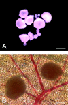
Figure 1.
Figure 1.
A: Follicles at different stages of development after mechanical microdissection in Dulbecco’s modified Eagle’s medium. A handpicking procedure guaranteed single connective tissue-free follicles for transplantation. B: Isolated follicles directly after transplantation into the hamster dorsal skinfold chamber. Scale bars: 800 μm (A), 400 μm (B).
Figure 2.
Figure 2.
H&E-stained cross-sections of formalin-fixed isolated follicular grafts. A: A tri- to quadrilaminar (arrow) follicle without any trace of thecal cell layers, indicating a secondary follicle of stage 3. B: A follicle of stage 6, exhibiting several layers of granulosa cells (arrow) with initiation of antral cavity formation. A well-developed multilayered thecal shell is clearly seen (double arrow). Scale bars: 20 μm (A), 50 μm (B).
Figure 3.
Figure 3.
Intravital fluorescence microscopic images of a follicular graft (borders are indicated by double arrows) directly after transplantation (A and B) as well as at day 5 (C) and day 10 (D) after transplantation into the hamster dorsal skinfold chamber. In contrast to the initial lack of nutritive capillaries within the freshly transplanted graft (A and B), on day 5 after transplantation (C) the newly formed microvessels begin to create a network of capillaries although substantial parts of the follicular graft still lack vascularization (asterisks). At day 10 after transplantation (D) the follicular graft exhibits a complete and fully developed glomerulum-like microvasculature (asterisk) that is supplied by feeding arterioles (arrows) and drained by a postcapillary venule (arrowhead). A, C, and D: Blue light epi-illumination with contrast enhancement by 5% FITC-labeled dextran 150,000 i.v. B: Ultraviolet epi-illumination of bisbenzimide-stained follicular tissue. Scale bars, 100 μm.
Figure 4.
Figure 4.
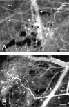
Intravital fluorescence microscopic images of the microvasculature of follicular grafts at day 14 after transplantation into hamster dorsal skinfold chambers. A: High magnification reveals the interaction of the newly formed microvessels with the microvasculature of the host tissue, demonstrating an arteriole (white arrows) that serves as vascular supply and multiple intercapillary anastomoses (black arrows) between the follicular capillaries (asterisk) and the striated muscle capillaries (arrowheads). B: Blood from the follicular graft is almost completely drained by a postcapillary vessel (arrowheads), which may function as a venule, but represents a former striated muscle capillary, as clearly indicated by the parallel arrangement with the other striated muscle capillaries (arrows). Blue light epi-illumination with contrast enhancement by 5% FITC-labeled dextran 150,000 i.v. Scale bars: 100 μm (A), 150 μm (B).
Figure 5.
Figure 5.
Quantification of vascularized area (%) (A) and microvessel density (cm/cm2) (B) of follicles after free transplantation into hamster dorsal skinfold chambers, as assessed by intravital fluorescence microscopy and computer-assisted image analysis. Follicles with diameters of 250 to 500 μm (filled squares) and diameters >500 μm (open squares) were harvested from hamsters at 48 hours after PMSG treatment and transplanted into synchronized animals. Follicles exhibiting diameters <250 μm, which were harvested from unstimulated animals or from animals at 48 hours after PMSG treatment and transplanted into unstimulated or synchronized animals, did not vascularize and are not displayed because of log-scaling of the y axis. Values are given as geometric means and 95% confidence intervals, which are based on a linear mixed model of log-transformed data. a, P < 0.05 versus day 3; b, P < 0.05 versus days 3 and 5; #, P < 0.05 versus follicles with diameters of 250 to 500 μm.
Figure 6.
Figure 6.
Quantification of vascularized area (%) (A) and microvessel density (cm/cm2) (B) of follicles after free transplantation into hamster dorsal skinfold chambers, as assessed by intravital fluorescence microscopy and computer-assisted image analysis. Follicles with diameters >500 μm were harvested from hamsters at 72 hours after either PMSG (open circles) or PMSG/LH treatment (open triangles) and transplanted into synchronized animals. Values are given as geometric means and 95% confidence intervals, which are based on a linear mixed model of log-transformed data. a, P < 0.05 versus day 3.
Figure 7.
Figure 7.
Intravital fluorescence microscopic images of a follicular microvascular network at day 14 after transplantation. Using green light epi-illumination with visualization of rhodamine 6G-stained white blood cells (A), signs of inflammatory reactions of the host tissue could not be found, as indicated by only few leukocytes (arrows, A) interacting with the endothelium of follicular capillaries (asterisk, B) and draining venules (arrowheads, B). Moreover, blue light epi-illumination with contrast enhancement using 5% FITC-labeled dextran 150,000 i.v. revealed absence of macromolecular leakage and edema formation (B). Scale bars, 200 μm.
Figure 8.
Figure 8.
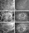
Intravital fluorescence microscopic images of follicles at day 7 after transplantation into hamster dorsal skinfold chambers. Note the dense microvascular network of the follicle (A, asterisk) with absence of cells displaying apoptotic chromatin condensation (B), whereas the follicle with only peripheral vascularization (C, asterisk) exhibits a small fraction of apoptotic cells (D, arrow). Lack of vascularization (asterisk) of a small follicle (<250 μm, follicular border is outlined, E) is associated with a marked incidence of cell apoptosis (F, arrows), supposedly indicating atretic regression of the graft. Intravital fluorescence microscopy with blue light epi-illumination and contrast enhancement using 5% FITC-labeled dextran 150,000 i.v. (A, C, E) and ultraviolet epi-illumination of bisbenzimide-stained follicles (B, D, F; asterisks indicate follicular oocytes). Scale bars: 50 μm (A–D), 30 μm (E and F).

References

    1. Folkman J: Tumor angiogenesis. Adv Cancer Res 1985, 43:174-203 - PubMed
    1. Folkman J: Angiogenesis in cancer, vascular, rheumatoid and other disease. Nat Med 1995, 1:27-31 - PubMed
    1. Greenburg GB, Hunt TK: Proliferative response in vitro of vascular endothelial and smooth muscle cells exposed to wound fluids and macrophages. J Cell Physiol 1978, 97:353-360 - PubMed
    1. Polverini PJ, Cotran PS, Gimbrone MA, Unanue ER: Activated macrophages induce vascular proliferation. Nature 1977, 269:804-806 - PubMed
    1. Banda MJ, Knighton DR, Hunt TK, Werb Z: Isolation of a nonmitogenic angiogenic factor from wound fluid. Proc Natl Acad Sci USA 1982, 79:7773-7777 - PMC - PubMed

Publication types

LinkOut - more resources