. 2001 Nov;159(5):1711-21.
doi: 10.1016/s0002-9440(10)63018-0.

Anti-inflammatory and chondroprotective effect of TSG-6 (tumor necrosis factor-alpha-stimulated gene-6) in murine models of experimental arthritis

Affiliations

Anti-inflammatory and chondroprotective effect of TSG-6 (tumor necrosis factor-alpha-stimulated gene-6) in murine models of experimental arthritis

T Bárdos et al. Am J Pathol. 2001 Nov.

Erratum in

  • Am J Pathol 2002 Mar;160(3):1193

Abstract

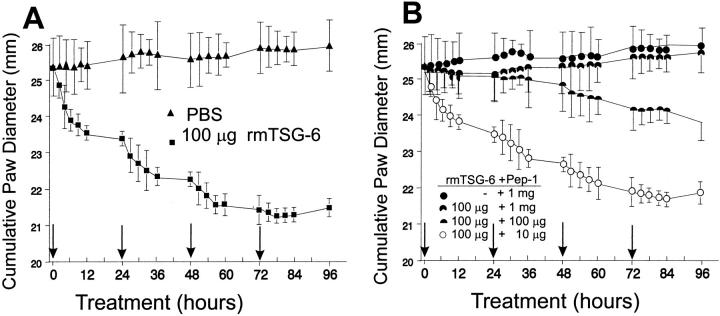
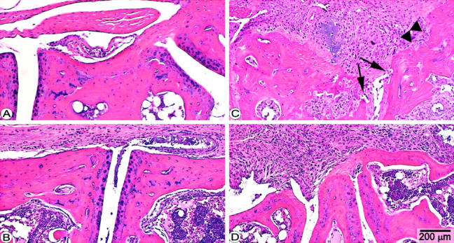
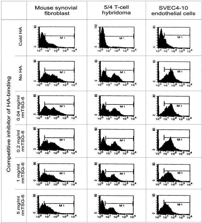
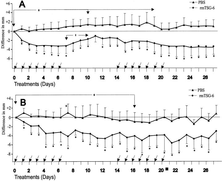
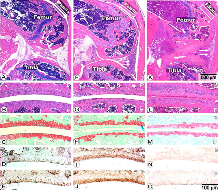
Tumor necrosis factor-alpha (TNF-alpha)-stimulated gene-6 (TSG-6) is up-regulated by various cytokines and growth factors. TSG-6 binds to hyaluronan in inflamed synovial tissue and forms a complex with a serine protease inter-alpha-trypsin inhibitor (IalphaI), increasing the protease inhibitory effect of IalphaI >100-fold. The TSG-6/IalphaI complex then blocks serine proteases, including the plasminogen-plasmin activation, probably the most important component in the activation processes of matrix metalloproteinases. To gain insight into the mechanisms of TSG-6 action in arthritis, we have used an autoimmune murine model (proteoglycan-induced arthritis) for systemic, and a monoarticular form of arthritis (antigen-induced arthritis) for local treatment of arthritis with recombinant mouse TSG-6 (rmTSG-6). Intravenous injection of rmTSG-6 induced a dramatic reduction of edema in acutely inflamed joints by immobilizing CD44-bound hyaluronan and, in long-term treatment, protected cartilage from degradation and blocked subchondral and periosteal bone erosion in inflamed joints. The intra-articular injection of a single dose (100 microg) of rmTSG-6 exhibited a strong chondroprotective effect for up to 5 to 7 days, preventing cartilage proteoglycan from metalloproteinase-induced degradation. In contrast, rmTSG-6 did not postpone the onset, nor reduce the incidence of arthritis. We were unable to detect any significant differences between control and rmTSG-6-treated animals when various serum markers (including pro- and anti-inflammatory cytokines, auto- and heteroantibody productions) or antigen-specific T-cell responses were compared, nor when the expressions of numerous cell surface receptors or adhesion molecules were measured. TSG-6 seems to play a critical negative regulatory feed-back function in inflammation, especially in arthritic processes.


Figures

Figure 1.
Figure 1.
Short-term effect of rmTSG-6 on joint swelling in PGIA mice. Animals were randomly grouped 3 to 7 days after the onset of arthritis (onset of swelling and redness) and each experimental group contained a comparable number of inflamed front and hind paws with approximately the same cumulative clinical scores of 8.2 to 8.4 ± 1.2 to 1.4 corresponding to 24.9 to 25.6 ± 1.1 to 1.3 mm cumulative joint diameter. Both PBS-injected (control; filled triangle) and 100 μg of rmTSG-6-injected (filled square) groups contained 16 animals in four independent experiments. Treatments with HA-binding Pep-1 (1 mg) (filled circle) or co-treatment of Pep-1 (10 μg to 1 mg) and rmTSG-6 (100 μg) (symbols of dose combinations are shown in figure) were repeated twice (a total of seven mice in each group). All injections were given intravenously in 100 μl of PBS (arrows). Joints were measured before each injection (early morning) and then every 2 to 3 hours after injections. Ankles and wrists were measured in frontal and sagittal planes, and the cumulative joint thickness of the four paws (eight measurements per animal) are shown.
Figure 2.
Figure 2.
Histopathology of tarsal joints of mice with PGIA. Panels represent sagittal sections of tarsal joints of a noninflamed paw (A) or sections of arthritic tarsal joints from PGIA mice 4 days (B) or 4 weeks after the onset of arthritis (C and D). Arthritic mice were treated with PBS (C) or with rmTSG-6 (B and D). Massive inflammation and synovial cell proliferation were characteristic of both PBS-treated (C) and TSG-6-treated (D) mice, but the articular cartilage remained histologically intact after 4 weeks in TSG-6-treated mice (D) (animals of Figure 3 ▶ ). In contrast, articular cartilage was essentially lost (arrows) from the joint surface and massive bone erosion was also present in nontreated (PBS-injected) mice (C). H&E staining; original magnifications, ×25.
Figure 3.
Figure 3.
Flow cytometry (fluorescence-activated cell sorting) analysis of competition assays for determining the ability of rmTSG-6 to compete with CD44 for HA binding. Mouse synovial fibroblasts and SVEC4-10 endothelial cells were digested with streptomyces hyaluronidase for 30 minutes at 37°C to remove endogenous HA and then cells were washed with PBS. All three cell lines (1 × 10⁶) were incubated with fluorescein-labeled HA (20 μg/ml; 100 μl final volume) and rmTSG-6 (0.04 to 5 mg/ml) or 100 μl of unlabeled HA (5 mg/ml) for 2 hours at 37°C. Cells were washed in PBS and analyzed by fluorescence-activated cell sorting. The y axis shows the cell number and the x axis the fluorescence intensity.
Figure 4.
Figure 4.
Long-term effect of repeated treatment with rmTSG-6 on PGIA mice. Arthritic animals were injected intravenously with 100 μl of PBS (n = 14) or 100 μg of rmTSG-6 (n = 15). Each mouse was injected every 24 hours for 7 days as indicated by arrows. Joints were measured before the injections and then twice per day. The treatment and cessation of therapy was repeated for another 14 days. Ankles and wrists were measured in frontal and sagittal planes and the change in cumulative joint thickness of the four paws (eight measurements) are shown. A shows the difference of cumulative joint thickness in mm between the PBS-injected and rmTSG-6-treated group (all paws), whereas B shows the differences of the inflamed joint thickness only (ie, paws were inflamed at the beginning of the TSG-6 treatment). The decrease of joint swelling in the rmTSG-6-treated group was significant within 24 hours (as shown in Figure 1 ▶ ), and the increased joint swelling in the PBS-injected group became significant (*, P < 0.05) throughout the first 10 to 12 days. Changes in joint swelling between PBS-injected and rmTSG-6-treated groups was statistically significant (*, P < 0.05 or smaller) from day 1 of the treatment and remained significant (comparing to day 0) throughout the entire experimental period.
Figure 5.
Figure 5.
Histology and immunohistochemistry of knee sections from C57BL/6j mice on day 5 of AIA. A–E represent sections from PBS-injected knees, F–J shows sections of knee joints injected intra-articularly with mBSA, and K–O are sections from knee joints injected with mBSA and rmTSG-6 together. A, B, F, G, K, and L are H&E-stained sections. White arrows in F and K show the cellular infiltration. Sections of C, H, and M are stained with safranin-O. D, I, and N are sections stained with antibody to -VDIPEN neoepitope (stromelysin-cleaved product of cartilage aggrecan). E, J, and O are sections stained with antibody to aggrecanase-generated -NITEGE neoepitope. Patella (P) and femoral condyle (F) are indicated. Scale bar on K indicates the scale for A, F, and K, whereas scale bar in O indicates scale for all other panels.

References

    1. Lee TH, Lee GW, Ziff EB, Vilcek J: Isolation and characterization of eight tumor necrosis factor-induced gene sequences from human fibroblasts. Mol Cell Biol 1990, 10:1982-1988 - PMC - PubMed
    1. Lee TH, Wisniewski H-G, Vilcek J: A novel secretory tumor necrosis factor-inducible protein (TSG-6) is a member of the family of hyaluronate binding proteins, closely related to the adhesion receptor CD44. J Cell Biol 1992, 116:545-557 - PMC - PubMed
    1. Wisniewski H-G, Maier R, Lotz M, Lee S, Klampfer L, Lee TH, Vilcek J: TSG-6: a TNF-, IL-1-, and LPS-inducible secreted glycoprotein associated with arthritis. J Immunol 1993, 151:6593-6601 - PubMed
    1. Fülöp C, Kamath RV, Li Y, Otto JM, Salustri A, Olsen BR, Glant TT, Hascall VC: Coding sequence, exon-intron structure and chromosomal localization of murine TNF-stimulated gene 6 that is specifically expressed by expanding cumulus cell-oocyte complexes. Gene 1997, 202:95-102 - PubMed
    1. Bayliss MT, Howat SLT, Dudhia J, Murphy JM, Barry FP, Edwards JCW, Day AJ: Up-regulation and differential expression of the hyaluronan-binding protein TSG-6 in cartilage and synovium in rheumatoid arthritis and osteoarthritis. Osteoarthr Cart 2001, 9:42-48 - PubMed

Publication types

MeSH terms