. 2001 Nov 12;155(4):637-48.
doi: 10.1083/jcb.200105081. Epub 2001 Nov 5.

alpha-Toxin is a mediator of Staphylococcus aureus-induced cell death and activates caspases via the intrinsic death pathway independently of death receptor signaling

Affiliations

alpha-Toxin is a mediator of Staphylococcus aureus-induced cell death and activates caspases via the intrinsic death pathway independently of death receptor signaling

H Bantel et al. J Cell Biol. .

Abstract

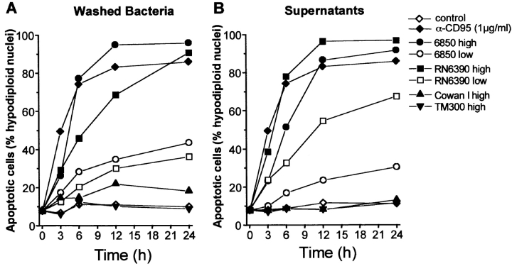
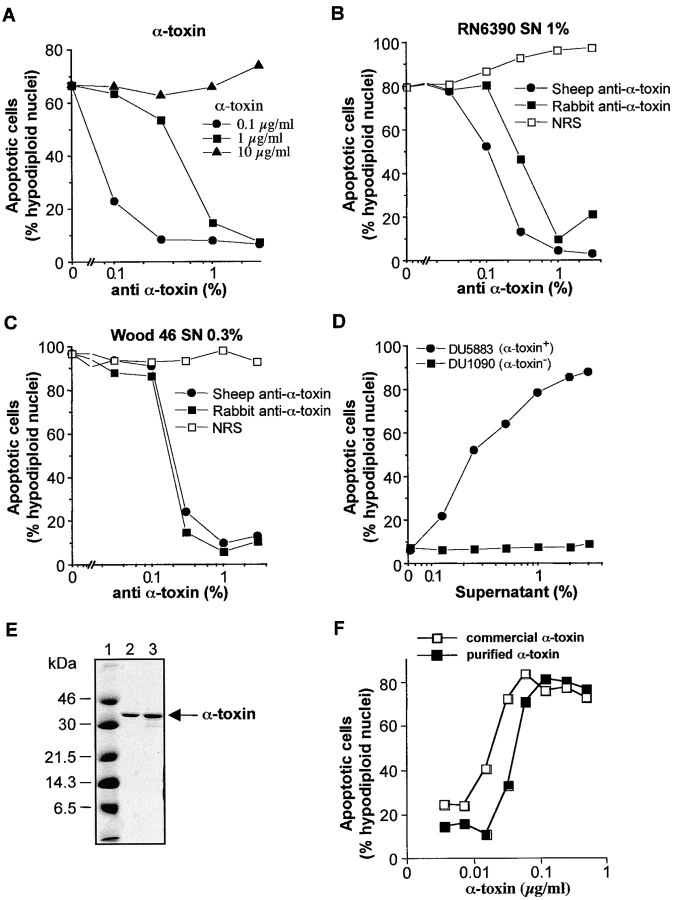
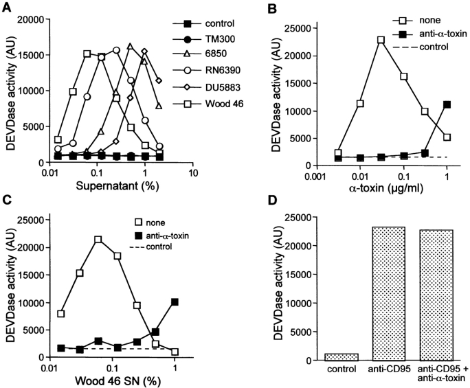
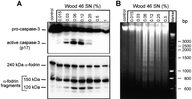
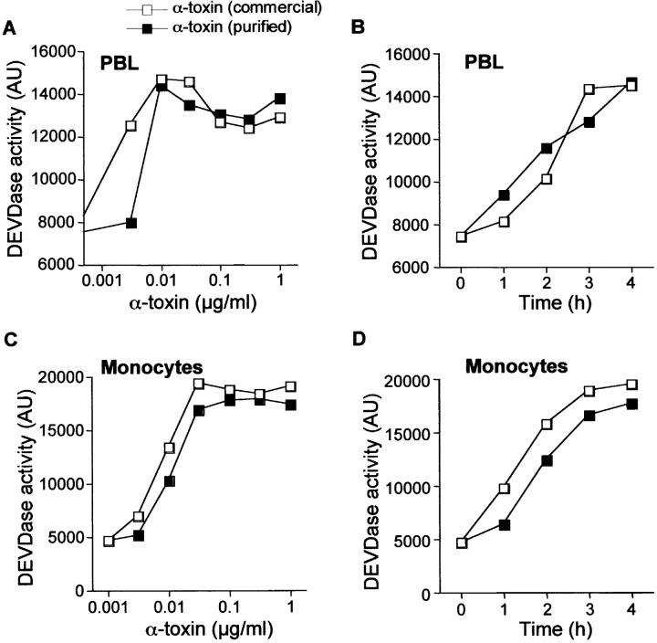
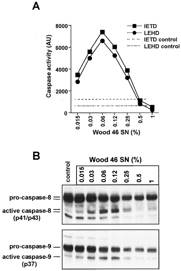
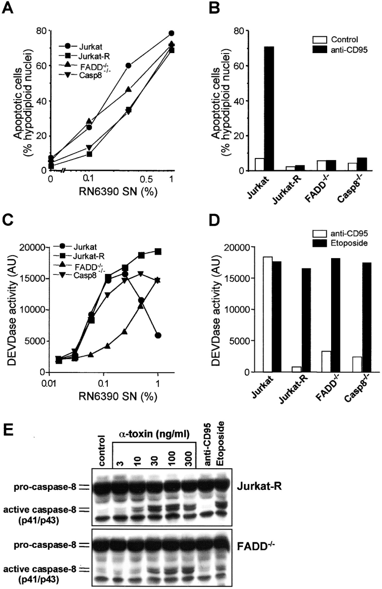
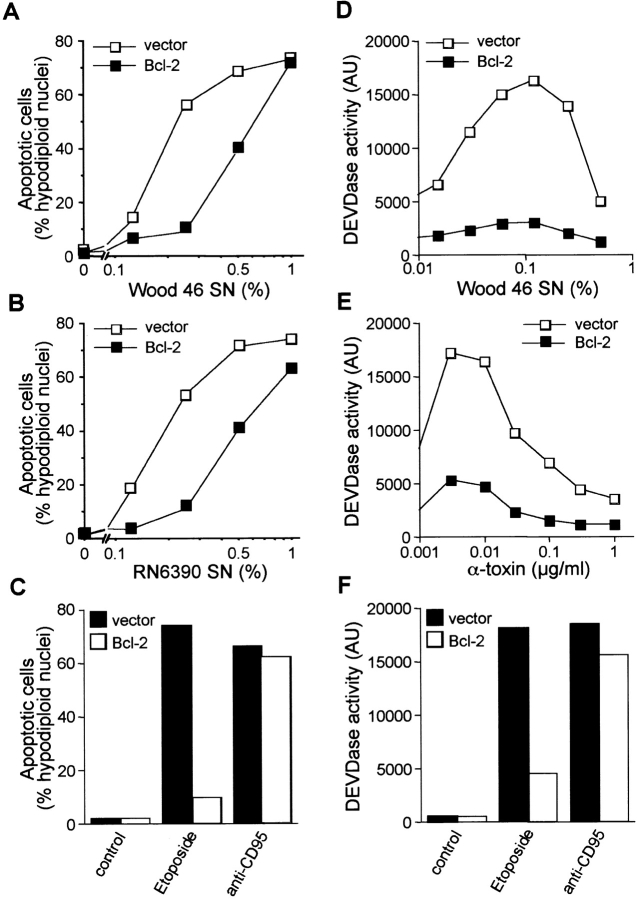
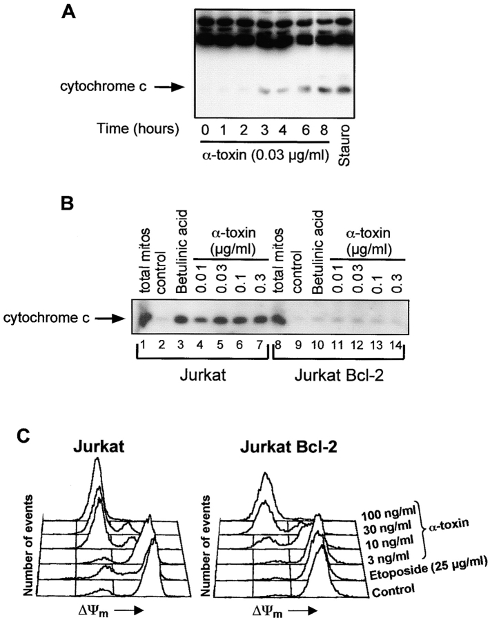
Infections with Staphylococcus aureus, a common inducer of septic and toxic shock, often result in tissue damage and death of various cell types. Although S. aureus was suggested to induce apoptosis, the underlying signal transduction pathways remained elusive. We show that caspase activation and DNA fragmentation were induced not only when Jurkat T cells were infected with intact bacteria, but also after treatment with supernatants of various S. aureus strains. We also demonstrate that S. aureus-induced cell death and caspase activation were mediated by alpha-toxin, a major cytotoxin of S. aureus, since both events were abrogated by two different anti-alpha-toxin antibodies and could not be induced with supernatants of an alpha-toxin-deficient S. aureus strain. Furthermore, alpha-toxin-induced caspase activation in CD95-resistant Jurkat sublines lacking CD95, Fas-activated death domain, or caspase-8 but not in cells stably expressing the antiapoptotic protein Bcl-2. Together with our finding that alpha-toxin induces cytochrome c release in intact cells and, interestingly, also from isolated mitochondria in a Bcl-2-controlled manner, our results demonstrate that S. aureus alpha-toxin triggers caspase activation via the intrinsic death pathway independently of death receptors. Hence, our findings clearly define a signaling pathway used in S. aureus-induced cytotoxicity and may provide a molecular rationale for future therapeutic interventions in bacterial infections.


Figures

Figure 1.
Figure 1.
Both intact S. aureus cells and bacterial supernatants induce T cell apoptosis. Jurkat cells were incubated with live washed bacteria (A) or sterile-filtered supernatants of the same bacterial cultures (B). After the indicated times, the proportion of apoptotic cells was determined by flow cytometry. (A) Fresh suspensions of the indicated bacterial strains were added to Jurkat cells, resulting in a MOI of 30 (low) and 120 (high). Cells were incubated on ice for 2 h to allow sedimentation and then shifted to 37°C. Lysostaphin (20 μg/ml) was added to lyse and kill staphylococci. Lysostaphin without bacteria served as a negative control, whereas agonistic anti-CD95 was used as a positive control. (B) Fresh bacterial supernatants were added to Jurkat cells, resulting in a final concentration (vol/vol) of 0.1% (low) and 1% (high).
Figure 2.
Figure 2.
S. aureusα-toxin is required for induction of apoptosis. (A) Effect of purified α-toxin. Jurkat cells were preincubated for 30 min with various dilutions of a sheep anti–α-toxin antiserum and then incubated with the indicated concentrations of the commercially available preparation of α-toxin. (B and C) Anti–α-toxin neutralizes the proapoptotic activity of S. aureus supernatants. Jurkat cells were preincubated with various dilutions of a sheep anti–α-toxin antiserum, a rabbit anti–α-toxin antiserum, or normal rabbit serum (NRS) for 30 min. Subsequently, sterile-filtered supernatants of RN6390 (B) or Wood 46 (C) were added at the indicated dilutions, and formation of hypodiploid nuclei was assessed. (D) α-Toxin–deficient S. aureus does not induce apoptosis. Jurkat cells were incubated with various concentrations of supernatants of the α-toxin–producing strain DU5883 or its α-toxin–deficient counterpart DU1090. (E) Coomassie-stained SDS-PAGE of a highly purified (lane 2) and the commercially available (lane 3) α-toxin preparation. The molecular sizes of the protein marker used in lane 1 are indicated on the left. (F) Jurkat cells were incubated with the indicated concentrations of the two α-toxin preparations, and cell death was assessed after 24 h.
Figure 3.
Figure 3.
S. aureus–induced caspase-3 activation is mediated by α-toxin. (A) DEVDase activity in extracts of Jurkat cells incubated for 4 h with the indicated supernatant dilutions of the various S. aureus strains. (B–D) DEVDase activity in extracts of Jurkat cells incubated for 4 h with the indicated concentrations of α-toxin (B), Wood 46 supernatant (SN) (C), or the agonistic anti-CD95 antibody (D) in the absence or presence of the neutralizing sheep anti– α-toxin antiserum (1:100). The dashed line (control) represents the DEVDase activity in untreated cells.
Figure 4.
Figure 4.
S. aureus–induced caspase-3 activation results in α-fodrin cleavage and DNA fragmentation. (A) Western blot analyses demonstrating caspase-3 processing (top) and α-fodrin cleavage (bottom) in Jurkat cells that were either left untreated (control) or incubated for 4 h with the indicated dilutions of Wood 46 supernatant (SN). (B) Agarose gel electrophoretic separation of DNA extracted from Jurkat cells that were either left untreated (control) or incubated for 6 h with the indicated dilutions of Wood 46 supernatant (SN). The positions of the DNA molecular size marker are indicated on the right.
Figure 5.
Figure 5.
S. aureusα-toxin induces caspase-3 activation in human PBL and monocytes. DEVDase activity in extracts of PBL (A and B) and monocytes (C and D). Cells were either incubated for 4 h with the indicated α-toxin concentrations (A and C) or with 30 ng/ml α-toxin for the indicated times (B and D).
Figure 6.
Figure 6.
S. aureus induces activation of the initiator caspases 8 and 9. (A) Jurkat cells were incubated with the indicated dilutions of Wood 46 supernatant (SN). After 4 h, cell extracts were prepared and analyzed for caspase activity using the fluorogenic substrates IETD AMC for caspase 8 (▪) and LEHD AMC for caspase 9 (•). The two dashed lines represent IETDase and LEHDase activity in untreated cells. (B) Western blot analysis of the status of caspase-8 (top) and caspase-9 (bottom) in Jurkat cells that were either left untreated (control) or incubated for 4 h with the indicated dilutions of Wood 46 supernatant (SN). The uncleaved proforms and the p43/p41 fragments of caspase-8 and the p37 fragment of caspase-9 are indicated.
Figure 7.
Figure 7.
S. aureusα-toxin signals cell death and caspase activation independently of death receptor pathways. Assessment of cell death (A and B) and DEVDase activity (C and D) in Jurkat cells (•), Jurkat-R cells (▪), FADD-deficient Jurkat cells (▴), and caspase-8–deficient Jurkat cells (▾). Cells were incubated with increasing concentrations of RN6390 supernatant (A and C), anti-CD95 (B and D), or etoposide (D). (E) Western blot analyses demonstrating the activation of caspase-8 in Jurkat-R cells (top) and FADD-deficient cells (bottom) that were either left untreated (control) or incubated for 4 h with the indicated concentrations of α-toxin. As a control, cells were treated for 4 h with anti-CD95 or etoposide. The blots show the uncleaved precursor forms and the p43/p41 intermediate fragments of caspase-8.
Figure 8.
Figure 8.
S. aureus mediates cell death and caspase activation via the Bcl-2–controlled mitochondrial death pathway. Assessment of cell death (A and B) and caspase-3–like activity (D and E) in Jurkat vector control cells (□) or cells overexpressing Bcl-2 (▪). Cells were incubated with increasing concentrations of Wood 46 supernatant (A and D), RN6390 supernatant (B), or α-toxin (E). Assessment of etoposide- or CD95-induced apoptosis (C) and DEVDase activity (F) in vector cells (filled bars) and Bcl-2 cells (open bars) served as controls. Cell death and DEVDase activity were determined after 24 and 4 h, respectively.
Figure 9.
Figure 9.
S. aureusα-toxin induces cytochrome c release in intact cells and isolated mitochondria in a Bcl-2–controlled manner. (A) Western blot analysis of cytosolic cytochrome c in Jurkat cells treated for the indicated times with 0.03 μg/ml α-toxin or for 8 h with 1 μg/ml staurosporine (Stauro). (B) Effect of α-toxin on isolated mitochondria. Isolated mitochondria from Jurkat (lanes 1–7) or from Bcl-2–overexpressing Jurkat cells (lanes 8–14) were incubated for 30 min with a buffer control (lanes 2 and 9), 10 μg/ml betulinic acid (lanes 3 and 10), or the indicated concentrations of α-toxin (lanes 4–7 and 11–14). The release of cytochrome c was detected by immunoblotting. Lanes 1 and 8 (total mitos) represent the complete amount of cytochrome c present in mitochondria from Jurkat and Jurkat Bcl-2 cells, respectively. (C) Measurement of the mitochondrial transmembrane potential (ΔΨm) in untreated (Control) Jurkat and Jurkat Bcl-2 cells or in cells treated for 4 h with etoposide or the indicated concentrations of α-toxin.

References

    1. Balwit, J.M., P. van Langevelde, J.M. Vann, and R.A. Proctor. 1994. Gentamicin-resistant menadione and hemin auxotrophic Staphylococcus aureus persist within cultured endothelial cells. J. Infect. Dis. 170:1033–1037. - PubMed
    1. Baran, J., K. Weglarczyk, M. Mysiak, K. Guzik, M. Ernst, H.D. Flad, and J. Pryjma. 2001. Fas (CD95)-Fas ligand interactions are responsible for monocyte apoptosis occurring as a result of phagocytosis and killing of Staphylococcus aureus. Infect. Immun. 69:1287–1297. - PMC - PubMed
    1. Bayles, K.W., C.A. Wesson, L.E. Liou, L.K. Fox, G.A. Bohach, and W.R. Trumble. 1998. Intracellular Staphylococcus aureus escapes the endosome and induces apoptosis in epithelial cells. Infect. Immun. 66:336–342. - PMC - PubMed
    1. Bossy-Wetzel, E., D.D. Newmeyer, and D.R. Green. 1998. Mitochondrial cytochrome c release in apoptosis occurs upstream of DEVD-specific caspase activation and independently of mitochondrial transmembrane depolarization. EMBO J. 17:37–49. - PMC - PubMed
    1. Boya, P., B. Roques, and G. Kroemer. 2001. Viral and bacterial proteins regulating apoptosis at the mitochondrial level. EMBO J. 20:4325–4331. - PMC - PubMed

Publication types

MeSH terms