

Monocyte/macrophage expression of ABCA1 has minimal contribution to plasma HDL levels
- PMID: 11696576
- PMCID: PMC209438
- DOI: 10.1172/JCI12810
Monocyte/macrophage expression of ABCA1 has minimal contribution to plasma HDL levels
Abstract
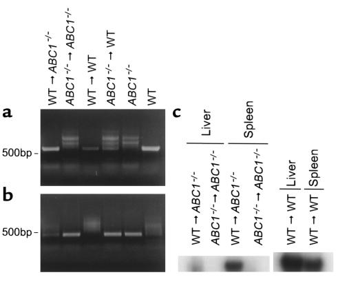
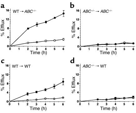
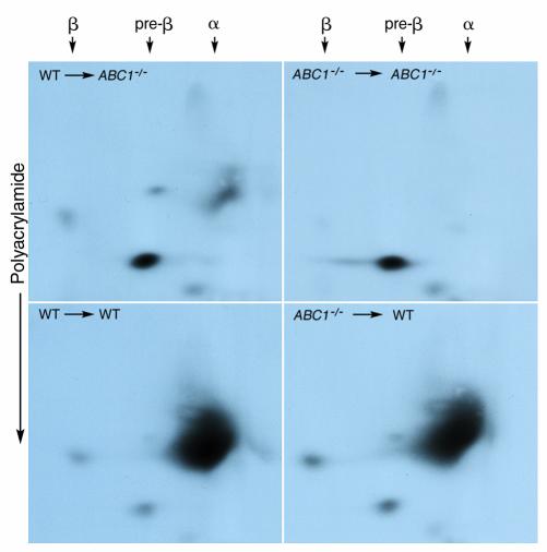
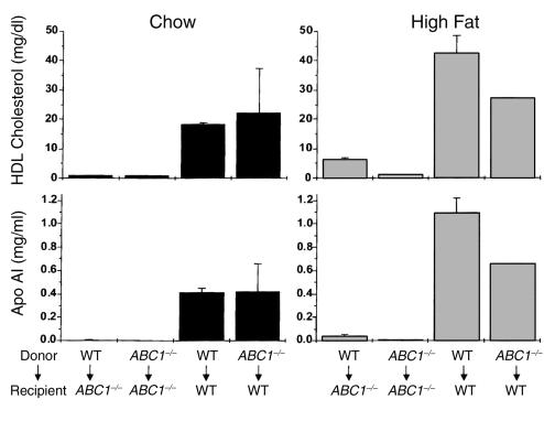
Excess accumulation of cholesterol in macrophages results in foam cell production and lesion development. Recent studies have demonstrated that ATP-binding cassette protein A1 (ABCA1) is highly regulated in macrophages and mediates the efflux of cholesterol and phospholipids to apolipoproteins, a process necessary for HDL formation. The goal of this study was to determine the contribution of monocyte/macrophage ABCA1 to HDL formation in vivo. We generated mice expressing ABCA1 in macrophages and mice with selected inactivation of ABCA1 in macrophages by bone marrow transplantation in ABCA1-deficient (ABC1(-/-)) and wild-type (WT) mice. At all times, the level of HDL in ABC1(-/-) recipient mice remained low relative to WT recipient mice irrespective of the genotype of the donor macrophage ABCA1 or high-fat feeding. Expression of WT macrophage ABCA1 in ABC1(-/-) mice resulted in a small but significant increase in apoA-I levels starting 2 weeks after transplantation. No further increase in apoAI was observed up to 14 weeks after transplantation. The increase in apoAI was accompanied by a small but significant increase in HDL cholesterol 6 weeks after transplantation. The HDL formed as a consequence of the expression of WT macrophage ABCA1 migrated to the alpha position in a two-dimensional gel electrophoresis. These results demonstrate that monocyte/macrophage ABCA1 contributes to HDL formation; however, the contribution to the overall plasma HDL levels is minimal.
Figures
Comment in
-
Is it time to modify the reverse cholesterol transport model?J Clin Invest. 2001 Nov;108(9):1273-5. doi: 10.1172/JCI14342. J Clin Invest. 2001. PMID: 11696569 Free PMC article. No abstract available.
Similar articles
-
HDL particle size is a critical determinant of ABCA1-mediated macrophage cellular cholesterol export.Circ Res. 2015 Mar 27;116(7):1133-42. doi: 10.1161/CIRCRESAHA.116.305485. Epub 2015 Jan 14. Circ Res. 2015. PMID: 25589556
-
Targeted inactivation of hepatic Abca1 causes profound hypoalphalipoproteinemia and kidney hypercatabolism of apoA-I.J Clin Invest. 2005 May;115(5):1333-42. doi: 10.1172/JCI23915. Epub 2005 Apr 7. J Clin Invest. 2005. PMID: 15841208 Free PMC article.
-
Inactivation of macrophage scavenger receptor class B type I promotes atherosclerotic lesion development in apolipoprotein E-deficient mice.Circulation. 2003 Nov 4;108(18):2258-63. doi: 10.1161/01.CIR.0000093189.97429.9D. Epub 2003 Oct 27. Circulation. 2003. PMID: 14581413
-
ABCA1-mediated transport of cellular cholesterol and phospholipids to HDL apolipoproteins.Curr Opin Lipidol. 2000 Jun;11(3):253-60. doi: 10.1097/00041433-200006000-00005. Curr Opin Lipidol. 2000. PMID: 10882340 Review.
-
Lipid efflux by the ATP-binding cassette transporters ABCA1 and ABCG1.Biochim Biophys Acta. 2006 Jul;1761(7):655-66. doi: 10.1016/j.bbalip.2006.04.012. Epub 2006 May 4. Biochim Biophys Acta. 2006. PMID: 16798073 Review.
Cited by
-
ATP binding cassette transporter A1--key roles in cellular lipid transport and atherosclerosis.Mol Cell Biochem. 2002 Aug;237(1-2):155-64. doi: 10.1023/a:1016506221047. Mol Cell Biochem. 2002. PMID: 12236582 Review.
-
Promoter-specific roles for liver X receptor/corepressor complexes in the regulation of ABCA1 and SREBP1 gene expression.Mol Cell Biol. 2003 Aug;23(16):5780-9. doi: 10.1128/MCB.23.16.5780-5789.2003. Mol Cell Biol. 2003. PMID: 12897148 Free PMC article.
-
Liver-X-receptor-mediated increase in ATP-binding cassette transporter A1 expression is attenuated by fatty acids in CaCo-2 cells: effect on cholesterol efflux to high-density lipoprotein.Biochem J. 2004 Feb 1;377(Pt 3):545-52. doi: 10.1042/BJ20030903. Biochem J. 2004. PMID: 14604434 Free PMC article.
-
TNF-alpha decreases ABCA1 expression and attenuates HDL cholesterol efflux in the human intestinal cell line Caco-2.J Lipid Res. 2010 Jun;51(6):1407-15. doi: 10.1194/jlr.M002410. Epub 2010 Jan 26. J Lipid Res. 2010. PMID: 20103810 Free PMC article.
-
Macrophage cholesterol efflux correlates with lipoprotein subclass distribution and risk of obstructive coronary artery disease in patients undergoing coronary angiography.Lipids Health Dis. 2009 Apr 6;8:14. doi: 10.1186/1476-511X-8-14. Lipids Health Dis. 2009. PMID: 19348677 Free PMC article.
References
-
- Tall AR, Blum CB, Forester GP, Nelson CA. Changes in the distribution and composition of plasma high density lipoproteins after ingestion of fat. J Biol Chem. 1982;257:198–207. - PubMed
-
- Rothblat GH, et al. Cell cholesterol efflux: integration of old and new observations provides new insights. J Lipid Res. 1999;40:781–796. - PubMed
-
- Ji Y, et al. Scavenger receptor BI promotes high density lipoprotein-mediated cellular cholesterol efflux. J Biol Chem. 1997;272:20982–20985. - PubMed
