. 2001 Nov 5;194(9):1195-205.
doi: 10.1084/jem.194.9.1195.

Combined allogeneic tumor cell vaccination and systemic interleukin 12 prevents mammary carcinogenesis in HER-2/neu transgenic mice

Affiliations

Combined allogeneic tumor cell vaccination and systemic interleukin 12 prevents mammary carcinogenesis in HER-2/neu transgenic mice

P Nanni et al. J Exp Med. .

Abstract

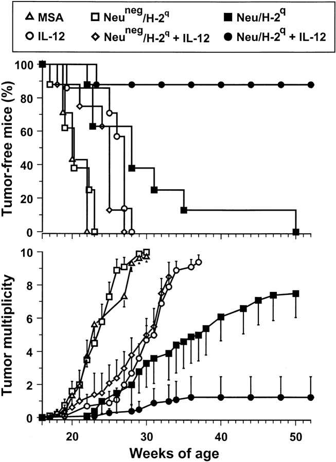
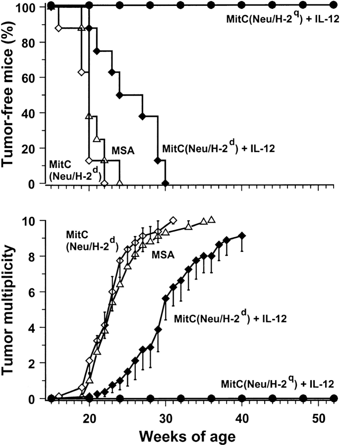
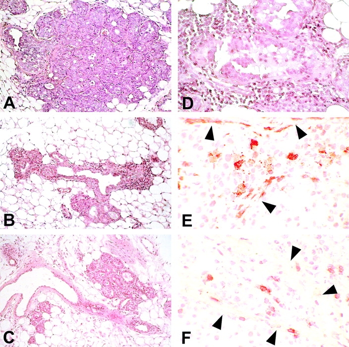
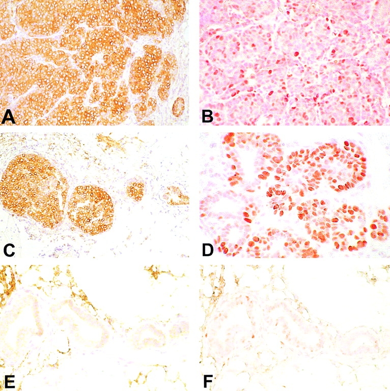
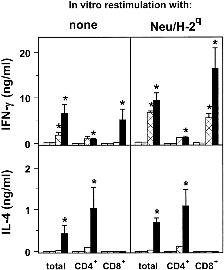
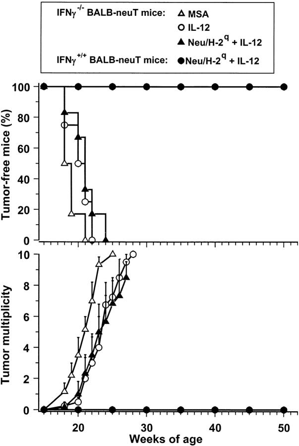
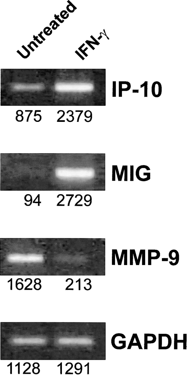
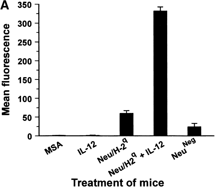
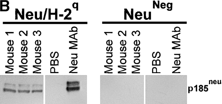
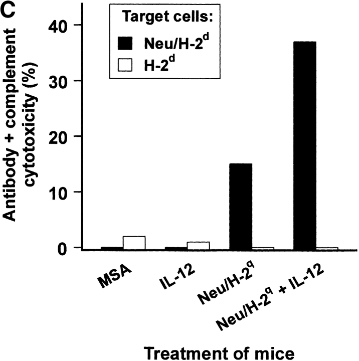
Transgenic Balb/c mice expressing the transforming rat HER-2/neu oncogene develop early and multifocal mammary carcinomas. Within the first 5 months of life the tissue-specific expression of HER-2/neu causes a progression in all their 10 mammary glands from atypical hyperplasia to invasive carcinoma. It was previously observed that chronic administration of interleukin (IL)-12 increased tumor latency, but every mouse eventually succumbed to multiple carcinomas. A significant improvement in tumor prevention was sought by administering allogeneic mammary carcinoma cells expressing HER-2/neu combined with systemic IL-12. This treatment reduced tumor incidence by 90% and more than doubled mouse lifetime. For the maximum prevention p185(neu) antigen must be expressed by allogeneic cells. IL-12 treatment strongly increased the cell vaccine efficacy. The mammary glands of mice receiving the combined treatment displayed a markedly reduced epithelial cell proliferation, angiogenesis, and HER-2/neu expression, while the few hyperplastic foci were heavily infiltrated by granulocytes, macrophages, and CD8(+) lymphocytes. Specific anti-HER-2/neu antibodies were produced and a nonpolarized activation of CD4(+) and CD8(+) cells secreting IL-4 and interferon (IFN)-gamma were evident. A central role for IFN-gamma in the preventive effect was proven by the lack of efficacy of vaccination in IFN-gamma gene knockout HER-2/neu transgenic Balb/c mice. A possible requirement for IFN-gamma is related to its effect on antibody production, in particular on IgG2a and IgG2b subclasses, that were not induced in IFN-gamma knockout HER-2/neu mice. In conclusion, our data show that an allogeneic HER-2/neu-expressing cell vaccine combined with IL-12 systemic treatment can prevent the onset of genetically determined tumors.


Figures

Figure 1.
Figure 1.
Cytofluorometric analysis of HER-2/neu and MHC class I glycoprotein expression in Neu/H-2q cells. Open profiles represent cells stained with secondary antibody alone; solid profiles represent cells incubated with the indicated antibodies. In each panel, the ordinate represents the number of cells. Data from an experiment representative of at least three similar experiments are shown.
Figure 2.
Figure 2.
Inhibition of mammary carcinogenesis in Balb-neuT female mice. Groups of 7–8 mice received the indicated treatments. Tumor-free survival curve of mice treated with Neu/H-2q cells plus IL-12 is significantly different from all other curves (P < 0.005 at least by the Mantel-Haenszel test); tumor-free survival curves of mice treated with IL-12 or Neu/H-2q cells are significantly different (P < 0.05 at least) from MSA controls. Tumor multiplicity is calculated as the cumulative number of incident tumors/total number of mice and is shown as mean ± SEM.
Figure 3.
Figure 3.
Inhibition of mammary carcinogenesis in Balb-neuT mice with MitC-treated allogeneic Neu/H-2q cells plus IL-12. Groups of eight mice received the indicated treatments. Tumor-free survival curves are significantly different (P < 0.0005 by the Mantel-Haenszel test). Tumor multiplicity is calculated as the cumulative number of incident tumors/total number of mice and is shown as mean ± SEM.
Figure 4.
Figure 4.
Morphologic and immunohistochemical analysis of mammary glands. Histology shows that at week 15 the mammary tissue of control mice (A) contains numerous foci of atypical hyperplasia with areas of lobular carcinoma in situ; the hyperplastic and neoplastic epithelial cells occupy and fill out all the ductal-lobular structures. The hyperplastic foci found in mice receiving IL-12 (B) or Neu/H-2q cells plus IL-12 (C) are less numerous and prosperous than in control mice and surrounded by an evident reactive infiltrate. At 27 wk in mice treated with Neu/H-2q cells plus IL-12 the few surviving foci of hyperplasia (D) are surrounded by an evident infiltrate composed of reactive cells that sometimes are found within epithelial cells after crossing or damaging the basal membrane. Immunohistochemistry reveals that granulocytes (E) and CD8+ lymphocytes (F) are present not only in the stroma but also inside the lobular structure after crossing the damaged basal membrane (arrowheads). A–C, original magnification: ×200; D, original magnification: ×400; E and F, original magnification: ×630.
Figure 5.
Figure 5.
Mammary tissue from untreated 27-wk-old mice (A and B), mice receiving IL-12 (C and D), or Neu/H-2q cells plus IL-12 (E and F). Immunohistochemistry with anti-p185neu antibody reveals that all the neoplastic cells express the p185neu in the cytoplasm and on their membrane (A). A similar p185neu expression pattern is evident in the mammary epithelial cells of the carcinoma in situ present in the IL-12–treated mouse (C). The cytoplasmic and membrane expression of the p185neu is associated with a marked positivity of PCNA (B and D). The mammary glands of mice treated with Neu/H-2q cells plus IL-12 are mainly composed of ductules lined by a single layer of epithelial cells without or with a slight p185neu expression mainly confined in the epithelial cell cytoplasm (E); the epithelial proliferation rate was low as assessed by PCNA positivity (F). A–F, original magnification: ×400.
Figure 6.
Figure 6.
Release of cytokines by spleen cells restimulated or not in vitro with Neu/H-2q cells. Spleen cells from untreated control mice (white bar); from mice treated with IL-12 (gray bar); from mice treated with Neu/H-2q cells (cross-hatched bar); and from mice treated with Neu/H-2q cells plus IL-12 (black bar). Asterisks denote a significant difference (P < 0.05 at least by Student's t test) with untreated controls. Mean ± SEM of groups of 3–5 Balb-neuT mice.
Figure 7.
Figure 7.
Lack of inhibition of mammary carcinogenesis in IFN-γ knockout Balb-neuT female mice. Transgenic mice received the indicated treatments. Tumor multiplicity was calculated as the cumulative number of incident tumors/total number of mice and is shown as mean ± SEM. Groups of 7–8 mice.
Figure 8.
Figure 8.
mRNA expression of angiogenic and antiangiogenic molecules determined by reverse transcriptase PCR in Neu/H-2d mammary carcinoma cells treated in vitro with IFN-γ. GAPDH, 20 cycles; MIG, 35 cycles; IP-10, 28 cycles; MMP-9, 28 cycles. Densitometric analysis was performed and values reported under each band.
Figure 9.
Figure 9.
Antibody production by Balb-neuT mice. (A) Cytofluorometric analysis of serum binding to Neu/H-2q cells. Each bar represents the mean ± SEM of sera (diluted 1:65) from three mice at the fifteenth week of age bled after the second course of the indicated treatment. (B) Neu/H-2q and Neuneg/H-2q cell extracts were immunoprecipitated by sera from three mice treated with Neu/H-2q cells plus IL-12, PBS as negative control, and anti–rat p185neu monoclonal antibody 7.16.4 as positive control (Neu mAb). Western blot analysis was performed using the antineu polyclonal antibody C18. (C) Complement dependent cytotoxicity against syngeneic Neu/H-2d cells and Balb-neuT lymphocytes, which do not express p185neu (H-2d). The percentage of viable lymphocytes was determined after a 30-min incubation with sera of mice bled after the second course of treatment (diluted 1:10) followed by a 30-min incubation with rabbit low-tox complement (diluted 1:10). Results are expressed as 100 − % viable lymphocytes.
Figure 9.
Figure 9.
Antibody production by Balb-neuT mice. (A) Cytofluorometric analysis of serum binding to Neu/H-2q cells. Each bar represents the mean ± SEM of sera (diluted 1:65) from three mice at the fifteenth week of age bled after the second course of the indicated treatment. (B) Neu/H-2q and Neuneg/H-2q cell extracts were immunoprecipitated by sera from three mice treated with Neu/H-2q cells plus IL-12, PBS as negative control, and anti–rat p185neu monoclonal antibody 7.16.4 as positive control (Neu mAb). Western blot analysis was performed using the antineu polyclonal antibody C18. (C) Complement dependent cytotoxicity against syngeneic Neu/H-2d cells and Balb-neuT lymphocytes, which do not express p185neu (H-2d). The percentage of viable lymphocytes was determined after a 30-min incubation with sera of mice bled after the second course of treatment (diluted 1:10) followed by a 30-min incubation with rabbit low-tox complement (diluted 1:10). Results are expressed as 100 − % viable lymphocytes.
Figure 10.
Figure 10.
Subclasses of antibodies induced in Balb-neuT mice and in IFN-γ knockout Balb-neuT mice. Cytofluorometric analysis of serum binding to Neu/H-2q cells with secondary anti–mouse isotype antibodies is reported. Each bar represents the mean ± SEM of sera (diluted 1:65) from three mice at the fifteenth week of age bled after the second course of the indicated treatment.

Similar articles

Cited by

References

    1. Noguchi, Y., A. Jungbluth, E.C. Richards, and L.J. Old. 1996. Effect of interleukin 12 on tumor induction by 3-methylcholanthrene. Proc. Natl. Acad. Sci. USA. 93:11798–11801. - PMC - PubMed
    1. Boggio, K., G. Nicoletti, E. Di Carlo, F. Cavallo, L. Landuzzi, C. Melani, M. Giovarelli, I. Rossi, P. Nanni, C. De Giovanni, et al. 1998. Interleukin 12-mediated prevention of spontaneous mammary adenocarcinomas in two lines of Her-2/neu transgenic mice. J. Exp. Med. 188:589–596. - PMC - PubMed
    1. Granziero, L., S. Krajewski, P. Farness, L. Yuan, M.K. Courtney, M.R. Jackson, P.A. Peterson, and A. Vitiello. 1999. Adoptive immunotherapy prevents prostate cancer in a transgenic animal model. Eur. J. Immunol. 29:1127–1138. - PubMed
    1. Cefai, D., B.W. Morrison, A. Sckell, L. Favre, M. Balli, M. Leunig, and C.D. Gimmi. 1999. Targeting HER-2/neu for active-specific immunotherapy in a mouse model of spontaneous breast cancer. Int. J. Cancer. 83:393–400. - PubMed
    1. Amici, A., F.M. Venanzi, and A. Concetti. 1998. Genetic immunization against neu/erbB2 transgenic breast cancer. Cancer Immunol. Immunother. 47:183–190. - PMC - PubMed

Publication types

MeSH terms