. 2001 Nov 5;194(9):1299-311.
doi: 10.1084/jem.194.9.1299.

The human immunodeficiency virus type 1 accessory protein Vpu induces apoptosis by suppressing the nuclear factor kappaB-dependent expression of antiapoptotic factors

Affiliations

The human immunodeficiency virus type 1 accessory protein Vpu induces apoptosis by suppressing the nuclear factor kappaB-dependent expression of antiapoptotic factors

H Akari et al. J Exp Med. .

Abstract

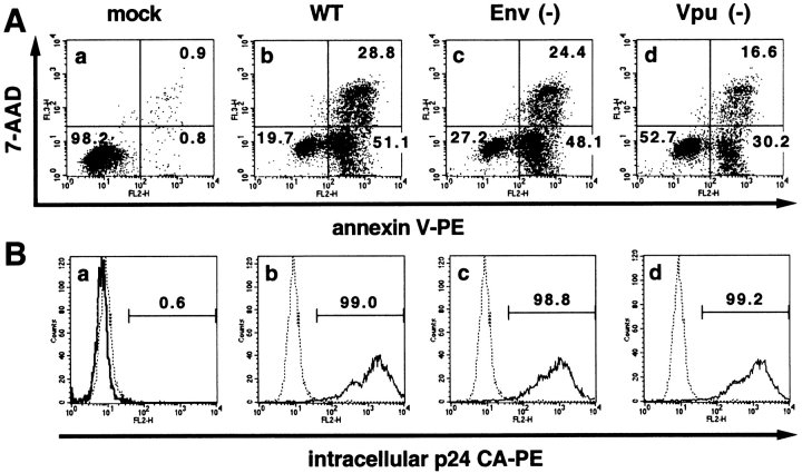
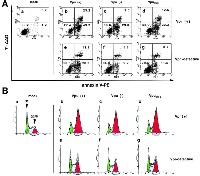
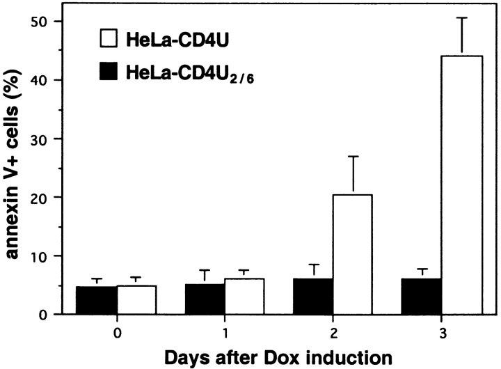
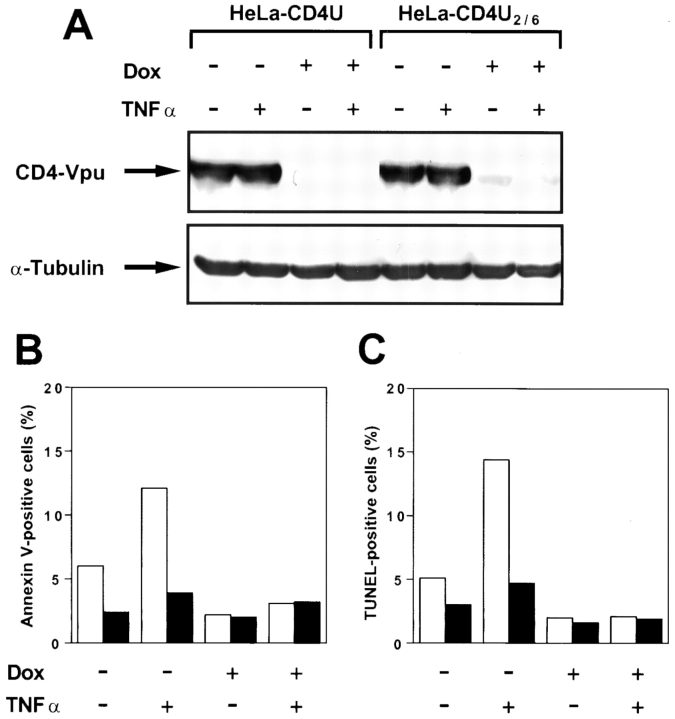
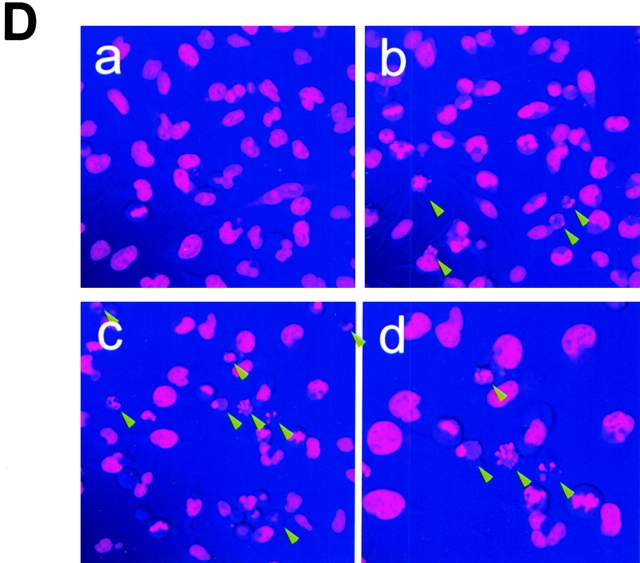
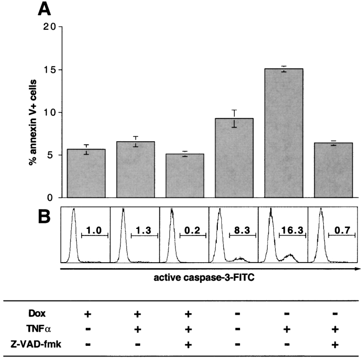
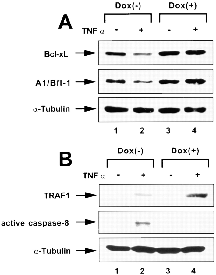
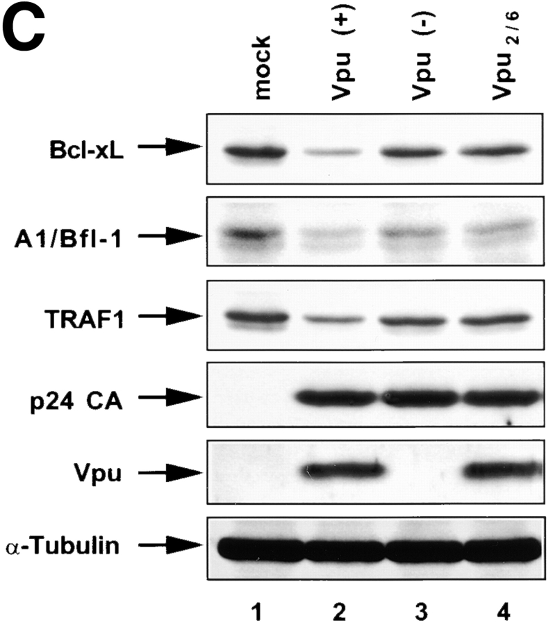
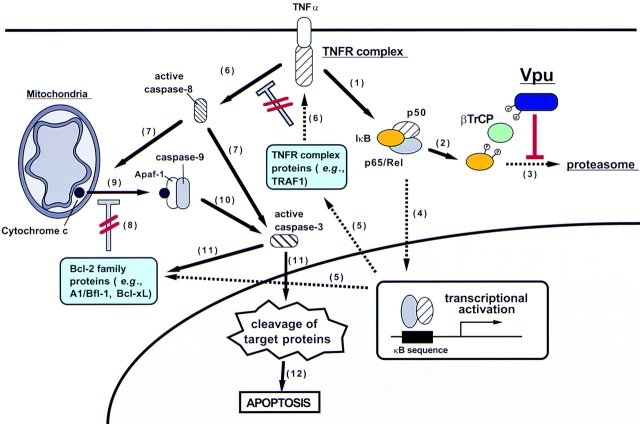
Human immunodeficiency virus (HIV) type 1 Vpu is an integral membrane protein with a unique affinity for betaTrCP (TrCP), a key member of the SkpI-Cullin-F-box E3 ubiquitin ligase complex that is involved in the regulated degradation of cellular proteins, including IkappaB. Remarkably, Vpu is resistant to TrCP-mediated degradation and competitively inhibits TrCP-dependent degradation of IkappaB, resulting in the suppression of nuclear factor (NF)-kappaB activity in Vpu-expressing cells. We now report that Vpu, through its interaction with TrCP, potently contributes to the induction of apoptosis in HIV-infected T cells. Vpu-induced apoptosis is specific and independent of other viral proteins. Mutation of a TrCP-binding motif in Vpu abolishes its apoptogenic property, demonstrating a close correlation between this property of Vpu and its ability to inhibit NF-kappaB activity. The involvement of NF-kappaB in Vpu-induced apoptosis is further supported by the finding that the levels of antiapoptotic factors Bcl-xL, A1/Bfl-1, and TNF receptor-associated factor (TRAF)1, all of which are expressed in an NF-kappaB-dependent manner, are reduced and, at the same time, levels of active caspase-3 are elevated. Thus, Vpu induces apoptosis through activation of the caspase pathway by way of inhibiting the NF-kappaB-dependent expression of antiapoptotic genes.


Figures

Figure 1.
Figure 1.
Vpu induces apoptosis in HIV-1–infected Jurkat cells. Uninfected Jurkat cells (a) or Jurkat cells infected with an m.o.i. of 5 with VSV-G-pseudotyped virus stocks of (b) wild-type (WT) NL4–3, (c) NL43-K1 (Env), or (d) NL4–3/Udel (Vpu) variants were used for this analysis. (A) Cultures were analyzed 48 h after infection for the presence of apoptotic cells by staining with 7-AAD and PE-conjugated annexin V, followed by flow cytometric analysis. Numbers represent the percentages of cells in the respective quadrants. (B) The same cultures were evaluated 24 h after infection for HIV-1 infection by intracellular p24 staining using PE-conjugated mouse mAb to HIV-1 p24 followed by flow cytometry. Numbers represent the percentages of p24-positive cells. The dotted lines in panels a–d represent p24-staining of mock-infected cells. The solid lines in panels a–d represent p24-staining of infected cells. The results shown are representative of three independent experiments.
Figure 2.
Figure 2.
Vpu-induced apoptosis is independent of HIV-1 Env or Vpr and requires a TrCP-binding motif. Jurkat cells were (a) mock-infected or infected with an m.o.i. of 5 with VSV-G-pseudotyped virus stocks of (b) NL43-K1 (Env), (c) NL43-K1/Udel (Env, Vpu), (d) NL43-K1/U2/6 (Env, Vpu2/6), (e) NL43-EcK1 (Env, Vpr), (f) NL43-EcK1/Udel (Env, Vpr, Vpu), or (g) NL43-EcK1/U2/6 (Env, Vpr, Vpu2/6). (A) Cultures were analyzed 48 h after infection for apoptotic cells as in Fig. 1 A. (B) The same cultures were examined 24 h after infection for their cell cycle status by propidium iodide staining followed by flow cytometry. Similar results were obtained from three independent experiments.
Figure 3.
Figure 3.
Expression of CD4U but not CD4U2/6 causes spontaneous apoptosis in HeLa cells. Inducible HeLa-CD4U and CD4U2/6 cell lines were cultured after the removal of Dox for the times indicated. The cells were then evaluated for induction of apoptosis by staining with PE-conjugated annexin V, followed by flow cytometry. Error bars reflect SDs from three independent experiments.
Figure 4.
Figure 4.
TNF-α treatment amplifies CD4U-induced apoptosis. CD4U and CD4U2/6 cell lines were cultured in complete DMEM medium in the presence or absence of Dox for 24 h TNF-α (20 ng/ml) was then added to the samples as indicated and cultures were incubated for an additional 16 h before analysis. (A) The expression of CD4U and CD4U2/6 (top) and α-tubulin (bottom) was determined by immunoblot analysis using a rabbit anti-Vpu polyclonal antibody (U2–3) and a mouse anti–α-tubulin mAb, respectively. (B and C) The cells were evaluated for induction of apoptosis by annexin V assay (B) or TUNEL assay (C), followed by flow cytometry. (D) Cultures were examined for apoptosis-related morphological changes of the nuclei by staining with PI, followed by confocal microscopic analysis. Panel a: Dox+/TNF-α2; panel b: Dox/TNF-α2; panels c and d: Dox/TNF-α1 by low and high power magnification, respectively. Arrowheads mark cells containing pyknotic apoptotic bodies.
Figure 4.
Figure 4.
TNF-α treatment amplifies CD4U-induced apoptosis. CD4U and CD4U2/6 cell lines were cultured in complete DMEM medium in the presence or absence of Dox for 24 h TNF-α (20 ng/ml) was then added to the samples as indicated and cultures were incubated for an additional 16 h before analysis. (A) The expression of CD4U and CD4U2/6 (top) and α-tubulin (bottom) was determined by immunoblot analysis using a rabbit anti-Vpu polyclonal antibody (U2–3) and a mouse anti–α-tubulin mAb, respectively. (B and C) The cells were evaluated for induction of apoptosis by annexin V assay (B) or TUNEL assay (C), followed by flow cytometry. (D) Cultures were examined for apoptosis-related morphological changes of the nuclei by staining with PI, followed by confocal microscopic analysis. Panel a: Dox+/TNF-α2; panel b: Dox/TNF-α2; panels c and d: Dox/TNF-α1 by low and high power magnification, respectively. Arrowheads mark cells containing pyknotic apoptotic bodies.
Figure 5.
Figure 5.
Vpu-induced apoptosis involves activation of the caspase pathway. CD4U and CD4U2/6 cell lines were cultured in complete DMEM medium in the presence or absence of Dox for 24 h. TNF-α (20 ng/ml) and z-VAD-fmk (50 μM) were then added where indicated and the cultures were incubated for an additional 16 h before analysis. (A) Cultures were evaluated for induction of apoptosis by annexin V staining. Error bars reflect SDs from three independent experiments. (B) The same cultures were analyzed by flow cytometry for the expression of the active form of caspase-3 using a FITC-conjugated rabbit antiactive caspase-3 polyclonal antibody. The numbers indicate percentages of FITC-positive cells.
Figure 6.
Figure 6.
Vpu affects the expression of antiapoptotic factors and induces caspase-8 activation. (A and B) HeLa-CD4U cells were cultured in complete DMEM medium in the presence or absence of Dox for 24 h. TNF-α (20 ng/ml) was then added to the cultures as indicated and incubation was continued for an additional 16 h. Cell lysates were analyzed by immunoblotting for the expression of Bcl-xL and A1/Bfl-1 (A) as well as TRAF1 and the active form of caspase-8 (B). Lysates were normalized for tubulin using an α-tubulin antibody. (C) Jurkat cells were single-cycle infected with VSV-G-pseudotyped NL43-K1, NL43-K1/Udel, or NL43-K1/U2/6. Cell lysates were analyzed 40 h after infection by immunoblotting to detect expression of Bcl-xL, A1/Bfl-1, TRAF1, p24 CA, Vpu, or α-tubulin.
Figure 6.
Figure 6.
Vpu affects the expression of antiapoptotic factors and induces caspase-8 activation. (A and B) HeLa-CD4U cells were cultured in complete DMEM medium in the presence or absence of Dox for 24 h. TNF-α (20 ng/ml) was then added to the cultures as indicated and incubation was continued for an additional 16 h. Cell lysates were analyzed by immunoblotting for the expression of Bcl-xL and A1/Bfl-1 (A) as well as TRAF1 and the active form of caspase-8 (B). Lysates were normalized for tubulin using an α-tubulin antibody. (C) Jurkat cells were single-cycle infected with VSV-G-pseudotyped NL43-K1, NL43-K1/Udel, or NL43-K1/U2/6. Cell lysates were analyzed 40 h after infection by immunoblotting to detect expression of Bcl-xL, A1/Bfl-1, TRAF1, p24 CA, Vpu, or α-tubulin.
Figure 7.
Figure 7.
Model for Vpu-induced apoptosis through activation of the caspase pathway. Details of the model are explained in the Discussion. Broken arrows symbolize inhibitory effects. Steps inhibited by Vpu are marked in red.

References

    1. Badley, A.D., A.A. Pilon, A. Landay, and D.H. Lynch. 2000. Mechanisms of HIV-associated lymphocyte apoptosis. Blood. 96:2951–2964. - PubMed
    1. Stewart, S.A., B. Poon, J.B. Jowett, and I.S. Chen. 1997. Human immunodeficiency virus type 1 Vpr induces apoptosis following cell cycle arrest. J. Virol. 71:5579–5592. - PMC - PubMed
    1. Yao, X.J., A.J. Mouland, R.A. Subbramanian, J. Forget, N. Rougeau, D. Bergeron, and E.A. Cohen. 1998. Vpr stimulates viral expression and induces cell killing in human immunodeficiency virus type 1-infected dividing Jurkat T cells. J. Virol. 72:4686–4693. - PMC - PubMed
    1. Hrimech, M., X.J. Yao, F. Bachand, N. Rougeau, and E.A. Cohen. 1999. Human immunodeficiency virus type 1 (HIV-1) Vpr functions as an immediate-early protein during HIV-1 infection. J. Virol. 73:4101–4109. - PMC - PubMed
    1. Strebel, K., T. Klimkait, F. Maldarelli, and M.A. Martin. 1989. Molecular and biochemical analyses of human immunodeficiency virus type 1 vpu protein. J. Virol. 63:3784–3791. - PMC - PubMed

Publication types

MeSH terms

Substances